. 2020 Oct 20;11(10):885.
doi: 10.1038/s41419-020-03103-7.

Selective knockdown of hexokinase 2 in rods leads to age-related photoreceptor degeneration and retinal metabolic remodeling

Affiliations

Selective knockdown of hexokinase 2 in rods leads to age-related photoreceptor degeneration and retinal metabolic remodeling

Rui Zhang et al. Cell Death Dis. .

Abstract

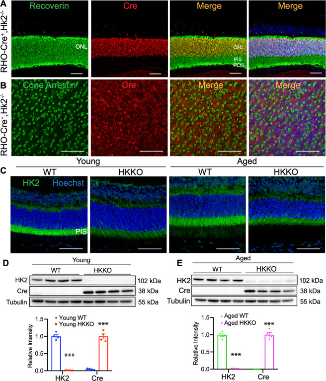
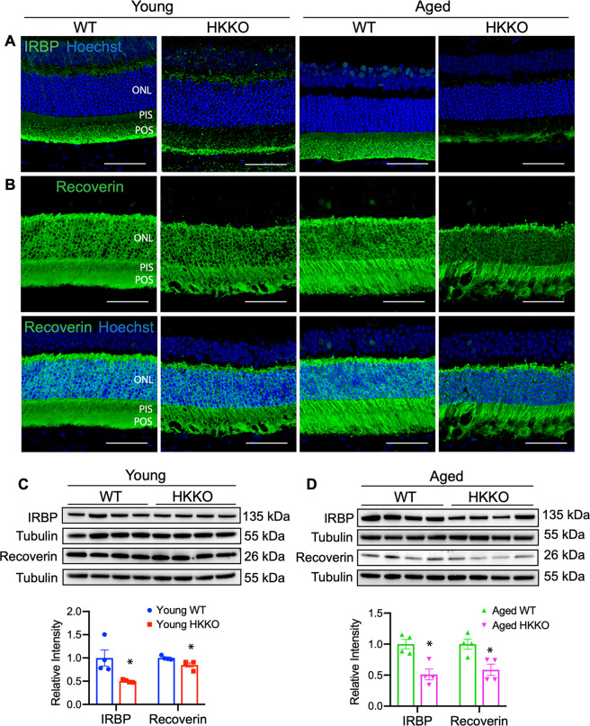
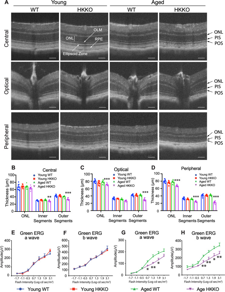
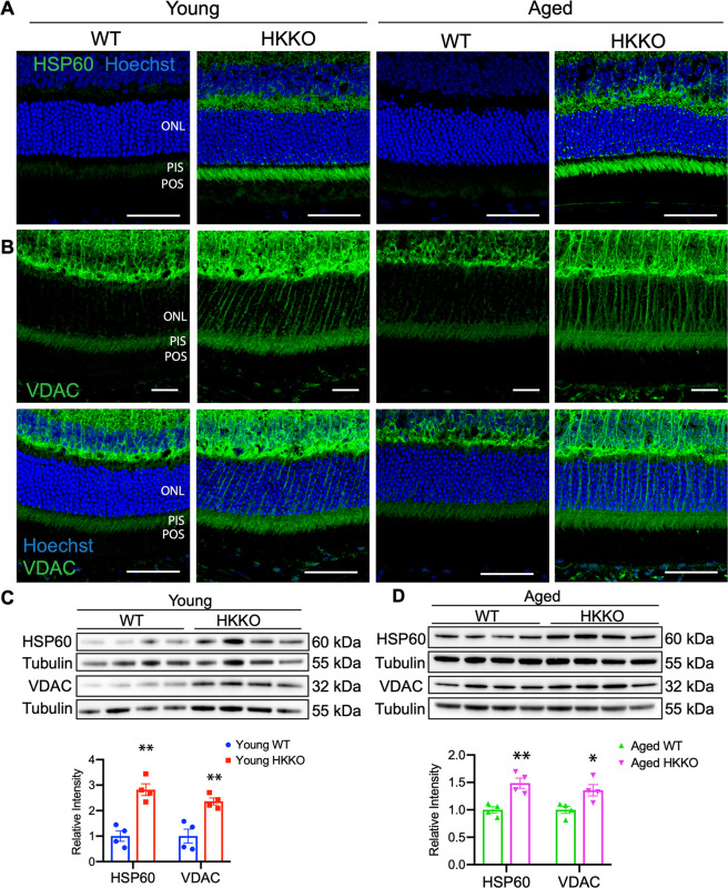
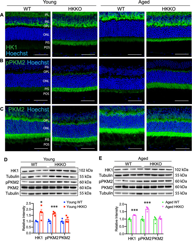
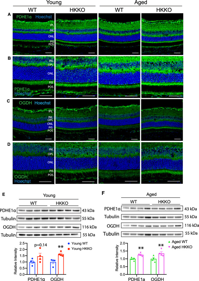
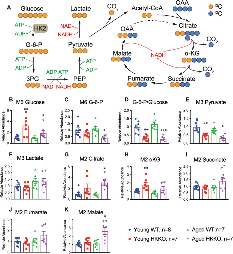
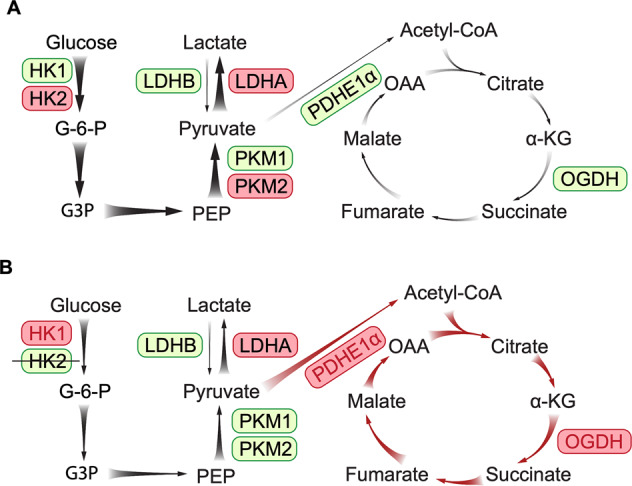
Photoreceptors, the primary site of phototransduction in the retina, require energy and metabolites to constantly renew their outer segments. They preferentially consume most glucose through aerobic glycolysis despite possessing abundant mitochondria and enzymes for oxidative phosphorylation (OXPHOS). Exactly how photoreceptors balance aerobic glycolysis and mitochondrial OXPHOS to regulate their survival is still unclear. We crossed rhodopsin-Cre mice with hexokinase 2 (HK2)-floxed mice to study the effect of knocking down HK2, the first rate-limiting enzyme in glycolysis, on retinal health and metabolic remodeling. Immunohistochemistry and Western blots were performed to study changes in photoreceptor-specific proteins and key enzymes in glycolysis and the tricarboxylic acid (TCA) cycle. Changes in retinal structure and function were studied by optical coherence tomography and electroretinography. Mass spectrometry was performed to profile changes in 13C-glucose-derived metabolites in glycolysis and the TCA cycle. We found that knocking down HK2 in rods led to age-related photoreceptor degeneration, evidenced by reduced expression of photoreceptor-specific proteins, age-related reductions of the outer nuclear layer, photoreceptor inner and outer segments and impaired electroretinographic responses. Loss of HK2 in rods led to upregulation of HK1, phosphorylation of pyruvate kinase muscle isozyme 2, mitochondrial stress proteins and enzymes in the TCA cycle. Mass spectrometry found that the deletion of HK2 in rods resulted in accumulation of 13C-glucose along with decreased pyruvate and increased metabolites in the TCA cycle. Our data suggest that HK2-mediated aerobic glycolysis is indispensable for the maintenance of photoreceptor structure and function and that long-term inhibition of glycolysis leads to photoreceptor degeneration.


Conflict of interest statement

The authors declare that they have no conflict of interest.

Figures

Fig. 1
Fig. 1. Deletion of HK2 in RHO-Cre mice crossed with HK2-floxed mice.
A Recoverin, a marker of rod receptors, was expressed in the outer nuclear layer (ONL) and photoreceptor inner and outer segments (PIS and POS). Cre recombinase was exclusively expressed in cell nuclei positive for recoverin in the ONL in RHO-Cre mice. B Double label IHC in retinal wholemounts from RHO-Cre mice indicated that Cre recombinase was not expressed in cone photoreceptors. CE Knockdown of HK2 in RHO-Cre mice crossed with HK-floxed mice (hereafter called HKKO mice). C IHC indicated that HK2 was dramatically reduced in photoreceptor inner segments (PIS) in HKKO mice compared with age-matched wild-type (WT) controls. Scale bars: 50μm in (AC). D, E Western blots indicated that the expression of HK2 was reduced by ~95% in the retina of HKKO mice compared with age-matched WT controls. N = 4–8/group. ***p < 0.001, analyzed by un-paired t-test.
Fig. 2
Fig. 2. Knockdown of HK2 in rods led to reduced expression of photoreceptor-specific protein.
A, B Immunostaining for interphotoreceptor retinoid-binding protein (IRBP) and recoverin in retinal sections from HKKO mice and age-matched WT controls. Scale bar: 50 μm in (A) and (B). C, D Western blots indicated that the expression of IRBP and recoverin was significantly reduced in HKKO mice compared with age-matched WT controls. N = 4/group. *p < 0.05, analyzed by un-paired t test.
Fig. 3
Fig. 3. Knockdown of HK2 in rods led to aged-related photoreceptor degeneration and impaired retinal function.
A Image-guided OCT was performed on the central retina in areas just above the optic nerve head and across the optic nerve head as well as on the peripheral retina. ONL outer nuclear layer, OLM outer limiting membrane, PIS photoreceptor inner segment, referring to the vertical length from the OLM to the outer border of the ellipsoid zone, POS photoreceptor outer segment, referring to the vertical length from the outer border of the ellipsoid zone to the apical side of the retinal pigment epithelium (RPE). BD Changes in thickness of the ONL, PIS, and POS in the three regions in young and aged mice after knocking down HK2 in rods. *P < 0.05, **P < 0.01, n = 8/group. EH Scotopic ERG measured under stimulation with a range of intensities of green (505 nm) light in young and aged HKKO mice and age-matched controls. E, F There were no significant changes in the amplitudes of a and b waves in young HKKO mice compared with age-matched WT controls. G, H The amplitudes of both a and b waves were significantly reduced in aged HKKO mice compared with age-matched WT controls. *P < 0.05, **P < 0.01, n = 8/group.
Fig. 4
Fig. 4. Knockdown of HK2 led to upregulation of mitochondrial proteins including heat-shock protein 60 (HSP60) and voltage-dependent-anion channel (VDAC).
A, B Immunostaining for HSP60 and VDAC in retinal sections from HKKO mice and WT controls. A Enhanced expression of HSP60 was observed in the photoreceptor inner segments (PIS) and the outer plexiform layer (OPL) in HKKO mice. B Increased expression of VDAC was observed in the outer retina of HKKO mice. The upper panel images are immunostaining for VDAC without Hoechst nuclear counterstaining. Scale bar: 50 μm in (A, B). C, D Western blots indicated that the expression of HSP60 and VDAC was significantly increased in HKKO mice compared with age-matched WT controls. *p < 0.05 and **p < 0.01, n = 4/group.
Fig. 5
Fig. 5. Loss of HK2 in rods led to increased expression of HK1 and phosphorylation of PKM2 at tyrosine residue 105 (p-PKM2).
AC Immunostaining for HK1, pPKM2, and PKM2 in retinal sections from HKKO mice and age-matched WT controls. Increased expression of HK1 (A) and pPKM2 (B) were observed in photoreceptor inner segments (PIS) in retinas of HKKO mice. Scale bar: 50 μm in (AC). D, E Western blots indicated that the expression of HK1 and pPKM2 was significantly increased while the level of PKM2 expression remained unchanged in HKKO mice compared with age-matched WT controls. *p < 0.05 and ***p < 0.001, n = 4/group.
Fig. 6
Fig. 6. Knocking down HK2 led to upregulation of enzymes in the TCA cycle.
AD Immunostaining for enzymes in the TCA cycle including pyruvate dehydrogenase E1α (PDHE1α, A and B) and oxoglutarate dehydrogenase (OGDH, C and D) in retinal sections from HKKO mice and age-matched WT controls. Loss of HK2 in rods led to increased expression of PDHE1α (A, B) and OGDH (C, D) in the photoreceptor inner segments (PIS). The panel of images in (B) and (D) are higher-power images of areas in (A) and (C) respectively. Scale bar: 50 μm in (AD). E, F Western blots indicated that the expression of PDHE1α (C) and OGDH (D) was significantly increased in HKKO mice compared with age-matched WT controls. *p < 0.05 and **p < 0.01, n = 4/group.
Fig. 7
Fig. 7. Knockdown of HK2 in rods led to retinal metabolic remodeling.
A Schematic diagram of labeled metabolites derived from 13C-glucose in glycolysis and the TCA cycle. BP Changes in the relative abundance of 13C-glucose (B) and its derivatives in glycolysis (CF) and the TCA cycle (GK) in HKKO mice compared with age-matched WT controls. The value of each metabolite was normalized to its abundance in young WT controls. G-6-P = glucose-6-phosphate (G-6-P), 3PG glyceraldehyde-3-phosphate, PEP phosphoenolpyruvate, OAA oxaloacetate (OAA), αKG Ketoglutarate. *p < 0.05 and **p < 0.01, n = 7~8/group.
Fig. 8
Fig. 8. Proposed mechanism of metabolic remodeling after inhibiting aerobic glycolysis resulted from the deletion of HK2 in rods.
A Glycolysis and mitochondrial oxidative phosphorylation (OXPHOS) in the normal retina. Glucose is mainly metabolized to pyruvate through glycolysis using enzymes including HK2, PKM2 and lactate dehydrogenase A (LDHA). A small amount of pyruvate is catalyzed by PDHE1α to produce acetyl-CoA for mitochondria OXPHOS in the TCA cycle. B Retinal metabolic remodeling after deletion of HK2 in rods. Loss of HK2 in rods leads to enhanced activities of HK1 and upregulation of pPKM2. The resultant production of pyruvate is mainly used for mitochondrial OXPHOS to adapt to the inhibition of aerobic glycolysis. The enzymes highlighted in red and the reactions indicated by thicker arrows represent the predominated enzymes and metabolic activities in normal and stressed retinas, while the enzymes highlighted in green and the reactions indicated by thinner arrows represent less-active enzymes and metabolic activities in each condition.

Similar articles

Cited by

References

    1. Nguyen-Legros J, Hicks D. Renewal of photoreceptor outer segments and their phagocytosis by the retinal pigment epithelium. Int. Rev. Cytol. 2000;196:245–313. - PubMed
    1. Chinchore Y, Begaj T, Wu D, Drokhlyansky E, Cepko CL. Glycolytic reliance promotes anabolism in photoreceptors. Elife. 2017;6:e25946. - PMC - PubMed
    1. Hurley JB, Lindsay KJ, Du J. Glucose, lactate, and shuttling of metabolites in vertebrate retinas. J. Neurosci. Res. 2015;93:1079–1092. - PMC - PubMed
    1. LaVail MM. Rod outer segment disk shedding in rat retina: relationship to cyclic lighting. Science. 1976;194:1071–1074. - PubMed
    1. Murakami Y, et al. Photoreceptor cell death and rescue in retinal detachment and degenerations. Prog. Retin Eye Res. 2013;37:114–140. - PMC - PubMed

Publication types

LinkOut - more resources