. 2020 Oct 20;10(1):17817.
doi: 10.1038/s41598-020-73659-z.

Targeting interleukin-17 receptor B enhances gemcitabine sensitivity through downregulation of mucins in pancreatic cancer

Affiliations

Targeting interleukin-17 receptor B enhances gemcitabine sensitivity through downregulation of mucins in pancreatic cancer

Lung-Hung Tsai et al. Sci Rep. .

Abstract

Pancreatic cancer is the fourth leading cause of death worldwide due to its poorest prognoses with a 7% 5-year survival rate. Eighty percent of pancreatic cancer patients relapse after chemotherapy and develop early metastasis and drug resistance. Resistance to nucleoside analog gemcitabine frequently used in first-line therapy is an urgent issue in pancreatic cancer treatment. Expression of mucin (MUC) glycoproteins has been shown to enhance chemoresistance via increased cell stemness. Here we show interlukine-17 receptor B (IL-17RB) expression is positively correlated with MUC1 and MUC4 expression in pancreatic cancer cells and tumor tissue. Moreover, IL-17RB transcriptionally up-regulates expression of MUC1 and MUC4 to enhance cancer stem-like properties and resistance to gemcitabine. These results suggest IL-17RB can be a potential target for pancreatic cancer therapy. Indeed, treatment with IL-17RB-neutralizing antibody has a synergistic effect in combination with gemcitabine for killing pancreatic cancer cells. Altogether, these findings provide feasible applications for IL-17RB-targeting therapy in pancreatic cancer treatment.


Conflict of interest statement

The authors declare no competing interests.

Figures

Figure 1
Figure 1
IL-17RB upregulates MUC1 and MUC4 expression in pancreatic cancer cells. (A) Pie chart of down-regulated genes (528 genes decreased in more than 50%, a, b and c) analyzed in IL-17RB-knockdown CFPAC1 cells. Cancer-related genes are listed by NCG5.0, n = 49, b and c. Resistance to chemotherapeutic drugs was listed in c. (B) Expression of IL-17RB (~ 65KD due to glycosylation), MUC1 (~ 140 KD due to glycosylation), and MUC4 (~ 130 KD due to glycosylation) in pancreatic cancer cell lines was evaluated by immunoblotting with α-tubulin (52 KD) used as a loading control. (CE) IL-17RB was knocked down by lentivirus-based shRNA in BxPC3 and CFPAC1 cells. Protein levels of IL-17RB, MUC1, and MUC4 were evaluated by immunoblotting (C). mRNA levels of MUC1 (D) and MUC4 (E) were analyzed by real-time RT-PCR. (FH), ectopic wildtype or ΔLBD of IL-17RB was overexpressed in SU.86.86 and HPAC cells. Protein levels of IL-17RB, MUC1, and MUC4 were evaluated by immunoblotting (F). mRNA levels of MUC1 (G) and MUC4 (H) were analyzed by real-time RT-PCR. The asterisk (*) represents a statistical significance with P value less than 0.05. The full blotting images were showed in Supplementary Figure 1.
Figure 2
Figure 2
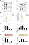
IL-17RB enhances cancer stem cell-like phenotype via upregulation of MUC1 and MUC4. (A) Expression of IL-17RB, MUC1, MUC4 and stemness markers SOX2 (40 KD), Nanog (42 KD), Oct-4 (45 KD) in IL-17RB-knockdown BxPC3 cells was measured by immunoblotting. GAPDH (36 KD) was included as a loading control. (B, C) CD44 expression was measured by flow cytometry, and the MFI (mean fluorescence intensity) was calculated by FlowJo 7.6, and presented in (C). (D) Sphere formation activity was evaluated by sphere formation assay and the diameter more than 100 µm was calculated. (E) Expression of IL-17RB, MUC1, MUC4 and stemness markers SOX2, Nanog, Oct-4 in IL-17RB-overexpressing SU.86.86 cells after transduction with shRNAs of MUC1 and MUC4. GAPDH was served as a loading control. (F, G) CD44 expression in IL-17RB-overexpressing SU.86.86 cells was measured by flow cytometry, and the MFI (mean fluorescence intensity) was calculated by FlowJo 7.6, and presented in (G). (H) IL-17RB-overexpressing SU.86.86 cells were transduced with shRNAs of MUC1 and MUC4. Colony formation activity was evaluated by sphere formation assay and the diameter more than 100 µm was calculated. The full blotting images were showed in Supplementary Figure 1.
Figure 3
Figure 3
IL-17RB enhances gemcitabine resistance via MUC1 and MUC4. (A) cell viability was measured by MTT assay in parental, scramble, and IL-17RB-knockdown BxPC3 cells treated with gemcitabine for 48 h. Statistical significance was calculated by comparison of shIL-17RB-1 or shIL-17RB-2 compared to Scramble. (B) The IC50 of gemcitabine toxicity was shown in mean ± SD. (C) Cell viability was measured by MTT assay in ectopic IL-17RB-overexpressed, MUC1-knockdown, MUC4-knockdown SU.86.86 cells. Statistical significance was calculated by comparison of IL-17RB OE with P. Ct., and OE/shMUC1 or OE/shMUC4 with OE/Scramble. The IC50 of gemcitabine toxicity was estimated in (D). Synergistic effect of MUC1 inhibitor Go-201 (5 μM) and gemcitabine (0.25 µM) on cell viability measured by MTT assay in BxPC3 and ectopic IL-17RB-overexpressing SU.86.86 cells (E). Asterisks (*, **, ***) indicated the statistical significance of P value less than 0.05, 0.01, or 0.001, respectively.
Figure 4
Figure 4
Inhibition of IL-17RB by neutralizing antibody suppresses stemness activity and gemcitabine resistance. (A) BxPC3 cells were treated with IgG or neutralizing antibody (D9) with indicated dosages for 48 h and harvested for protein extraction. The expression of MUC1, MUC4, sox2, nanog, and oct-4 was measured by immunoblotting. (B) Surface CD44 expression was measured by flow cytometry in BxPC3 with 10 µg/ml IgG or D9 for 48 h. MFI was calculated by FlowJo in (C). (D) Sphere formation activity was evaluated by sphere formation assay in BxPC3 with 10 µg/ml IgG or D9. (E) Cell viability was estimated by MTT following treatment with 0.1 µM doxorubicin, 0.25 µM gemcitabine, or 25 µM etoposide for 48 h in IgG or D9-treated BxPC3. F, cell viability curves were plotted and measured by MTS assay with indicated dosages of D9 and gemcitabine treatment for 24 h in BxPC3 cells. Each cell viability (%) of combined D9 and gemcitabine was shown in the Table. (G) normalized isobologram for combination of D9 and gemcitabine treatment in a non-constant ratio was plotted by CompuSyn. The point on the upper-right or lower-left of the line of additivity indicates an antagonistic or synergistic effect, respectively. Combination index (CI) values were calculated by CompuSyn. The full blotting images were showed in Supplementary Figure 1.
Figure 5
Figure 5
Expression of IL-17RB, MUC1, and MUC4 in pancreatic cancers. Representative IHC staining images of membrane-bound IL-17RB, MUC1, and MUC4 were shown in high expressed tumors at 5× or 20× objective lens, in low expressed tumors at 5× objective lens, and in normal tissues at 5× objective lens.

Similar articles

Cited by

References

    1. Siegel RL, Miller KD, Jemal A. Cancer statistics, 2017. CA Cancer J. Clin. 2017;67:7–30. doi: 10.3322/caac.21387. - DOI - PubMed
    1. Vincent A, Herman J, Schulick R, Hruban RH, Goggins M. Pancreatic cancer. Lancet. 2011;378:607–620. doi: 10.1016/S0140-6736(10)62307-0. - DOI - PMC - PubMed
    1. Tseng CM, Huang SP, Liao WC, Chiang CJ, Yang YW, Chang CY, et al. Incidence and mortality of pancreatic cancer on a rapid rise in Taiwan, 1999–2012. Cancer Epidemiol. 2017;49:75–84. doi: 10.1016/j.canep.2017.05.011. - DOI - PubMed
    1. Ryan DP, Hong TS, Bardeesy N. Pancreatic adenocarcinoma. N. Engl. J. Med. 2014;371:2140–2141. doi: 10.1056/NEJMra1404198. - DOI - PubMed
    1. Bond-Smith G, Banga N, Hammond TM, Imber CJ. Pancreatic adenocarcinoma. BMJ. 2012;344:e2476. doi: 10.1136/bmj.e2476. - DOI - PubMed

Publication types

MeSH terms