. 2020 Oct 20;10(1):17730.
doi: 10.1038/s41598-020-74610-y.

Neuron type-specific expression of a mutant KRAS impairs hippocampal-dependent learning and memory

Affiliations

Neuron type-specific expression of a mutant KRAS impairs hippocampal-dependent learning and memory

Hyun-Hee Ryu et al. Sci Rep. .

Abstract

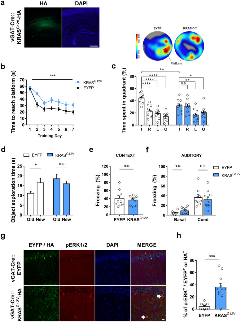
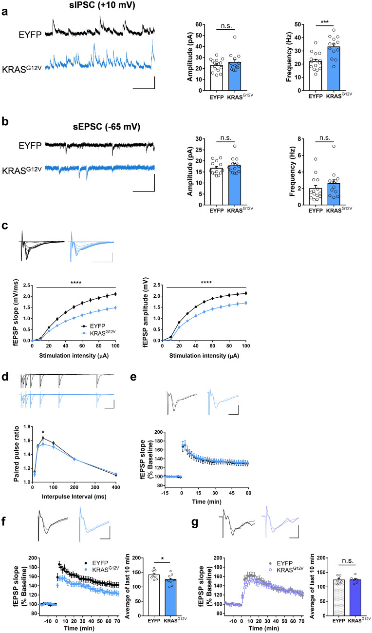
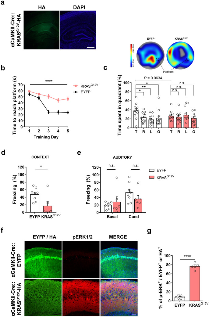
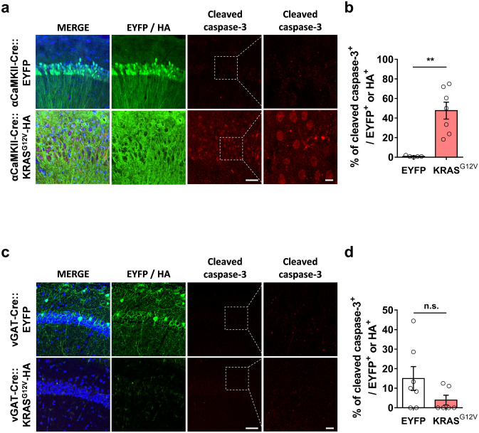
KRAS mutations are associated with rare cases of neurodevelopmental disorders that can cause intellectual disabilities. Previous studies showed that mice expressing a mutant KRAS have impaired the development and function of GABAergic inhibitory neurons, which may contribute to behavioural deficits in the mutant mice. However, the underlying cellular mechanisms and the role of excitatory neurons in these behavioural deficits in adults are not fully understood. Herein, we report that neuron type-specific expression of a constitutively active mutant KRASG12V in either excitatory or inhibitory neurons resulted in spatial memory deficits in adult mice. In inhibitory neurons, KRASG12V induced ERK activation and enhanced GABAergic synaptic transmission. Expressing KRASG12V in inhibitory neurons also impaired long-term potentiation in the hippocampal Shaffer-collateral pathway, which could be rescued by picrotoxin treatment. In contrast, KRASG12V induced ERK activation and neuronal cell death in excitatory neurons, which might have contributed to the severe behavioural deficits. Our results showed that both excitatory and inhibitory neurons are involved in mutant KRAS-associated learning deficits in adults via distinct cellular mechanisms.


Conflict of interest statement

The authors declare no competing interests.

Figures

Figure 1
Figure 1
Ectopic expression of KRASG12V in inhibitory neurons causes spatial memory deficits and ERK hyperactivation. (a) Expression of adeno-associated virus (AAV) expressing KRASG12V-HA in the dorsal CA regions of adult vGAT-Cre mouse. 4′,6-diamidino-2-phenylindole (DAPI) staining was used to identify nuclei. Scale bars, 200 μm. (b) Learning curve showing that the latency to find the hidden platform during the Morris water maze (MWM) training trials was significantly increased in vGAT-Cre::KRASG12V mice as compared to vGAT-Cre::EYFP. vGAT-Cre::EYFP, n = 10; vGAT-Cre::KRASG12V, n = 10; Two-way repeated measures ANOVA, F 1, 18 = 21.53, ***P = 0.0002. (c) Lower panel, time spent in each quadrant during the probe test. vGAT-CRE:: KRASG12V mice spent significantly less time in the target quadrant compared to vGAT-Cre::EYFP mice (comparison of target quadrant occupancy, unpaired t-test, **P = 0.0063). Moreover, vGAT-Cre::EYFP mice selectively searched for the platform in the target quadrant, while KRASG12V did not (vGAT-Cre::EYFP, n = 10; One-Way ANOVA, followed by Dunnett’s post hoc test, ****P < 0.0001; vGAT-Cre::KRASG12V, n = 10; one-way ANOVA, followed by Dunnett’s post hoc test, P = 0.9982, **P = 0.0069, *P = 0.0493). T, target quadrant, R, right to target, L, left to target, O, opposite to target. Upper panel, representative heat map summary of MWM probe test. Platform position during the training trials is indicated by the dotted circle. (d) In object-place recognition test, vGAT-Cre::EYFP mice showed significant preference to the relocated (new) object, while vGAT-Cre::KRASG12V mice showed similar preference between old and new object. vGAT-Cre::EYFP, n = 26; paired t-test, *P = 0.0112; vGAT-Cre::KRASG12V, n = 27; paired t-test, P = 0.1223; New, new location; old, object location used in training session. (e, f) In contextual (e) and cued (f) fear memory test, two groups showed comparable levels of freezing. Contextual test, vGAT-Cre::EYFP, n = 10, vGAT-Cre::KRASG12V, n = 11, unpaired t-test, P = 0.534; Cued test, vGAT-Cre::EYFP, n = 9; vGAT-Cre::KRASG12V, n = 11, unpaired t-test, P = 0.1157 for basal freezing before tone presented, P = 0.5690 for freezing in response to tone. (g) Representative image of HA and pErk staining of EYFP or KRASG12Vexpressing hippocampal slices from vGAT-Cre mice. Slices were immunostained for p-ERK1/2 (red), HA (green), and DAPI (blue). White arrows indicate pERK1/2 and HA-KRASG12Vdouble labelled cells, Scale bar = 20 μm. (h) Percentage of pERK-positive neurons was significantly higher in the KRASG12Vexpressing hippocampal slices. vGAT-Cre::EYFP, n = 9 slices from 3 hippocampi, vGAT-Cre::KRASG12V, n = 11 slices from 4 hippocampi. ***P = 0.0002. Data are expressed as the mean ± SEM.
Figure 2
Figure 2
Ectopic KRASG12V expression in the inhibitory neurons increases inhibitory synaptic transmission and impairs LTP. (a) Ectopic KRASG12V expression in the inhibitory neurons increased the frequency of spontaneous inhibitory postsynaptic current (sIPSC). The amplitudes of the sIPSCs in the vGAT-Cre::KRASG12V mice were comparable to that from vGAT- Cre::EYFP mice (EYFP, n = 16 cells from 3 mice; KRASG12V, n = 13 cells from 3 mice, unpaired t-test, P = 0.2883). The frequencies of the sIPSCs in the vGAT-Cre::KRASG12V mice were higher than that in the vGAT-Cre::EYFP mice (EYFP, n = 16 cells from 3 mice; KRASG12V, n = 13 cells from 3 mice, unpaired t-test, ***P = 0.0003). Vertical bar, 50 pA; horizontal bar, 200 ms. (b) Ectopic expression of KRASG12V in the inhibitory neurons did not affect either the frequency or the amplitude of spontaneous excitatory postsynaptic current (sEPSC). The amplitudes of the sEPSCs were comparable between vGAT-Cre::KRASG12V and vGAT- Cre::EYFP; (EYFP, n = 14 cells from 3 mice; KRASG12V, n = 13 cells from 3 mice, unpaired t-test, P = 0.3528). The frequency of the sEPSCs from the vGAT-Cre::KRASG12V were similar with that from vGAT-Cre::EYFP; KRASG12V (EYFP, n = 14 cells from 3 mice; KRASG12V, n = 13 cells from 3 mice, unpaired t-test, P = 0.3044). Vertical bar, 50 pA; horizontal bar, 200 ms. (c) Ectopic KRASG12V expression in the inhibitory neurons significantly decreased the input–output relationship of the field excitatory postsynaptic potential (fEPSP) at CA3-CA1 synapse. Outputs were measured either by initial fEPSP slope (left) or peak amplitude (right). vGAT-Cre::EYFP, n = 20 slices from 10 mice; vGAT-Cre::KRASG12V, n = 23 slices from 10 mice; Two-way repeated measures ANOVA, effect of virus, ****P < 0.0001 for both slope and amplitude. Vertical bar, 1 mV; horizontal bar, 10 ms. (d) Paired pulse facilitation ratio was significantly decreased at 50 ms interpulse interval in vGAT-Cre::KRASG12Vmice. vGAT-Cre::EYFP, n = 20 slices from 10 mice; vGAT-Cre::KRASG12V, n = 23 slices from 10 mice, unpaired t-test, *P = 0.0403. Vertical bar, 1 mV; horizontal bar, 50 ms. (e) Ectopic KRASG12V expression in inhibitory neurons did not affect long-term potentiation (LTP) induced by weak stimulation protocol (4 bursts of 4 100 Hz pulses, 5 Hz inter-burst interval). vGAT-Cre::EYFP, n = 10 slices from 5 mice; vGAT-Cre::KRASG12V, n = 12 slices from 5 mice. Traces represent the average of fEPSPs at the baseline (-15 min – 0 min) and after LTP induction (50 – 60 min). Vertical bar, 1 mV; horizontal bar, 5 ms. (f) Ectopic KRASG12V expression in inhibitory neurons significantly impaired LTP induced by the stronger stimulation protocol (10 bursts of 4 100 Hz pulses, repeated 4 times with 10 s interval). vGAT-Cre::EYFP, n = 8 slices from 5 mice; vGAT-Cre::KRASG12V, n = 10 slices from 5 mice. The averages fEPSP slope of 61 to 70 min after LTP induction were compared. Unpaired t test, *P = 0.0422. Traces represent the average of fEPSPs at the baseline (-20 min – 0 min) and after LTP induction (61 – 70 min). Vertical bar, 1 mV; horizontal bar, 5 ms. (g) Picrotoxin (10 μM) treatment rescued the strong TBS-induced LTP deficit at CA3-CA1 synapse in vGAT-Cre::KRASG12V mice. vGAT-Cre::EYFP, n = 7 slices from 3 mice; vGAT-Cre::KRASG12V, n = 7 slices from 3 mice. The averages fEPSP slope of 61 to 70 min after LTP induction were compared. Unpaired t test, P = 0.9887. Traces represent the average of fEPSPs at the baseline (-20 min – 0 min) and after LTP induction (61 – 70 min). Vertical bar, 1 mV; horizontal bar, 5 ms. Data are expressed as the mean ± SEM.
Figure 3
Figure 3
Ectopic expression of KRASG12V in excitatory neurons impairs spatial memory and induces ERK activation. (a) HA staining of KRASG12V-expressing hippocampal slices from αCaMKII-Cre mice. 4′,6-diamidino-2-phenylindole (DAPI) staining was used to identify nuclei. Scale bars, 200 μm. (b) Learning is significantly slower in KRASG12V expressing mice compared to EYFP controls. αCaMKII-Cre::EYFP, n = 12; αCaMKII-Cre::KRASG12V, n = 11; Two-way repeated measures ANOVA, F1, 21 = 69.63, ****P < 0.001 (c) Lower panel, time spent in each quadrant during the probe test. αCaMKII-Cre::EYFP mice selectively searched for the platform in the target quadrant, while KRASG12V did not. αCaMKII-Cre::EYFP, n = 12, One-Way ANOVA, followed by a Dunnett’s post hoc test, *P = 0.0292, **P = 0.0030, *P = 0.0156; αCaMKII-Cre::KRASG12V, n = 11, One-way ANOVA, followed by Dunnett’s post hoc test, P = 0.9510, P = 0.9743, P = 0.7006. αCaMKII-Cre::KRASG12V mice tend to spend less time in the target quadrant compared to EYFP mice (unpaired t-test, P = 0.0634). T, target quadrant, R, right to target, L, left to target, O, opposite to target. Upper panel, representative heat map summary of MWM probe test. Platform position during the training trials is indicated by the dotted circle. (d) In contextual fear memory test, αCaMKII-Cre::KRASG12V mice showed significantly reduced freezing behaviour compared to αCaMKII-Cre::EYFP mice. αCaMKII-Cre::EYFP, n = 8; αCaMKII-Cre::KRASG12V, n = 6; unpaired t-test, *P = 0.0247. (e) The freezing level of αCaMKII-Cre::KRASG12V in response to the conditioned tone (cue) was not statistically different from that of αCaMKII-Cre::EYFP mice. αCaMKII-Cre::EYFP, n = 8; αCaMKII-Cre::KRASG12V, n = 6; unpaired t-test, P = 0.5026 for basal freezing level before tone played, P = 0.2178 for cued freezing. (f) Representative images of immunohistochemistry from slices expressing EYFP or KRASG12V in excitatory neurons. Slices were immunostained for HA (green), p-ERK1/2 (red), and DAPI (blue). Scale bar = 80 μm. (g) The percentage of pERK-positive neurons was significantly higher in KRASG12Vexpressing hippocampal slices. αCaMKII-Cre::EYFP, n = 6 slices from 3 hippocampi, αCaMKII-Cre::KRASG12V, n = 5 slices from 3 hippocampi, unpaired t-test, ****P < 0.0001. Data are expressed as the mean ± SEM.
Figure 4
Figure 4
KRASG12V induced neuronal cell death only in excitatory neurons. (a) αCaMKII-Cre mice were injected with either AAV expressing EYFP or HA-KRASG12V. Slices were immunostained for cleaved caspase-3 (red), HA (green), and DAPI (blue), Scale bar = 50 μm; high magnification, Scale bar = 20 μm. (b) The percentage of cleaved caspase-3 positive neurons was significantly higher in the KRASG12Vexpressing hippocampal slices. αCaMKII-Cre::EYFP, n = 5 slices from 3 hippocampi, αCaMKII-Cre::KRASG12V, n = 7 slices from 3 hippocampi, unpaired t-test, **P = 0.0012. (c) vGAT-Cre mice were injected with either AAV expressing EYFP or HA- KRASG12V. Slices were immunostained for cleaved caspase-3 (red), HA (green), and DAPI (blue), Scale bar = 50 μm, high magnification; Scale bar = 20 μm. (d) The percentage of cleaved caspase-3 positive neurons was comparable between vGAT-CRE::EYFP and vGAT-Cre::KRASG12Vmice; vGAT-CRE::EYFP, n = 6 slices from 3 hippocampi, vGAT-Cre::KRASG12V, n = 6 slices from 4 hippocampi, unpaired t-test, n.s., not significant, P = 0.2488. Data are expressed as the mean ± SEM.

Similar articles

Cited by

References

    1. Samuels IS, Saitta SC, Landreth GE. MAP'ing CNS development and cognition: an ERKsome process. Neuron. 2009;61:160–167. doi: 10.1016/j.neuron.2009.01.001. - DOI - PMC - PubMed
    1. Molina JR, Adjei AA. The Ras/Raf/MAPK pathway. J. Thorac. Oncol. 2006;1:7–9. doi: 10.1016/S1556-0864(15)31506-9. - DOI - PubMed
    1. Aoki Y, Niihori T, Inoue S, Matsubara Y. Recent advances in RASopathies. J. Hum. Genet. 2016;61:33–39. doi: 10.1038/jhg.2015.114. - DOI - PubMed
    1. Rauen KA. The rasopathies. Annu. Rev. Genom. Hum. Genet. 2013;14:355–369. doi: 10.1146/annurev-genom-091212-153523. - DOI - PMC - PubMed
    1. Tajan M, Paccoud R, Branka S, Yart A, Edouard T. The RASopathy family: consequences of germline activation of the RAS/MAPK pathway. Endocr. Rev. 2018;39:676–700. doi: 10.1210/er.2017-00232. - DOI - PubMed

Publication types

LinkOut - more resources