. 2020 Oct 20;10(1):17853.
doi: 10.1038/s41598-020-74944-7.

Interleukin-11 is important for vascular smooth muscle phenotypic switching and aortic inflammation, fibrosis and remodeling in mouse models

Affiliations

Interleukin-11 is important for vascular smooth muscle phenotypic switching and aortic inflammation, fibrosis and remodeling in mouse models

Wei-Wen Lim et al. Sci Rep. .

Abstract

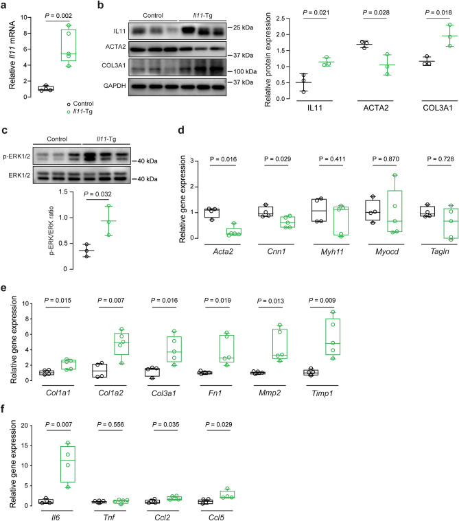
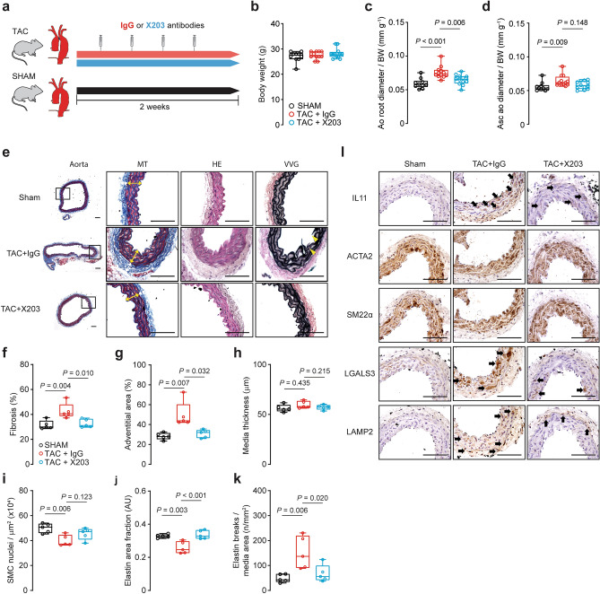
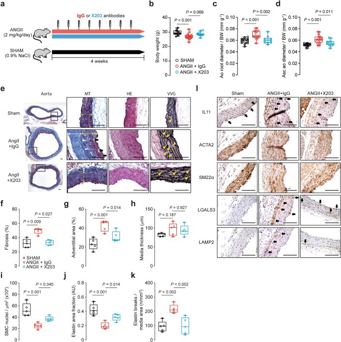
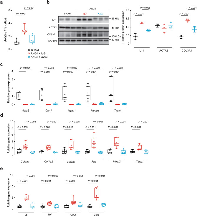
Transforming growth factor beta-1 (TGFβ1) is a major driver of vascular smooth muscle cell (VSMC) phenotypic switching, an important pathobiology in arterial disease. We performed RNA-sequencing of TGFβ1-stimulated human aortic or arterial VSMCs which revealed large and consistent upregulation of Interleukin 11 (IL11). IL11 has an unknown function in VSMCs, which highly express the IL11 receptor alpha, suggestive of an autocrine loop. In vitro, IL11 activated ERK signaling, but inhibited STAT3 activity, and caused VSMC phenotypic switching to a similar extent as TGFβ1 or angiotensin II (ANGII) stimulation. Genetic or therapeutic inhibition of IL11 signaling reduced TGFβ1- or ANGII-induced VSMC phenotypic switching, placing IL11 activity downstream of these factors. Aortas of mice with Myh11-driven IL11 expression were remodeled and had reduced contractile but increased matrix and inflammatory genes expression. In two models of arterial pressure loading, IL11 was upregulated in the aorta and neutralizing IL11 antibodies reduced remodeling along with matrix and pro-inflammatory gene expression. These data show that IL11 plays an important role in VSMC phenotype switching, vascular inflammation and aortic pathobiology.


Conflict of interest statement

S.A.C. and S.S. are co-inventors of the patent applications ‘Treatment of fibrosis’ (WO/2017/103108) and ‘IL-11 antibodies’ (WO/2018/109174). S.A.C., S.S., W.W.L. and B.N are co-inventors of the patent application ‘Treatment of SMC mediated disease’ (WO/2019/073057). S.A.C. and S.S. are co-founders and shareholders of Enleofen Bio PTE LTD, a company (which S.A.C. is a director of) that develops anti-IL11 therapeutics. All other authors declare no competing interests.

Figures

Figure 1
Figure 1
IL11 is secreted from VSMCs stimulated with TGFβ1 and causes VSMC phenotypic switching. Volcano plots of RNA-seq analysis showing the transcriptomic response of (a) aortic and (b) arterial VSMCs to TGFβ1 stimulation (Aortic, n = 10; left internal mammary artery (LIMA), n = 22). FDR: false discovery rate. (c) ELISA of IL11 in the supernatant of VSMC cultures following TGFβ1 stimulation (n = 5–8 biological replicates). (d) From left to right, IL6R and IL11RA gene expression in SMCs (n = 13, FANTOM5 database), RNA-seq data from unstimulated aortic (n = 10) and arterial (n = 22) VSMCs. 1 outlier was removed from the FANTOM5 dataset by the 2% ROUT method. Data presented as violin plots with quartiles indicated by dotted lines and median indicated by full lines. TPM: transcripts per million. (e) Flow cytometry forward scatter (FSC) plots of arterial VSMCs at baseline demonstrated IL11RA to be highly expressed in VSMCs whereas IL6R expression is scant. (f) Representative immunofluorescence staining of VSMCs for IL11RA and IL6R showing high IL11RA and undetectable IL6R expression. Stimulation of human VSMCs with IL11 (n = 5 biological replicates) increases both (g) TIMP1 and (h) MMP2 secretion. (i) Proliferation (EdU+ve cells) and COL1A1 expression by human VSMCs following IL11, TGFβ1 or ANGII stimulation (representative immunostaining images and Operetta assay results from 4 independent experiments). (j) Collagen secretion as measured by Sirius red collagen detection assay in culture medium following cytokine stimulation (n = 4). (k) VSMC migration measured by scratch wound assay following cytokine stimulation (n = 4). (l) Matrigel invasion indices of VSMCs induced by IL11 (1, 5, and 10 ng/ml), TGFβ1 or ANGII (n = 4). FC: fold change. Scale bars in representative images for f represent 100 µm and i and k represent 200 µm. Statistical analyses by two-tailed paired t-tests or one-way ANOVA with Sidak post hoc tests; data either presented as median ± IQR with whiskers demarcating minimum and maximum values or mean ± SD. Cytokines were applied for 24 h at doses of 5 ng/ml for IL11 and TGFβ1 and 100 nM for ANGII, unless otherwise stated.
Figure 2
Figure 2
Inhibition of VSMC phenotypic switching with neutralizing IL11 antibodies or ERK inhibition. (a) Proliferation (EdU+ve staining) and COL1A1 expression after TGFβ1 or ANGII stimulation in the presence of isotype control (IgG) or IL11 (X203) antibodies. Representative immunostaining images and Operetta assay results from 4 independent experiments. (b) Total collagen secretion (Sirius Red) following TGFβ1 or ANGII stimulation with either IgG or X203 antibody (n = 4). (c) Cytokine-induced VSMC migration (scratch wound assay) in the presence of IgG or X203 (n = 4). (d) Matrigel invasion indices of TGFβ1-stimulated VSMC in the presence of X203 or IgG antibody (n = 3). (e) Representative immunoblots of IL11-induced ERK1/2 and STAT3 phosphorylation in human VSMCs. Samples were derived from the same experiment and blots were processed in parallel. Extended blots are presented in Supplementary Fig. S12 online. Collated densitometry for phosphorylated ERK1/2 and STAT3 normalized to total ERK1/2 and STAT3 respectively (n = 9). P-values stated in comparison to 0 min time point. (f) Representative immunoblots of ERK1/2 activation and extracellular matrix proteins fibronectin (FN1) and collagen 3 (COL3A1) in human VSMCs stimulated with TGFβ1, or ANGII in the presence of anti-IL11 (X203) or IgG control antibodies. Samples were derived from the same experiment and blots were processed in parallel. Extended blots are presented in Supplementary Fig. S12 online. Collated densitometry for phosphorylated ERK1/2 normalized to total ERK1/2, FN1 and COL3A1 normalized to GAPDH (n = 5). (g) Proliferation (EdU+ve cells) and COL1A1 expression after IL11 stimulation and either MEK/ERK inhibitor U0126 (10 µM) or dimethyl sulfoxide (DMSO) vehicle. Representative immunostaining images and Operetta assay results from 4 independent experiments. Scale bars in representative images for a represent 200 µm and g represent 100 µm. Statistical analyses by one-way ANOVA with Sidak post hoc tests or two-tailed paired t-test; data presented as mean ± SD. Cytokines were applied for 24 h at doses of 5 ng/ml for IL11 and TGFβ1 and 100 nM for ANGII and antibodies at 2 µg/ml.
Figure 3
Figure 3
Smooth muscle cell-restricted IL11 expression induces aortic remodeling, fibrosis, and inflammation. (a) Schematic diagram featuring induction protocol of smooth muscle-specific Il11 overexpression with 3 doses of 50 mg/kg tamoxifen (TAM) treatment in Myh11CreERT2:Rosa26IL11/+ (Il11-Tg) or corn oil vehicle-treated (control) littermates. Animals were sacrificed for studies at 8 weeks of age following a week of washout. (b) Body weights and aortic echocardiography for (c) aortic root diameter and (d) ascending aortic diameter normalized against individual body mass in Il11-Tg mice as compared with controls (n = 8/group). (e) Representative photomicrographs captured at 100X and 400X magnification of transverse aortic sections in Il11-Tg and control mice stained with Masson’s Trichrome (MT), hematoxylin & eosin (HE) and Verhoeff Van Gieson (VVG) stains. The white and yellow double-headed arrows demarcate the tunica adventitia and media layers respectively. Red arrows indicate media fibrosis and yellow arrowheads indicate lamellar elastin breaks. Histological analyses of (f) fibrosis, (g) adventitial area, (h) media thickness, (i) smooth muscle cell (SMC) nuclei, (j) elastin area and (k) elastic lamella breaks (n = 5–6 animals per group). Statistical analyses by unpaired t-test; data presented as median ± IQR, whiskers define the minimum and maximum values. (l) Representative photomicrographs (400X mag) of aortic sections in Il11-Tg and control mice immunostained for IL11, ACTA2, SM22α, LGALS3 and LAMP2 (n = 5/group). Black arrows indicate labeled VSMCs within the media. Scale bars represent 100 µm.
Figure 4
Figure 4
Smooth muscle cell-restricted IL11 expression induces molecular changes of aortic remodeling. (a) RT-qPCR of Il11 mRNA expression normalized to Gapdh expression in aortas of Il11-Tg and control mice (n = 5/group). Representative immunoblots for aortic expression of (b) IL11, ACTA2, COL3A1 and GAPDH protein and (c) phosphorylated (p) and total ERK1/2 protein expression in Il11-Tg mice as compared with controls (n = 3/group). Collated densitometry for IL11, ACTA2 and COL3A1 normalized to GAPDH and p-ERK1/2 was normalized to total ERK1/2 protein expression. Samples were derived from the same experiment and blots were processed in parallel. Extended blots are presented in Supplementary Fig. S13 online. Data expressed in mean ± SD. RT-qPCR of (d) contractile genes (Acta2*, Cnn1, Myh11, Myocd, and Tagln), (e) ECM genes (Col1a1, Col1a2, Col3a1*, Fn1, Mmp2, and Timp1), and (f) inflammatory genes (Il6, Tnf*, Ccl2 and Ccl5*) normalized to Gapdh expression in aortas of Il11-Tg and control mice (n = 4–5/group). Statistical analyses by unpaired t-test unless data deviated from normal, in which case a Mann-Whitey test was performed (denoted by *). Data presented as median ± IQR, whiskers define the minimum and maximum values.
Figure 5
Figure 5
Antibody-mediated inhibition of IL11 reduces aortic constriction-induced aortic remodeling. (a) Schematic diagram depicting TAC experimental protocol. Wildtype C57BL/6 J mice were injected with X203 or IgG control antibodies (20 mg/kg IP twice per week) starting from 24 h post-TAC surgery. (b) Body weights* and aortic echocardiography for (c) aortic root diameter and (d) ascending aortic diameter* in Sham, TAC + IgG or TAC + X203 treated animals (n = 10–12/group). Aortic diameters were normalized to individual body mass. (e) Representative photomicrographs captured at 100X and 400X magnification of transverse aortic sections in sham, TAC + IgG, and TAC + X203 mice stained with Masson’s Trichrome (MT), hematoxylin and eosin (HE) and Verhoeff Van Gieson (VVG) stains. The white and yellow double-headed arrows demarcate the tunica adventitia and media layers respectively. Yellow arrowheads indicate lamellar elastin breaks. Histological analyses of (f) fibrosis, (g) adventitial area*, (h) media thickness, (i) SMC nuclei, (j) elastin area and (k) elastic lamella breaks (n = 5/group). Statistical analyses by one-way ANOVA with Sidak multiple comparisons, unless data deviated significantly from normal, in which case a Kruskal–Wallis test with Dunn’s multiple comparisons was performed (denoted by *). Data presented as median ± IQR, whiskers define the minimum and maximum values. (l) Representative photomicrographs captured at 400X magnification of transverse aortic sections in sham, TAC + IgG, and TAC + X203 mice immunostained with anti-IL11, ACTA2, SM22α, LGALS3, and LAMP2 (n = 3/group). Black arrows indicate labeled VSMCs within the media. Scale bars represent 100 µm.
Figure 6
Figure 6
Anti-IL11 antibodies reduce TAC-induced molecular changes in the mouse aorta. (a) RT-qPCR of Il11 mRNA expression normalized to Gapdh expression in aortas of Sham, TAC + IgG or TAC + X203 mice (n = 4/group). (b) Representative immunoblots of IL11, ACTA2, COL3A1, and GAPDH in the aorta of Sham, TAC + IgG or TAC + X203 treated animals (n = 3/group). Collated densitometry for IL11, ACTA2 and COL3A1 normalized to GAPDH protein expression. Samples were derived from the same experiment and blots were processed in parallel. Extended blots are presented in Supplementary Fig. S14 online. Data expressed in mean ± SD. RT-qPCR of (c) contractile genes (Acta2, Cnn1, Myh11, Myocd*, and Tagln), (d) ECM genes (Col1a1, Col1a2, Col3a1, Fn1, Mmp2, and Timp1), and (e) inflammatory genes (Il6, Tnf, Ccl2 and Ccl5) normalized to Gapdh expression in Sham, TAC + IgG or TAC + X203 mice (n = 3–4/group). Statistical analyses by one-way ANOVA with Sidak multiple comparisons unless data deviated from normal, in which case a Kruskal–Wallis test was performed (denoted by *). Data presented as median ± IQR, whiskers demarcate the minimum and maximum values.
Figure 7
Figure 7
Administration of IL11 antibodies prevents ANGII-induced aortic remodeling. (a) Schematic diagram depicting ANGII experimental protocol. Wildtype C57BL/6 J mice were injected with X203 or IgG control antibodies (20 mg/kg via IP twice per week) starting from 24 h after being implanted with osmotic minipumps containing ANGII. (b) Body weights and aortic echocardiography for (c) aortic root diameter and (d) ascending aortic diameter in Sham, ANGII + IgG, ANGII + X203 animals (n = 11–17/group). (e) Representative photomicrographs captured at 100X and 400X magnification of transverse aortic sections in sham, ANGII + IgG, and ANGII + X203 mice stained with Masson’s Trichrome (MT), hematoxylin and eosin (HE) and Verhoeff Van Gieson (VVG) stains. The white and yellow double-headed arrow demarcates the tunica adventitia and media layers respectively. Black arrows indicate cell-free foci of proteoglycan-rich matrix, white and yellow arrowheads indicate replacement fibrosis in the tunica media and lamellar elastin breaks respectively. Histological analyses of (f) fibrosis*, (g) adventitial area, (h) media thickness, (i) SMC nuclei, (j) elastin area and (k) elastic lamella breaks (n = 5/group). Statistical analyses by one-way ANOVA with Sidak multiple comparisons, unless data deviated significantly from normal, in which case a Kruskal–Wallis test with Dunn’s multiple comparisons was performed (denoted by *). Data presented as median ± IQR, whiskers define the minimum and maximum values. (l) Representative photomicrographs captured at 400X magnification of transverse aortic sections in sham, ANGII + IgG, and ANGII + X203 mice immunostained with anti-IL11, ACTA2, SM22α, LGALS3, and LAMP2 (n = 3/group). Black arrows indicate labeled VSMCs within the media. Scale bars represent 100 µm.
Figure 8
Figure 8
Anti-IL11 treatment prevents ANGII-induced histological changes in the mouse aorta. (a) RT-qPCR of Il11 gene expression normalized to Gapdh expression (n = 5–8/group). (b) Representative immunoblots of IL11, ACTA2, COL3A1, and GAPDH in the aortas from Sham, ANGII + IgG or ANGII + X203 animals (n = 3/group). Collated densitometry for IL11, ACTA2 and COL3A1 normalized to GAPDH protein expression. Samples were derived from the same experiment and blots were processed in parallel. Extended blots are presented in Supplementary Fig. S14 online. Data expressed in mean ± SD. RT-qPCR of (c) contractile genes (Acta2, Cnn1*, Myh11*, Myocd*, and Tagln*), (d) ECM genes (Col1a1*, Col1a2, Col3a1*, Fn1, Mmp2, and Timp1), and (e) inflammatory genes (Il6, Tnf, Ccl2* and Ccl5) normalized to Gapdh expression in the aortas of ANGII mice treated with IgG or X203 antibodies compared with sham controls (n = 4–8/group). Statistical analyses by one-way ANOVA with Sidak multiple comparisons, unless data deviated significantly from normal, in which case a Kruskal–Wallis test with Dunn’s multiple comparisons was performed (denoted by *). Data presented as median ± IQR, whiskers demarcate the minimum and maximum values.

Similar articles

Cited by

References

    1. Owens GK, Kumar MS, Wamhoff BR. Molecular regulation of vascular smooth muscle cell differentiation in development and disease. Physiol. Rev. 2004;84:767–801. doi: 10.1152/physrev.00041.2003. - DOI - PubMed
    1. van Varik BJ, et al. Mechanisms of arterial remodeling: lessons from genetic diseases. Front. Genet. 2012;3:290. - PMC - PubMed
    1. Alexander MR, Owens GK. Epigenetic control of smooth muscle cell differentiation and phenotypic switching in vascular development and disease. Annu. Rev. Physiol. 2012;74:13–40. doi: 10.1146/annurev-physiol-012110-142315. - DOI - PubMed
    1. Kuang S-Q, et al. Aortic remodeling after transverse aortic constriction in mice is attenuated with AT1 receptor blockade. Arterioscler. Thromb. Vasc. Biol. 2013;33:2172–2179. doi: 10.1161/ATVBAHA.113.301624. - DOI - PMC - PubMed
    1. Elia L, et al. UHRF1 epigenetically orchestrates smooth muscle cell plasticity in arterial disease. J. Clin. Investig. 2018;128:2473–2486. doi: 10.1172/JCI96121. - DOI - PMC - PubMed

Publication types