. 2020 Oct 20;10(1):17745.
doi: 10.1038/s41598-020-74749-8.

Novel and potent antimicrobial effects of caspofungin on drug-resistant Candida and bacteria

Affiliations

Novel and potent antimicrobial effects of caspofungin on drug-resistant Candida and bacteria

Makoto Sumiyoshi et al. Sci Rep. .

Abstract

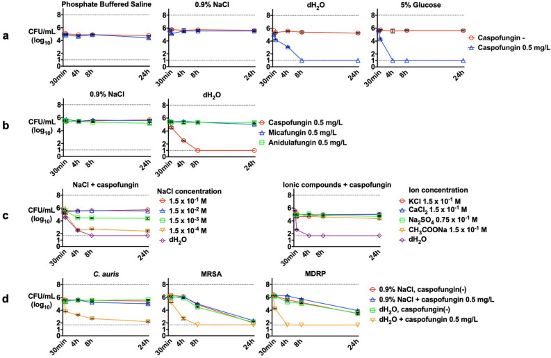
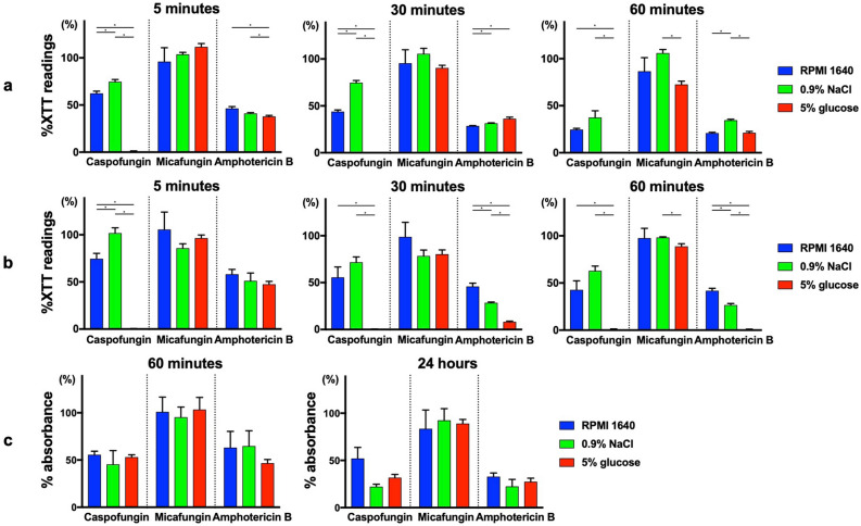
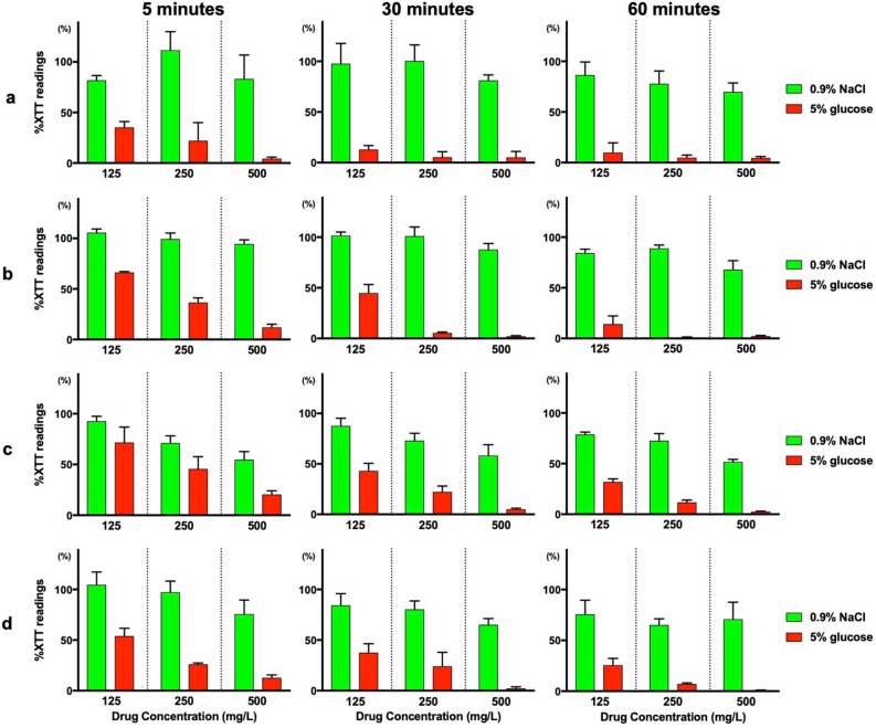
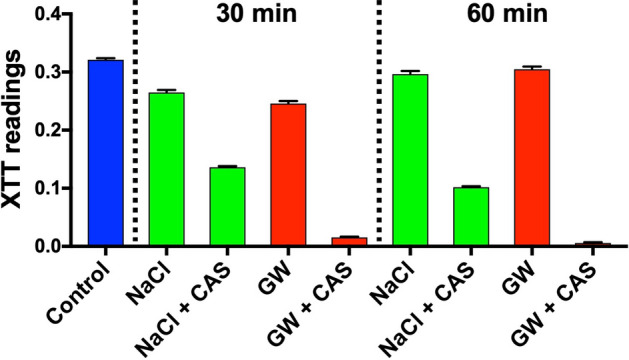
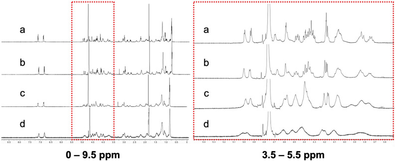
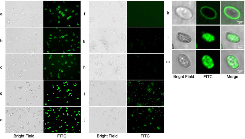
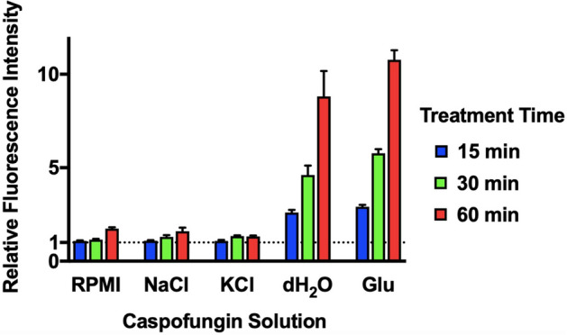
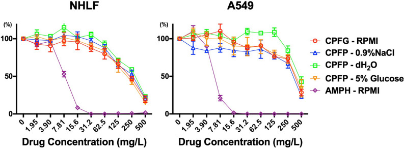
Echinocandins, including caspofungin, micafungin, and anidulafungin, are first-line antifungal agents for the treatment of invasive candidiasis. They exhibit fungicidal activity by inhibiting the synthesis of β-1,3-D-glucan, an essential component of the fungal cell wall. However, they are active only against proliferating fungal cells and unable to completely eradicate fungal cells even after a 24 h drug exposure in standard time-kill assays. Surprisingly, we found that caspofungin, when dissolved in low ionic solutions, had rapid and potent antimicrobial activities against multidrug-resistant (MDR) Candida and bacteria cells even in non-growth conditions. This effect was not observed in 0.9% NaCl or other ion-containing solutions and was not exerted by other echinocandins. Furthermore, caspofungin dissolved in low ionic solutions drastically reduced mature biofilm cells of MDR Candida auris in only 5 min, as well as Candida-bacterial polymicrobial biofilms in a catheter-lock therapy model. Caspofungin displayed ion concentration-dependent conformational changes and intracellular accumulation with increased reactive oxygen species production, indicating a novel mechanism of action in low ionic conditions. Importantly, caspofungin dissolved in 5% glucose water did not exhibit increased toxicity to human cells. This study facilitates the development of new therapeutic strategies in the management of catheter-related biofilm infections.


Conflict of interest statement

The authors declare no competing interests.

Figures

Figure 1
Figure 1
Time-kill assay of Candida and bacterial cells using echinocandin drugs under various conditions. Antifungal effects of (a) caspofungin under non-growth conditions, (b) caspofungin, micafungin, and anidulafungin under non-growth conditions, (c) caspofungin in the presence of different salt concentrations, and (d) caspofungin against C. auris, MRSA, and MDRP. Each data point represents the mean (± SD). Broken lines represent the limits of quantification at the upper (108 CFU/mL) and lower limits (50 CFU/mL).
Figure 2
Figure 2
XTT assay and crystal violet assay of Candida biofilm cells treated with antifungals in different media and solutions. XTT assay of (a) C. albicans and (b) C. auris biofilm cells after 5 min, 30 min, and 60 min of antifungal treatment, respectively. Crystal violet assay of (c) C. albicans biofilm cells after 60 min and 24 h of antifungal treatment. Values are expressed as average percent readings (± SD) relative to control wells containing antifungal-free solution. *p < 0.0056, Bonferroni adjustment.
Figure 3
Figure 3
XTT reduction assay of bacterial and polymicrobial biofilm cells treated with caspofungin. XTT reduction assay of (a) MSSA, (b) MRSA, (c) C. albicans + MSSA, and (d) C. albicans + MRSA biofilm cells after treatment with caspofungin dissolved in 0.9% NaCl and 5% glucose water at the indicated concentrations for 5 min, 30 min, or 60 min, respectively. Values are expressed as average percent readings (± SD) relative to control wells containing antifungal-free solution.
Figure 4
Figure 4
XTT reduction assay using clinically used central venous catheters. C. auris biofilm cells were grown in 14 G central venous catheter and treated with 0.9% NaCl (NaCl) and 5% glucose water (GW), with and without 125 mg/L caspofungin (CAS) for 30 min and 60 min, respectively. The catheters were then injected with XTT solution for spectrophotometric analysis at 492 nm.
Figure 5
Figure 5
1H NMR spectra (500 MHz) of caspofungin in dH2O with and without NaCl. Caspofungin was dissolved in (a) dH2O, (b) NaCl (28 mM), (c) NaCl (125 mM), or (d) NaCl (417 mM). The left panel shows the scale at 0–9.5 ppm, and the right panel shows the red dotted squared section scaled to 3.5–5.5 ppm.
Figure 6
Figure 6
Fluorescent imaging of FITC-caspofungin accumulation patterns in C. glabrata and MRSA. Observed with a digital microscope, C. glabrata was exposed to FITC-caspofungin dissolved in (a) RPMI-1640, (b) 0.9% NaCl, (c) 0.9% KCl, (d) dH2O, and (e) 5% glucose water, MRSA was exposed to FITC-caspofungin dissolved in (f) RPMI-1640, (g) 0.9% NaCl, (h) 0.9% KCl, (i) dH2O, and (j) 5% glucose water. Observed with a confocal laser microscope, C. glabrata was exposed to FITC-caspofungin dissolved in (k) RPMI-1640, (l) dH2O, and (m) 5% glucose water.
Figure 7
Figure 7
Intracellular ROS level of C. glabrata. C. glabrata cells that were treated with 50 μM H2DCFDA and exposed to 50 mg/L caspofungin dissolved in RMPI-1640, 0.9% NaCl, 0.9% KCl, dH2O, and 5% glucose water, respectively. Caspofungin-free solutions were used as controls. The intracellular ROS level was determined by measuring H2DCFDA conversion to fluorescent dichloroluorescein. Fluorescence intensity (FI) was measured at 15 min, 30 min, and 60 min. Relative FI (± SD) was calculated as caspofungin treated FI/control FI.
Figure 8
Figure 8
Toxicity evaluation of caspofungin solutions on human cell lines. NHLF and A549 cells were exposed to caspofungin dissolved in RPMI-1640, 0.9% NaCl, dH2O, and 5% glucose water, and amphotericin B dissolved in RPMI-1640 at various drug concentrations. Toxicity was evaluated by MTT cell viability assay.

Similar articles

Cited by

References

    1. Brown, G. D. et al. Hidden killers: human fungal infections. Sci. Transl. Med.4, 165rv113 (2012). - PubMed
    1. Kullberg BJ, Arendrup MC. Invasive candidiasis. N. Engl. J. Med. 2015;373:1445–1456. doi: 10.1056/NEJMra1315399. - DOI - PubMed
    1. Patil A, Majumdar S. Echinocandins in antifungal pharmacotherapy. J. Pharm. Pharmacol. 2017;69:1635–1660. doi: 10.1111/jphp.12780. - DOI - PubMed
    1. Onishi J, et al. Discovery of novel antifungal (1,3)-β-d-glucan synthase inhibitors. Antimicrob. Agents Chemother. 2000;44:368–377. doi: 10.1128/AAC.44.2.368-377.2000. - DOI - PMC - PubMed
    1. Perlin DS. Echinocandin resistance in Candida. Clin. Infect. Dis. 2015;61:S612–S617. doi: 10.1093/cid/civ791. - DOI - PMC - PubMed

Publication types