. 2021 Jan;17(1):113-121.
doi: 10.1038/s41589-020-00679-1. Epub 2020 Oct 20.

Bi-paratopic and multivalent VH domains block ACE2 binding and neutralize SARS-CoV-2

Collaborators, Affiliations

Bi-paratopic and multivalent VH domains block ACE2 binding and neutralize SARS-CoV-2

Colton J Bracken et al. Nat Chem Biol. 2021 Jan.

Abstract

Neutralizing agents against SARS-CoV-2 are urgently needed for the treatment and prophylaxis of COVID-19. Here, we present a strategy to rapidly identify and assemble synthetic human variable heavy (VH) domains toward neutralizing epitopes. We constructed a VH-phage library and targeted the angiotensin-converting enzyme 2 (ACE2) binding interface of the SARS-CoV-2 Spike receptor-binding domain (Spike-RBD). Using a masked selection approach, we identified VH binders to two non-overlapping epitopes and further assembled these into multivalent and bi-paratopic formats. These VH constructs showed increased affinity to Spike (up to 600-fold) and neutralization potency (up to 1,400-fold) on pseudotyped SARS-CoV-2 virus when compared to standalone VH domains. The most potent binder, a trivalent VH, neutralized authentic SARS-CoV-2 with a half-maximal inhibitory concentration (IC50) of 4.0 nM (180 ng ml-1). A cryo-EM structure of the trivalent VH bound to Spike shows each VH domain engaging an RBD at the ACE2 binding site, confirming our original design strategy.


Conflict of interest statement

Competing Interests:

The authors declare no competing interests.

Figures

Extended Data Fig. 1
Extended Data Fig. 1. CDR loop design of VH-phage library
Schematic of CDR amino acid composition as compared to parental template. Positions in pink highlight CDR H1 charged amino acid insertions. Positions in blue highlight the insertion of “X” synthetic amino acid mixture. Positions in gray remain unchanged from template.
Extended Data Fig. 2
Extended Data Fig. 2. Specificity of lead VH domain binders
Bio-layer interferometry (BLI) binding traces of VH domains at 150nM to RBD and two decoy antigens expressed as biotinylated Fc-fusions: programmed cell death protein 1 (PD-1), and platelet glycoprotein 4 (CD36). Respective VH show successful binding to RBD, but not to either of the decoy antigens.
Extended Data Fig. 3
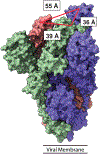
Extended Data Fig. 3. Measurements of inter-RBD distance on Spike trimer
Structure of SARS-CoV-2 Spike trimer (PDB: 6VSB). RBD colored in blue is in the “up” position while RBDs colored in green and pink are in the “down” position. Distance between the mid-points of the ACE2 binding interface (PDB: 6M17) on respective RBDs was measured in Pymol.
Extended Data Fig. 4
Extended Data Fig. 4. ELISA of VH binders to Spike-RBD
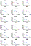
ELISA of VH binders against Spike-RBD. Data presented show the average and standard deviation from three independent experiments. Data were fit to a non-linear, four-parameter variable slope regression model using Prism 8 to obtain EC50 values for each binder.
Extended Data Fig. 5
Extended Data Fig. 5. Pseudotyped virus neutralization by VH Binders
Pseudotyped virus neutralization assays of VH binders. Data represent average and standard deviation of two biological replicates. Data were fit to a non-linear, four-parameter variable slope regression model using Prism 8 to obtain IC50 values.
Extended Data Fig. 6
Extended Data Fig. 6. Cryo-EM reconstruction of SARS-CoV-2 Spike trimer
The SARS-CoV-2 Spike trimer + VH3 B01 cryo-EM reconstruction from non-uniform refinement in cryoSPARC at two different thresholds colored by resolution in the range from 3 Å to 10 Å. At high threshold the core S2 clearly displays high resolution features but the periphery of the molecule is closer to 6–7 Å.
Extended Data Fig. 7
Extended Data Fig. 7. pH dependent binding of lead multivalent and bi-paratopic VH
BLI assay of lead constructs at 25 nM to Secto and RBD at pH 7.4 or pH 4.5. Binding of VH leads is only observed with Secto at pH 7.4 and RBD at pH 4.5.
Figure 1:
Figure 1:. Design and validation of VH-phage library
(A) 3D surface representation (left) of the VH-4D5 parental scaffold (PDB:1FVC) and a cartoon diagram (right) where individual CDRs are annotated in color with the designed loop length variations according to Kabat nomenclature. (B) NGS analysis of the longest H3 loop (X=16) shows that expected global amino acid frequencies are comparable to designed frequencies. Gray region denotes the 95% confidence interval. (C) Representative NGS analysis of the longest H3 loop (X=16) shows positional frequency distribution matches designed frequencies. Position 1 refers to residue 95 (Kabat definition). Data for the other CDR H3 lengths are reported in Fig. S2. (D) NGS analysis of unique clones shows that all H3 lengths are represented in the pooled VH-phage library.
Figure 2:
Figure 2:. Identification of VH domains that bind Spike-RBD at two unique epitopes by phage display
(A) Diagram illustrating phage selection strategy to isolate VH-phage that bind at the ACE2 binding interface. Red indicates clearance of the phage pool by Spike-RBD-Fc/ACE2-Fc complex, green indicates positive selection against Spike-RBD-Fc alone. To increase stringency, successively lower concentrations of Spike-RBD-Fc were used, and after 4 rounds of selection, individual phage clones were analyzed by phage ELISA. BLI of (B) VH A01 (C) VH B01 and (D) VH B02 against Spike-RBD. (E) BLI-based epitope binning of VH A01 and VH B01, (F) VH A01 and VH B02, (G) VH B01 and VH B02. The antigen loaded onto the sensor tip was Spike-RBD. (H) Diagram of the two different epitope bins targeted by VH domains.
Figure 3:
Figure 3:. In vitro characterization of multivalent and bi-paratopic VH binders
(A) Cartoon depiction of engineered VH binders generated by linking VH domains via Fc-fusion or a 20-aa Gly-Ser linker. BLI traces of lead VH binders, (B) VH-Fc B01, (C) VH2 A01-B01, (D) VH3 B01 against RBD (upper panel) or Secto (lower panel). (E) Sequential BLI binding experiments that measured binding of ACE2-Fc to Secto pre-blocked with our VH binders show that multivalent VH binders can block ACE2-Fc binding to Secto. (F) Competition serology ELISA with convalescent patient sera indicates that VH-Fc binders can compete with patient antibodies. P1-P9 are sera from patients with a history of prior SARS-CoV-2 infection. C1-C2 are two donor sera collected before the SARS-CoV-2 outbreak. Individual data points represent technical replicates (n=2) from the serum of the same patient and are shown as black circles.
Figure 4:
Figure 4:. Multivalent and bi-paratopic VH binders neutralize pseudotyped and authentic SARS-CoV-2
(A) Pseudotyped virus IC50 of VH binders. Neutralization potency improves when VH domains are engineered into multivalent and bi-paratopic constructs. (B) Correlation of in vitro binding affinity (KD) and pseudotyped virus neutralization (IC50) of VH binders. Data were fit to a log-log linear extrapolation. (C) Pseudotyped virus neutralization curves of multi-site VH2 in comparison to single-site VH2 demonstrate that the multi-site VH2 have a more cooperative neutralization curve. (D) Pseudotyped virus neutralization curves of mono-, bi-, and tri-valent formats of VH B01 demonstrate potency gains driven by valency. (E) Authentic SARS-CoV-2 neutralization curves for the most potent VH formats were determined via qPCR of viral genome in cellular RNA. All pseudoviral neutralization data were repeated as n=2 independent replicates. Authentic virus neutralization data were repeated as n=2 independent replicates. Data represent the average and standard deviation of replicates.
Figure 5:
Figure 5:. Cryo-EM reveals trivalent VH binding at the ACE2 binding interface of RBD
(A) Side and top views of cryo-EM 3D reconstructions of VH3 B01 + Secto (PDB: 7JWB) are shown with individual VH domain densities of VH3 B01 fit with PDB: 3P9W (VH scaffold; orange cartoon). A total of three VH domains, each bound to an RBD of the Spike trimer, are resolved. 3D model of Secto was fit with reference structure (PDB:6X2B with additional rigid body fit of the individual RBDs; blue cartoon) and shows RBDs in a distinct two “up”, one “down” conformation. Cryo-EM map was low-pass filtered to 6 Å. (B) View of the epitope (Site B) of one VH domain from VH3 B01. Site B overlays directly with the ACE2 binding site (yellow surface; contacts defined as RBD residues within 8 Å of an ACE2 residue from PDB:6M0J).

Update of

Similar articles

Cited by

References

    1. Yan R et al.Structural basis for the recognition of SARS-CoV-2 by full-length human ACE2. Science 367, 1444–1448 (2020). - PMC - PubMed
    1. Abraham J Passive antibody therapy in COVID-19. Nat. Rev. Immunol 20, 401–403 (2020). - PMC - PubMed
    1. Robbiani DF et al.Convergent antibody responses to SARS-CoV-2 in convalescent individuals. Nature 584, 437–442 (2020). - PMC - PubMed
    1. Pinto D et al.Cross-neutralization of SARS-CoV-2 by a human monoclonal SARS-CoV antibody. Nature 583, 290–295 (2020). - PubMed
    1. Rogers TF et al.Isolation of potent SARS-CoV-2 neutralizing antibodies and protection from disease in a small animal model. Science 7520, eabc7520 (2020). - PMC - PubMed

Publication types

MeSH terms