Volume-Staged Radiosurgery for Large Arteriovenous Malformation
- PMID: 33082766
- PMCID: PMC7548954
- DOI: 10.1159/000508943
Volume-Staged Radiosurgery for Large Arteriovenous Malformation
Abstract
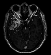
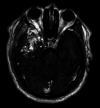
Large arteriovenous malformations (AVMs) are challenges in management because of outcomes and adverse affects. Volume-staged radiosurgery has been an appropriate approach when removal resection and embolization are not recommended. A 53-year-old gentleman was diagnosed with a large intracranial AVM with persistent headache and short-term seizure. Brain magnetic resonance and angiograph showed a bulky volume of AVM nidus. Removal resection and embolization were not recommended because of high risk of adverse affects. The patient was treated by volume-staged radiosurgery. One year post-treatment, obliteration for right internal carotid artery was completed. Volume-staged radiosurgery is a potential treatment option for large AVM with controlled and obliteration efficacy, especially to AVMs which are not appropriate for removal surgery and embolization.
Keywords: Arteriovenous malformation; Digital subtraction angiography; Volume-staged radiosurgery.
Copyright © 2020 by S. Karger AG, Basel.
Conflict of interest statement
The authors declare no financial disclosures or conflicts of interest.
Figures


References
-
- Deruty R, Pelissou-Guyotat I, Morel C, Bascoulergue Y, Turjman F. Reflections on the management of cerebral arteriovenous malformations. Surg Neurol. 1998 Sep;50((3)):245–55. - PubMed
-
- Chapter 10. In. Kalash R, Engh Johnathan A, Amankulor N. Brain Radiosurgery. Stereotactic Radiosurgery and Stereotactic Body Radiation Therapy. SBRT; 2018.
-
- Stefani MA, Porter PJ, terBrugge KG, Montanera W, Willinsky RA, Wallace MC. Large and deep brain arteriovenous malformations are associated with risk of future hemorrhage. Stroke. 2002 May;33((5)):1220–4. - PubMed
-
- Flickinger JC, Kondziolka D, Maitz AH, Lunsford LD. Analysis of neurological sequelae from radiosurgery of arteriovenous malformations: how location affects outcome. Int J Radiat Oncol Biol Phys. 1998 Jan;40((2)):273–8. - PubMed
-
- Lawton MT, Project UB, UCSF Brain Arteriovenous Malformation Study Project Spetzler-Martin Grade III arteriovenous malformations: surgical results and a modification of the grading scale. Neurosurgery. 2003 Apr;52((4)):740–8. - PubMed
Publication types
LinkOut - more resources
Full Text Sources

