

Green-Synthesized Silver Nanoparticles Induced Apoptotic Cell Death in MCF-7 Breast Cancer Cells by Generating Reactive Oxygen Species and Activating Caspase 3 and 9 Enzyme Activities
- PMID: 33082906
- PMCID: PMC7559220
- DOI: 10.1155/2020/1215395
Green-Synthesized Silver Nanoparticles Induced Apoptotic Cell Death in MCF-7 Breast Cancer Cells by Generating Reactive Oxygen Species and Activating Caspase 3 and 9 Enzyme Activities
Abstract
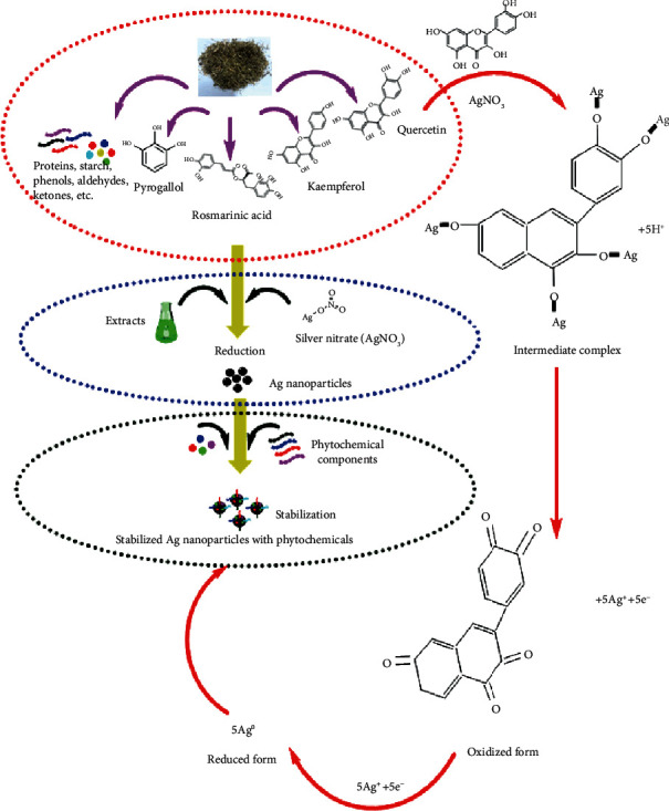
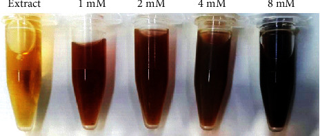
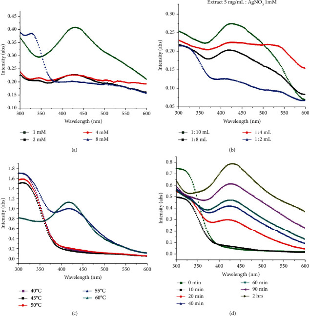
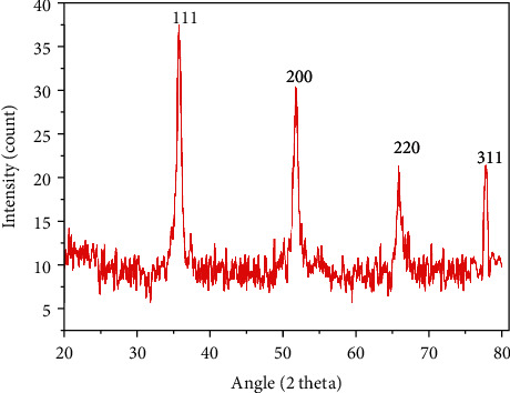
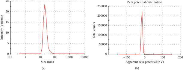
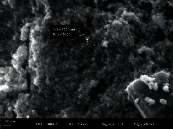
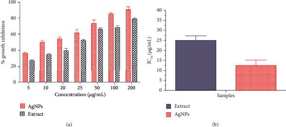
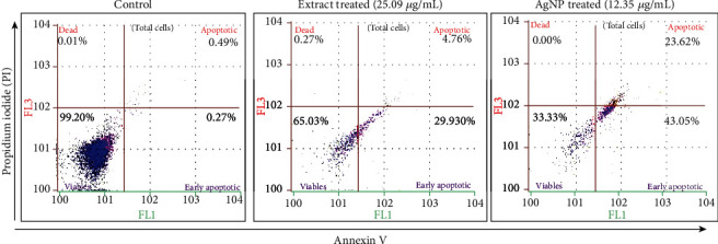
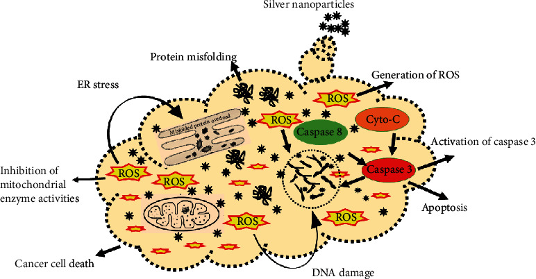
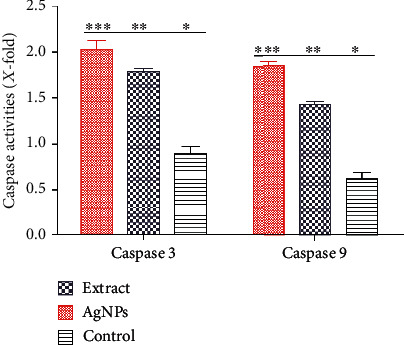
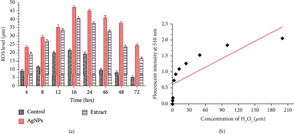
Silver nanoparticles are among the most significant diagnostic and therapeutic agents in the field of nanomedicines. In the current study, the green chemistry approach was made to optimize a cost-effective synthesis protocol for silver nanoparticles from the aqueous extract of the important anticancer plant Fagonia indica. We investigated the anticancer potential and possible involvement of AgNPs in apoptosis. The biosynthesized AgNPs are stable (zeta potential, -16.3 mV) and spherical with a crystal size range from 10 to 60 nm. The MTT cell viability assay shows concentration-dependent inhibition of the growth of Michigan Cancer Foundation-7 (MCF-7) cells (IC50, 12.35 μg/mL). In addition, the fluorescent microscopic analysis shows activation of caspases 3 and 9 by AgNPs that cause morphological changes (AO/EB assay) in the cell membrane and cause nuclear condensation (DAPI assay) that eventually lead to apoptotic cell death (Annexin V/PI assay). It was also observed that AgNPs generate reactive oxygen species (ROS) that modulate oxidative stress in MCF-7 cells. This is the first study that reports the synthesis of a silver nanoparticle mediated by Fagonia indica extract and evaluation of the cellular and molecular mechanism of apoptosis.
Copyright © 2020 Ikram Ullah et al.
Conflict of interest statement
The authors declare that they have no conflict of interest.
Figures

References
-
- Özmen V. Breast cancer in the world and Turkey. Journal of Breast Health. 2008;4:7–12.
-
- Khalil A. T., Ovais M., Ullah I., et al. Sageretia thea (Osbeck.) modulated biosynthesis of NiO nanoparticles and their in vitro pharmacognostic, antioxidant and cytotoxic potential. Artificial Cells, Nanomedicine, and Biotechnology. 2018;46(4):838–852. - PubMed
-
- Khalil A. T., Ovais M., Ullah I., Ali M., Shinwari Z. K., Maaza M. Biosynthesis of iron oxide (Fe2O3) nanoparticles via aqueous extracts ofSageretia thea(Osbeck.) and their pharmacognostic properties. Green Chemistry Letters and Reviews. 2017;10(4):186–201. doi: 10.1080/17518253.2017.1339831. - DOI
-
- Thema F. T., Manikandan E., Dhlamini M. S., Maaza M. Green synthesis of ZnO nanoparticles via Agathosma betulina natural extract. Materials Letters. 2015;161:124–127. doi: 10.1016/j.matlet.2015.08.052. - DOI
MeSH terms
Substances
LinkOut - more resources
Full Text Sources
Research Materials
Miscellaneous
