. 2020 Oct 2:9:171.
doi: 10.1038/s41377-020-00403-7. eCollection 2020.

Miniscope3D: optimized single-shot miniature 3D fluorescence microscopy

Affiliations

Miniscope3D: optimized single-shot miniature 3D fluorescence microscopy

Kyrollos Yanny et al. Light Sci Appl. .

Erratum in

Abstract

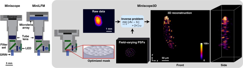
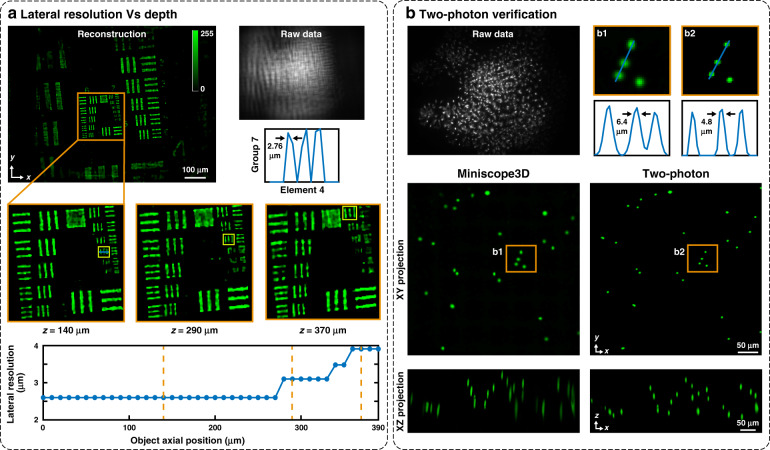
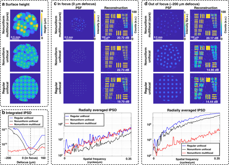
Miniature fluorescence microscopes are a standard tool in systems biology. However, widefield miniature microscopes capture only 2D information, and modifications that enable 3D capabilities increase the size and weight and have poor resolution outside a narrow depth range. Here, we achieve the 3D capability by replacing the tube lens of a conventional 2D Miniscope with an optimized multifocal phase mask at the objective's aperture stop. Placing the phase mask at the aperture stop significantly reduces the size of the device, and varying the focal lengths enables a uniform resolution across a wide depth range. The phase mask encodes the 3D fluorescence intensity into a single 2D measurement, and the 3D volume is recovered by solving a sparsity-constrained inverse problem. We provide methods for designing and fabricating the phase mask and an efficient forward model that accounts for the field-varying aberrations in miniature objectives. We demonstrate a prototype that is 17 mm tall and weighs 2.5 grams, achieving 2.76 μm lateral, and 15 μm axial resolution across most of the 900 × 700 × 390 μm3 volume at 40 volumes per second. The performance is validated experimentally on resolution targets, dynamic biological samples, and mouse brain tissue. Compared with existing miniature single-shot volume-capture implementations, our system is smaller and lighter and achieves a more than 2× better lateral and axial resolution throughout a 10× larger usable depth range. Our microscope design provides single-shot 3D imaging for applications where a compact platform matters, such as volumetric neural imaging in freely moving animals and 3D motion studies of dynamic samples in incubators and lab-on-a-chip devices.

Keywords: Imaging and sensing; Microscopy.


Conflict of interest statement

Conflict of interestThe authors declare that they have no conflict of interest.

Figures

Fig. 1
Fig. 1. Miniscope3D system overview.
Compared with previous Miniscope and MiniLFM designs, our Miniscope3D is lighter weight and more compact. We remove the Miniscope’s tube lens and place a 55 μm thick optimized phase mask at the aperture stop (Fourier plane) of the GRIN objective lens. A sparse set (64 per depth) of calibration point spread functions (PSFs) is captured by scanning a 2.5 μm green fluorescent bead throughout the volume. We use this data set to pre-compute an efficient forward model that accurately captures field-varying aberrations. The forward model is then used to iteratively solve an inverse problem to reconstruct 3D volumes from single-shot 2D measurements. The 3D reconstruction here is of a freely swimming fluorescently tagged tardigrade
Fig. 2
Fig. 2. Experimental characterization.
a Reconstructions of a fluorescent USAF target at different axial positions to determine the depth-dependent lateral resolution. We recover a 2.76 μm resolution across most of the 390 μm range of depths, with the worst case of 3.9 μm (dashed orange lines mark the inset locations, and yellow boxes in the insets indicate the smallest resolved groups). Note that the resolution target has discrete levels of resolution that result in jumps in the data and that resolution here refers to the gap between bars, not the line-pair width. b Reconstruction of a 160 μm thick sample of 4.8 μm fluorescent beads compared with a two-photon 3D scanning image (maximum intensity projections in the yx and zx planes are shown). Our system detects the same features, with a slightly larger lateral spot size
Fig. 3
Fig. 3. Experimental 3D reconstructions.
a GFP-tagged neurons in two different samples of 100 μm thick fixed mouse brain tissue. b a 300-μm thick optically cleared mouse brain slice. We clearly resolve dendrites running across the volume axially (see Video 1). All mouse brain volume reconstructions are 790 × 617 × 210 μm3. c Maximum intensity projections from several frames of the reconstructed 3D videos of two different samples of freely moving tardigrades captured at a maximum of 40 frames per second (see Videos 2 and 3)
Fig. 4
Fig. 4. Each 3D voxel maps to a different PSF.
a As a point source translates axially, the PSF scales, and different spots come into focus. b As a point source translates laterally, the PSF shifts and incurs field-varying aberrations that destroy shift invariance. c When a shift-invariant approximation is made, reconstructions of a fluorescent resolution target (at z = 250 μm) display worse resolution (6.2 μm resolution) and more artefacts than when our field-varying model is used (2.76 μm resolution)
Fig. 5
Fig. 5. Simulations to motivate our phase mask design, comparing our proposed nonuniform multifocal design with regular unifocal and nonuniform unifocal designs.
a Surface height profiles. b Sum of each design’s PSF inverse power spectral density (IPSD) versus object depth (up to the designed cutoff frequency, where lower is better). c PSFs and simulated reconstructions in-focus (at the native focus of the unifocal arrays), with the reconstruction peak signal-to-noise ratio (PSNR) listed. The measurement is corrupted with 100 e−1 (peak) Poisson noise. In focus, the nonuniform unifocal design has a slightly better PSNR and resolution than those of our design, while the regular unifocal design performs worse. The radially averaged IPSD (lower is better) matches this trend. d Imaging 200 μm off-focus, both unifocal designs produce blurry PSFs, which result in a significantly worse PSNR and resolution in the reconstruction compared with those of our design. This result is also seen in the much higher inverse power spectra curves for the unifocal designs
Fig. 6
Fig. 6. Phase mask parameterized by the point-wise maximum of convex spheres.
Each sphere is outlined by a dashed line, and the final optic is shaded blue (not to scale)
Fig. 7
Fig. 7. Comparison of our optimized phase mask with random multifocal and regular microlens arrays.
a Phase mask surface height maps for all three cases, including the designed aberrations that were added in our optimized phase mask. b Axial cross-coherence matrices for all three cases and the target matrix: each entry is the maximum cross-correlation between the PSFs at the depths indicated by the row and column labels. The ideal system, which is not feasible, would be close to an identity matrix. c xz slices from the 3D reconstructions of a test object consisting of differently spaced point sources (x-spacings of 3.5 μm and 7 μm, z-spacings of 19.4 μm and 38 μm). We add Poisson noise with 1000 peak counts to each measurement. Both nonuniform multifocal designs do significantly better than the regular unifocal array, and our optimized design performs slightly better than the random version, particularly near the edges of the depth
Fig. 8
Fig. 8. Phase mask fabrication with nanoscribe.
a Rectangular stitching leads to seams (black lines) going through the many microlenses, whereas adaptive stitching puts the seams at the boundaries of the microlenses to mitigate artefacts. b Comparison between designed and experimental PSFs at a few sample depths, showing good agreement, with slight degradation at the edge of the volume

References

    1. Ghosh KK, et al. Miniaturized integration of a fluorescence microscope. Nat. Methods. 2011;8:871–878. doi: 10.1038/nmeth.1694. - DOI - PMC - PubMed
    1. Liberti WA, III, et al. J. An open source, wireless capable miniature microscope system. J. Neural Eng. 2017;14:045001. doi: 10.1088/1741-2552/aa6806. - DOI - PMC - PubMed
    1. Jacob AD, et al. A compact head-mounted endoscope for in vivo calcium imaging in freely behaving mice. Curr. Protoc. Neurosci. 2018;84:e51. doi: 10.1002/cpns.51. - DOI - PubMed
    1. De Groot A, et al. NINscope, a versatile miniscope for multi-region circuit investigations. eLife. 2020;9:e49987. doi: 10.7554/eLife.49987. - DOI - PMC - PubMed
    1. UCLA. Miniscope. at https://miniscope.org.