. 2020 Sep 28:5:91.
doi: 10.1038/s41541-020-00243-x. eCollection 2020.

Ad26 vector-based COVID-19 vaccine encoding a prefusion-stabilized SARS-CoV-2 Spike immunogen induces potent humoral and cellular immune responses

Affiliations

Ad26 vector-based COVID-19 vaccine encoding a prefusion-stabilized SARS-CoV-2 Spike immunogen induces potent humoral and cellular immune responses

Rinke Bos et al. NPJ Vaccines. .

Abstract

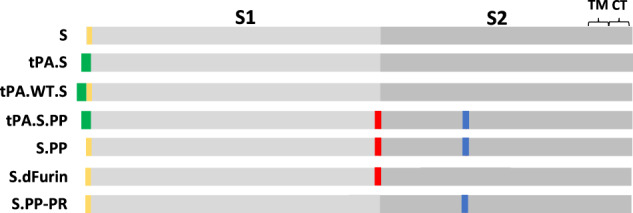
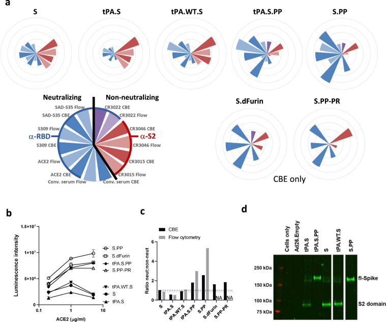
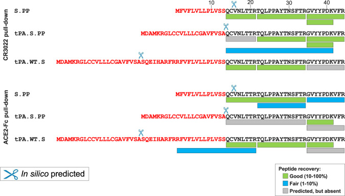
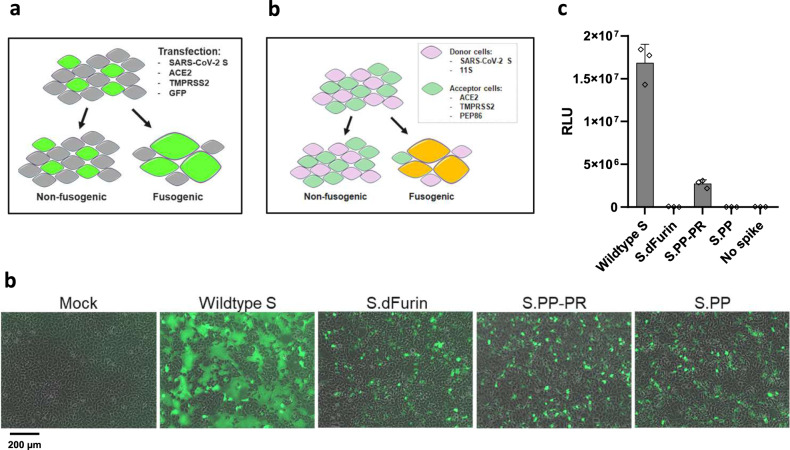
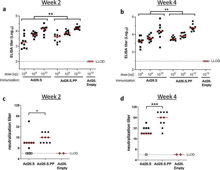
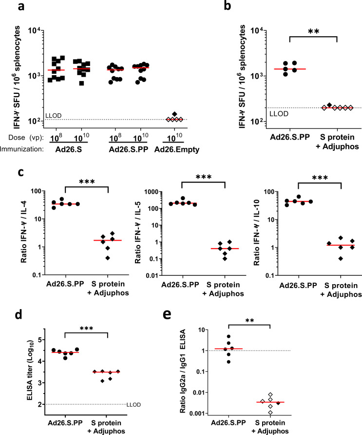
Development of effective preventative interventions against SARS-CoV-2, the etiologic agent of COVID-19 is urgently needed. The viral surface spike (S) protein of SARS-CoV-2 is a key target for prophylactic measures as it is critical for the viral replication cycle and the primary target of neutralizing antibodies. We evaluated design elements previously shown for other coronavirus S protein-based vaccines to be successful, e.g., prefusion-stabilizing substitutions and heterologous signal peptides, for selection of a S-based SARS-CoV-2 vaccine candidate. In vitro characterization demonstrated that the introduction of stabilizing substitutions (i.e., furin cleavage site mutations and two consecutive prolines in the hinge region of S2) increased the ratio of neutralizing versus non-neutralizing antibody binding, suggestive for a prefusion conformation of the S protein. Furthermore, the wild-type signal peptide was best suited for the correct cleavage needed for a natively folded protein. These observations translated into superior immunogenicity in mice where the Ad26 vector encoding for a membrane-bound stabilized S protein with a wild-type signal peptide elicited potent neutralizing humoral immunity and cellular immunity that was polarized towards Th1 IFN-γ. This optimized Ad26 vector-based vaccine for SARS-CoV-2, termed Ad26.COV2.S, is currently being evaluated in a phase I clinical trial (ClinicalTrials.gov Identifier: NCT04436276).

Keywords: SARS-CoV-2; Vaccines; Viral infection.


Conflict of interest statement

Competing interestsThe authors declare no competing financial interests. R.B., L.R., M.J.G.B., F.W., D.Z., and J.P.L. are co-inventors on related vaccine patents. R.B., L.R., J.E.M.vd.L., M.J.G.B., G.H., F.W., D.Z., A.H.d.W., A.K., A.V., D.v.M., T.K., R.V., J.V., J.P.M.L., R.C.Z, J.C., and H.S. are employees of Janssen Vaccines & Prevention BV. R.B., L.R., F.W., D.Z., D.v.M., T.K., R.V., J.V., J.P.M.L., R.C.Z, J.C., and H.S. hold stock of Johnson & Johnson.

Figures

Fig. 1
Fig. 1. S immunogen designs.
Seven plasmids encoding variants of the SARS-CoV-2 S protein were produced: (1) native full-length S (S), (2) full-length S in which the wt SP is replaced by tissue plasminogen activator SP (tPA.S), (3) full-length S in which tPA SP is added upstream of the wt SP (tPA.WT.S), (4) full-length S in which SP is replaced by tPA and in which the furin cleavage site mutations and proline substitutions (K986P, V987P) have been introduced (tPA.S.PP), (5) full-length S in which furin cleavage site mutations and proline substitutions have been introduced (S.PP), (6) full-length S with only the furin cleavage site mutations (S.dFurin), and (7) full-length S with only the proline substitutions (S.PP-PR), which is therefore processed (PR) at the furin cleavage site. Green bars represent tPA SP, yellow bars represent wild-type SP, red vertical lines represent furin cleavage site mutations, and blue vertical lines represent proline substitutions.
Fig. 2
Fig. 2. S protein antigenicity.
a Radar plots showing luminescence intensities measured with cell-based ELISA (CBE) and median fluorescence intensity (MFI) measured with flow cytometry (“Flow”). For CBE, cells were transfected with DNA constructs, whereas for flow cytometry cells were transduced with Ad26 vectors. Luminescence and MFI were calculated as an average of a duplicate and the MFI were normalized (by multiplying with a factor 342) to have the highest intensity the same as the highest CBE luminescence intensity. The outer ring of the circles represents a value of 10,000,000. Conv. serum stands for convalescent serum. S.dFurin and S.PP-PR were measured only with CBE. b ACE2 binding measured with CBE (n = 2). Data are represented as mean + s.d. c Average luminescence intensities measured with CBE or MFI measured with flow cytometry of neutralizing ligands and antibodies divided by that of non-neutralizing antibodies. The black horizontal dashed line indicates the height of the neut:non-neut ratio for S in CBE and the gray one for S in flow cytometry. d Western blot analysis for expression from Ad26 vaccine vectors encoding tPA.S, tPA.S.PP, S, tPA.WT.S, and S.PP in MRC-5 cell lysates under non-reduced conditions using a human monoclonal antibody (CR3046). Ad26.Empty was included as negative control.
Fig. 3
Fig. 3. Signal peptide analysis of membrane-bound S proteins.
LC-MS/MS analysis of the N-terminus of S.PP, tPA.S.PP, and tPA.WT.S after either ACE2-Fc or CR3022 pull-down from cell membranes. Sequences are shown of the wt SP, tPA SP and the tPA SP linked to the wt SP (in red letters) upstream of the mature S protein sequence (in black letters). Cleavage events as predicted by SignalP-5.0 are indicated with scissors. The recovery rates of the different expected peptides are color-coded. LOD lower limit of detection.
Fig. 4
Fig. 4. Effect of stabilizing mutations on fusogenicity of S protein.
a S protein fusogenicity as measured in a cell–cell fusion assay in HEK293 cells by co-transfection of plasmids encoding S protein, ACE2, TMPRSS2, and GFP. b Overlay of GFP and brightfield channels 24 h after transfection, as in the setup of a. The different S protein constructs are indicated; mock is an untransfected monolayer. The scale bar is indicating 200 μm. c Quantitative cell–cell fusion assay setup. d Luciferase signal shown as relative light units (RLU) measured at 4 h post mixing of donor and acceptor cells, as in the setup of c. The error bars represent one s.d.
Fig. 5
Fig. 5. Immunization with Ad26-based vaccine constructs induce humoral immune responses in immunized mice.
Naïve mice (C57BL/6, N = 5 per group) were immunized with either 108, 109, or 1010 vp of Ad26-based vaccine candidates, or with 1010 vp of an Ad26 vector without gene insert as control (Ad26.Empty). Four weeks and six weeks after the immunization, S protein-specific binding and NAb titers were determined. a + b Spike protein-specific antibody binding titers were measured by ELISA. c + d SARS-CoV-2 NAb titers were measured by wt VNA determining the inhibition of the cytopathic effect (CPE) of virus isolate Leiden1 (L-0001) on Vero-E6 cells. Mice immunized with Ad26.Empty were taken along as two separate pools. Median responses per group are indicated with horizontal lines. Dotted lines indicate the LLOD per assay. Animals with a response at or below the LLOD were put on LLOD and are shown as open symbols. Statistical differences as determined by Cochran–Mantel–Haenszel tests (a, b) or Exact Wilcoxon rank-sum test (c, d) are indicated by asterisks; *p < 0.05. LLOD lower limit of detection, vp virus particles.
Fig. 6
Fig. 6. Significant higher S protein binding antibody titers and SARS-CoV-2 neutralization titers induced by Ad26.S.PP compared to Ad26.S.
Naïve mice (BALB/c, N = 10 per group) were immunized with either 108, 109, or 1010 vp of Ad26.S.PP, Ad26.S or with Ad26.Empty (N = 5). Serum was sampled 2 and 4 weeks post immunization and splenocytes were analyzed 4 weeks post immunization. a + b S protein-specific binding antibody titers were measured by ELISA. c + d SARS-CoV-2 NAb titers were measured by wt VNA. Mice immunized with Ad26.empty were taken along as two separate pools in VNA. Median responses per group are indicated with horizontal lines. Dotted lines indicate the LLOD per assay. Animals with a response at or below the LLOD were put on LLOD and are shown as open symbols. Statistically significant differences between groups per dose, or across doses (indicated by brackets), as determined by t-test from ANOVA, are indicated by asterisks; *p < 0.05, **p < 0.01; ***p < 0.001. LLOD lower limit of detection, vp virus particles.
Fig. 7
Fig. 7. Analysis of IFN-γ secretion and Th1/Th2 skewing in Ad26.S.PP immunized mice.
a Naïve BALB/c mice were immunized with either 108 or 1010 vp of Ad26.S.PP, Ad26.S (N = 10 per group) or with Ad26.Empty (N = 5). Number of IFN-γ producing cells per million splenocytes 4 weeks after immunization as measured by ELISpot. Median responses per group are indicated with horizontal lines. Dotted line indicates the LLOD. Animals with a response at or below the LLOD were put on LLOD and are shown as open symbols. be Naïve mice (BALB/c, N = 6 per group) were immunized with either 1010 vp of Ad26.S.PP or 50 µg of S protein adjuvanted with 100 µg Adjuphos (Adju-Phos®). Two weeks after immunization samples were analyzed for antibody and cellular responses. b Number of IFN-γ producing cells per million splenocytes as measured by ELISpot. One mouse excluded due to high background in peptide pool 1 stimulation. Dotted line indicates the LLOD. c Th1 (IFN-γ) over Th2 (IL-4, IL-5, and IL-10) cytokine ratios measured by multiplex ELISA after stimulation of splenocytes with SARS-CoV-2 S protein peptides. d S protein-specific IgG binding antibody titers were measured by ELISA. e Ratio of IgG2a/IgG1 S protein-specific antibody titers as measured by IgG2a and IgG1 ELISA. Dotted line indicates an IgG2a/IgG1 ratio of 1. Animals with a response at or below the LLOD in the IgG2a ELISA are shown as open symbols. Horizontal lines denote group medians. Statistically significant differences as determined by t-test from ANOVA (a), z-test from Tobit ANOVA (c, d), or Mann–Whitney U test (b, e) are indicated by asterisks **p < 0.01; ***p < 0.001. LLOD lower limit of detection, vp virus particles.

Similar articles

Cited by

References

    1. Coronaviridae Study Group of the International Committee on Taxonomy of, V. The species Severe acute respiratory syndrome-related coronavirus: classifying 2019-nCoV and naming it SARS-CoV-2. Nat. Microbiol. 2020;5:536–544. doi: 10.1038/s41564-020-0695-z. - DOI - PMC - PubMed
    1. Sternberg A, Naujokat C. Structural features of coronavirus SARS-CoV-2 spike protein: targets for vaccination. Life Sci. 2020;257:118056. doi: 10.1016/j.lfs.2020.118056. - DOI - PMC - PubMed
    1. Krarup A, et al. A highly stable prefusion RSV F vaccine derived from structural analysis of the fusion mechanism. Nat. Commun. 2015;6:8143. doi: 10.1038/ncomms9143. - DOI - PMC - PubMed
    1. Rutten L, et al. A universal approach to optimize the folding and stability of prefusion-closed HIV-1 envelope trimers. Cell Rep. 2018;23:584–595. doi: 10.1016/j.celrep.2018.03.061. - DOI - PMC - PubMed
    1. Sanders RW, et al. Stabilization of the soluble, cleaved, trimeric form of the envelope glycoprotein complex of human immunodeficiency virus type 1. J. Virol. 2002;76:8875–8889. doi: 10.1128/JVI.76.17.8875-8889.2002. - DOI - PMC - PubMed

Associated data