. 2020 Oct 12:5:94.
doi: 10.1038/s41541-020-00240-0. eCollection 2020.

Optimising T cell (re)boosting strategies for adenoviral and modified vaccinia Ankara vaccine regimens in humans

Affiliations

Optimising T cell (re)boosting strategies for adenoviral and modified vaccinia Ankara vaccine regimens in humans

Stefania Capone et al. NPJ Vaccines. .

Abstract

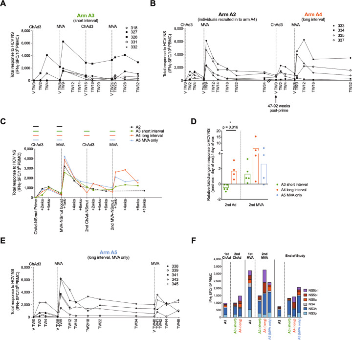
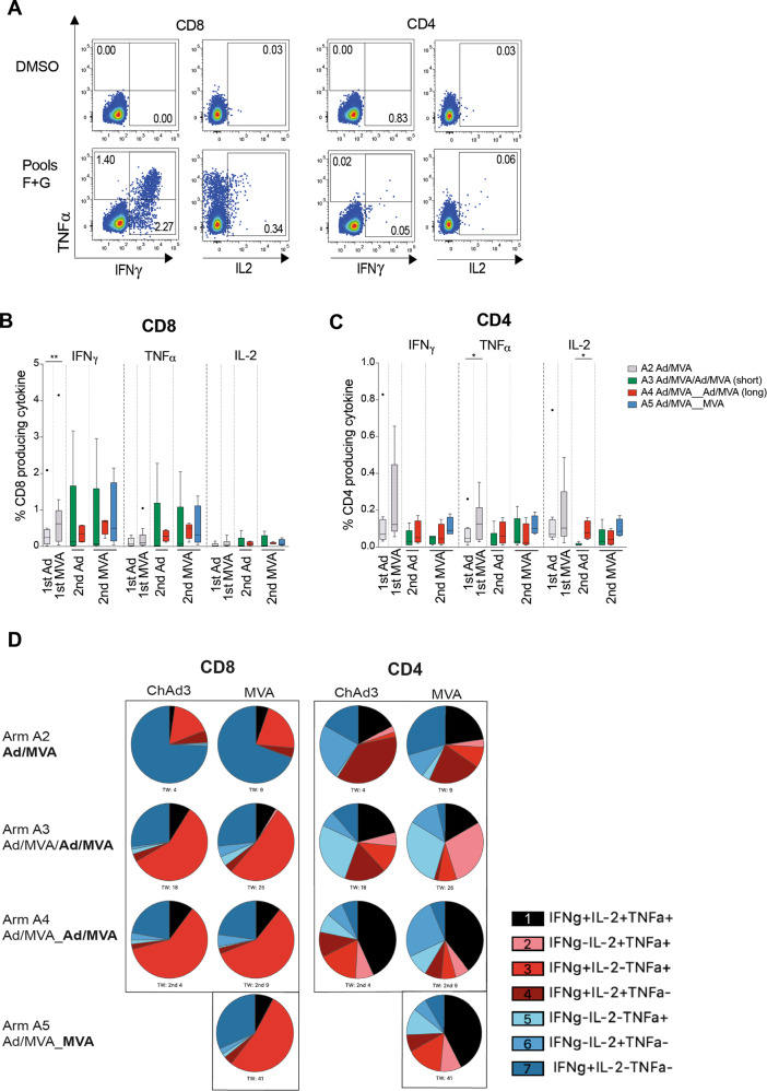
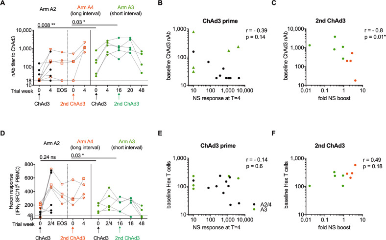
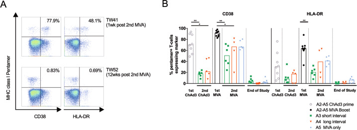
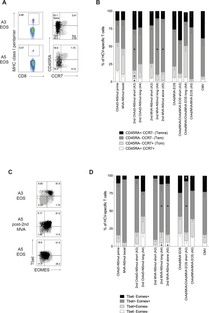
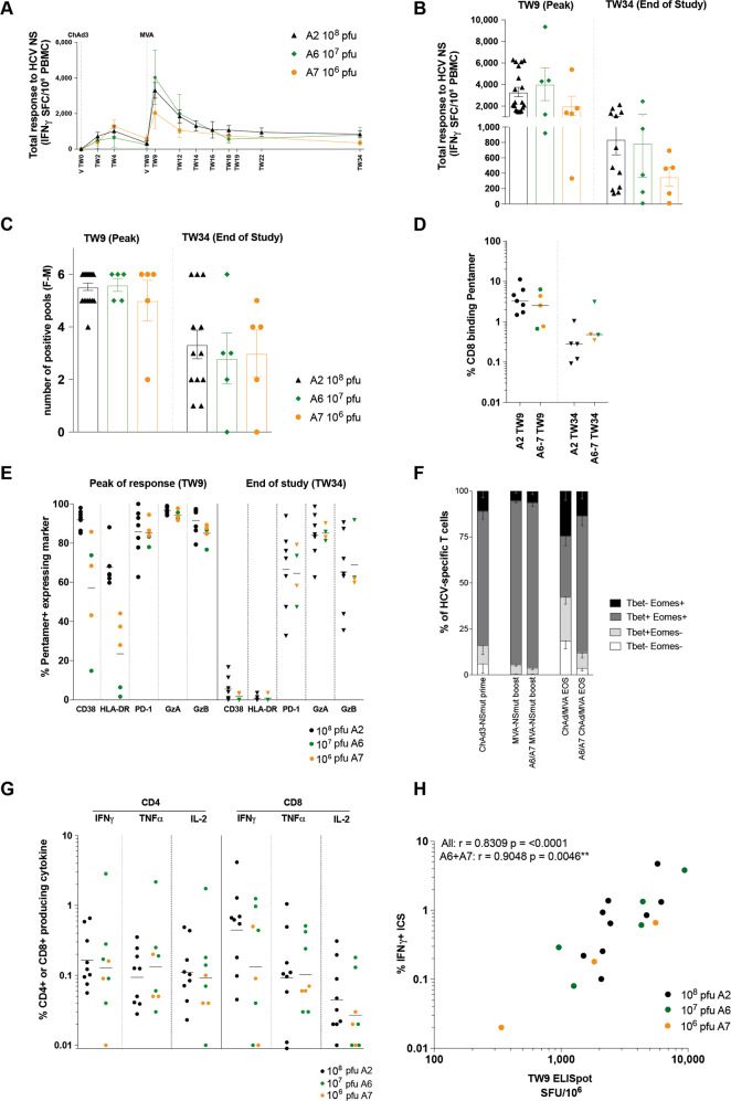
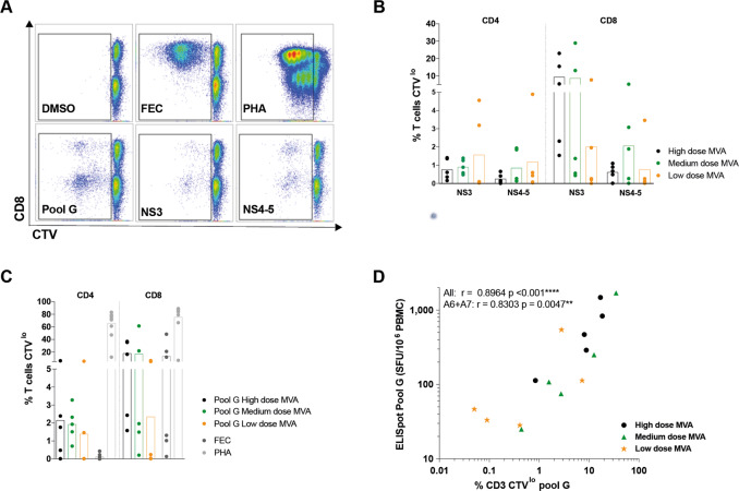
Simian adenoviral and modified vaccinia Ankara (MVA) viral vectors used in heterologous prime-boost strategies are potent inducers of T cells against encoded antigens and are in advanced testing as vaccine carriers for a wide range of infectious agents and cancers. It is unclear if these responses can be further enhanced or sustained with reboosting strategies. Furthermore, despite the challenges involved in MVA manufacture dose de-escalation has not been performed in humans. In this study, healthy volunteers received chimpanzee-derived adenovirus-3 and MVA vaccines encoding the non-structural region of hepatitis C virus (ChAd3-NSmut/MVA-NSmut) 8 weeks apart. Volunteers were then reboosted with a second round of ChAd3-NSmut/MVA-NSmut or MVA-NSmut vaccines 8 weeks or 1-year later. We also determined the capacity of reduced doses of MVA-NSmut to boost ChAd3-NSmut primed T cells. Reboosting was safe, with no enhanced reactogenicity. Reboosting after an 8-week interval led to minimal re-expansion of transgene-specific T cells. However, after a longer interval, T cell responses expanded efficiently and memory responses were enhanced. The 8-week interval regimen induced a higher percentage of terminally differentiated and effector memory T cells. Reboosting with MVA-NSmut alone was as effective as with ChAd3-NSmut/MVA-NSmut. A ten-fold lower dose of MVA (2 × 10⁷pfu) induced high-magnitude, sustained, broad, and functional Hepatitis C virus (HCV)-specific T cell responses, equivalent to standard doses (2 × 10⁸ pfu). Overall, we show that following Ad/MVA prime-boost vaccination reboosting is most effective after a prolonged interval and is productive with MVA alone. Importantly, we also show that a ten-fold lower dose of MVA is as potent in humans as the standard dose.

Keywords: Adaptive immunity; Live attenuated vaccines; Vaccines.


Conflict of interest statement

Competing interestsS.Co., A.F., R.C., and A.N. are named inventors on patent applications covering HCV-vectored vaccines and chimpanzee adenovirus vectors [WO 2006133911 (A3) hepatitis C virus nucleic acid vaccine, WO 2005071093 (A3) chimpanzee adenovirus vaccine carriers, WO 03031588 (A2) hepatitis C virus vaccine]. V.V. is an employee of GSK group of companies. The rest of the authors declare that there are no competing interests.

Figures

Fig. 1
Fig. 1. Reboosting vaccination schedules.
A chimpanzee-derived adenovirus 3 (ChAd3) and a modified vaccinia Ankara (MVA) were engineered to express the non-structural region (NS3-5b, BK strain genotype 1b, NSmut) of hepatitis C virus. Healthy volunteers were given a prime-boost vaccine regimen (ChAd3-NSmut at trial week 0 [TW0], MVA-NSmut boost at TW8) followed by reboosting vaccination with a second round of ChAd3-NSmut/MVA-NSmut after a short interval (all vaccines 8 weeks apart, arm A3), after a long interval (TW47-92, arm A4; volunteers re-recruited from arm A2), or were reboosted with MVA-NSmut alone after a long interval (TW40, arm A5). Vaccine dose 2.5 × 10¹⁰ viral particles (vp) for ChAd3-NSmut and 2 × 10⁸ plaque forming units (pfu) for MVA-NSmut.
Fig. 2
Fig. 2. HCV-specific T cell responses are expanded by reboosting after a long interval with a second round of ChAd3-NSmut and MVA-NSmut or with MVA-NSmut alone.
ac The total ex vivo T cell response to the non-structural (NS) region of HCV encoded within the vaccine is shown over time (IFNγ ELISpot; spot forming cells per 106 PBMC; calculated by summing the responses to positive peptide pools corrected for background; materials and methods). Individual healthy volunteers are shown for arms A3 (a), arm A2/A4 (b) and overlaid group means (c). Colored horizontal lines above the graph indicate when a vaccine was given. d Fold change in T cell response to HCV NS (post-vaccination response–pre-vaccination response/pre-vaccination response). Post-vaccination response was taken as +2 weeks for ChAd3, +1 week for MVA. Bars at group mean. e The total ex vivo T cell response to the NS region of HCV over time for individual volunteers for arm A5. ac, e Vertical dashed lines indicate time of vaccination. chAd3, chimpanzee-derived adenovirus 3; MVA, modified vaccina Ankara. f The ex vivo T cell response to HCV by antigen at the peak of the T cell response post-vaccination and at the end of the study (2–4 weeks post-ChAd3-NSmut and 1–4 weeks post-MVA-NSmut for peak response and 14–40 weeks post vaccination for end of study; group mean per peptide pool by IFNγ ELISpot, n = 4–9; all pools background subtracted).
Fig. 3
Fig. 3. Cytokine-producing HCV-specific T cells are induced by reboosting after a long interval.
ac Comparison of ex vivo cytokine production by T cells at the time of peak magnitude of response (peak NS response by IFNγ ELISpot) to each vaccination (1–4 weeks post-ChAd3-NSmut and MVA-NSmut). a Example plots showing TNFα/IFNγ and IL-2/TNFα after ICS for CD4+ and CD8+ T cells stimulated with pools F+G or DMSO control (volunteer 343 arm A5 TW41, 2nd MVA; see Supplementary Fig. 4 for full gating strategy). The percentage of total CD8+ (b) or CD4+ (c) T cells producing IFNγ, TNFα, or IL-2 after overnight stimulation with peptides covering the non-structural region of HCV within the vaccines (Pools F+G, H+I, L+M summed after subtracting values for paired DMSO wells from each stimulation) are shown. d The proportion of the transgene-specific CD4+ or CD8+ T cells producing different combinations of IFNγ, TNFα, and IL-2 are presented as pie charts (base mean) at the peak of the T cell response post vaccination (samples with a transgene-specific response <0.025% of CD4+ or CD8+ T cells were excluded from polyfunctionality analysis; n = 4–9 per arm). b, c Box–whisker Tukey, outliers shown as dots (n = 9 A2, n = 5 arm A3, n = 4 arms A4, A5). Wilcoxon paired t test 1st Ad vs. 1st MVA, Mann–Whitney unpaired t test for 2nd Ad A3 vs. A4, Kruskal–Wallis one-way ANOVA with Dunn’s correction for 2nd MVA A3, A4, A5 per cytokine. Ad, adenovirus; MVA, modified vaccina Ankara. *P < 0.05; **P < 0.005.
Fig. 4
Fig. 4. Anti-vector immunity.
a Titer of neutralising antibodies (nAbs) against ChAd3 vector (dotted line at nAb titer of 18, limit of detection for the assay). Volunteers who received ChAd3-NSmut prime and MVA-NSmut boost as part of arm A2 which were then reboosted in arm A4 are indicated by red symbols. Trial week indicates weeks since first vaccination for arm A2 and A3 and time of second ChAd3 vaccination for Arm A4. b Anti-ChAd3 nAb titer at baseline vs. peak magnitude of T cell responses to NS region of HCV post-vaccination (IFNγ ELISpot; spot forming cells per 106 PBMC). c Anti-ChAd3 nAb titer at time of second ChAd3-NSmut vaccination vs. fold change in magnitude of T cell responses to HCV NS post-vaccination. Fold change (FC) was calculated as (total NS response 2 weeks post-vaccination—NS response on day of second ChAd3-NSmut vaccination)/NS response pre-vaccination. d Ex vivo T cell response to Ad5 hexon (IFNγ ELISpot; horizontal dotted line at 48, positivity cut-off for the assay). Trial week indicates weeks since first vaccination for arm A2 and A3 and time of second ChAd3 vaccination for Arm A4. e T cell response to Ad5 hexon at baseline vs. peak magnitude of T cell responses to NS region of HCV post-vaccination. f T cell response to Ad5 hexon at time of second ChAd3-NSmut vaccination vs. fold change in magnitude of T cell responses to HCV NS post vaccination (FC calculated as above). a, d Paired Wilcoxon t test. b, c, e, f Spearman rank correlation. ChAd3, chimpanzee-derived adenovirus 3; EOS, end of study. NS, non-structural; MVA, modified vaccina Ankara.
Fig. 5
Fig. 5. T cell activation is strongest after MVA vaccination but it is not affected by the length of the interval since last NS encoding vaccination.
a Example plots (CD8+ T cells) showing pentamer vs. activation marker (arm A5) 1 week and 12 weeks post second MVA-NSmut vaccination (see Supplementary Fig. 4 for full gating strategy). Percentage of pentamer+ cells expressing the activation marker is shown. b Ex vivo MHC class I pentamer staining was performed on all HLA-A*01+ or A*02+ individuals (NS31435–1443, NS31406–1415, NS3−1073–1081, see methods) from arms A2-A5. PBMC were co-stained with individual pentamers and antibodies against the CD8+ T cell surface activation markers CD38 and HLA-DR. The percentage of pentamer+ cells expressing these markers is shown, bars at median. b Kruskal–Wallis one-way ANOVA with Dunn’s correction comparing all group means for 1st and 2nd ChAd3-NSmut vaccination, and for 1st and second MVA vaccination for CD38 and for HLA-DR, significant differences shown. *P < 0.05; **P < 0.005.
Fig. 6
Fig. 6. Reboosting alters the proportions of T cell memory subsets.
(ad) PBMC from HLA-A*01+ or A*02+ individuals were co-stained ex vivo with individual MHC class I pentamers and antibodies against surface markers CD45RA, CCR7 (a, b), or intranuclear transcription factors Tbet and Eomes (c, d). a Example FACS plots (pentamer gating and overlay of total CD8+ in grey and pentamer+ in black; see Supplementary Fig. 4 for full gating strategy). b Stacked bars showing the proportion of pentamer+ T cells with a given phenotype at the peak of the T cell response (1–4 weeks post vaccination, according to IFNγ ELISpot) after each vaccination and at the end of the study (EOS; n = 4–13 per pie). Where CMV responses were detected by IFNγ ELISpot in HLA-A*02+ individuals an immunodominant CMV pentamer was used in parallel (n = 10). c Example FACS plots (overlay of total CD8+ in grey and pentamer+ in black). d Stacked bars showing the proportion of pentamer+ T cells co-expressing Tbet and Eomes at the peak of the response (1–4 weeks post vaccination, according to IFNγ ELISpot) after each vaccination and at the end of the study (n = 4–13 per pie). The proportion of CMV-specific T cells showing each subset phenotype is also shown (n = 6). b, d Mean, bars at standard error of mean. Kruskal–Wallis unpaired non-parametric Anova comparing ChAd3-NSmut prime vs. 2nd ChAd-NSmut short (A3) or long (A4), MVA-NSmut boost vs. 2nd MVA-NSmut short (A3) or long (A4) or alone (A5), ChAd3-NSmut/MVA-NSmut EOS vs. ChAd/MVA/CMVA EOS short (A3) or ChAd/MVA/MVA (A5). *P < 0.05.
Fig. 7
Fig. 7. The magnitude, breadth, functionality, and phenotype of vaccine-induced HCV-specific T cells are unchanged when using a medium MVA-NSmut dose when compared to high dose.
ah Healthy volunteers receiving ChAd3-NSmut prime vaccination were boosted with high (108 pfu; black dots, arm A6), medium (107 pfu; green dots, arm A7), or low dose (106 pfu; orange dots) MVA-NSmut vaccination 8 weeks later (Supplementary Table 1). a, b: the summed ex vivo IFNγ ELISpot response to HCV NS encoded in the vaccine. a Kinetics of the HCV-specific T cell response across the vaccine trial (group mean). be Comparison of peak (1-week post-MVA-NSmut, TW9) and memory (end of study [EOS], TW34) (b) HCV-specific T cell response, (c) breadth of the HCV-specific T cell response (number of positive pools, see methods), (d) percentage of CD8+ T cells binding MHC class I pentamers ex vivo (NS31435–1443, NS31406–1415), and (e) percentage of HCV-specific pentamer+ T cells expressing CD38, HLA-DR, PD-1, granzyme A (GzA) or granzyme B (GzB). f The percentage of pentamer+ T cells co-expressing Tbet and Eomes at the peak of the T cell response after ChAd3-NSmut prime (TW2-4), after MVA-NSmut (TW9) and at EOS (arms A6 and A7 combined; TW34). g The percentage of CD4+ or CD8+ T cells producing IFNγ, TNFα or IL2 at the peak of the T cell response (TW9). h Correlation between the magnitude of HCV-specific T cell response induced by vaccination as measured by response to peptide pool G by ELISpot and percentage pentamer+ (immunodominant epitope in pool G, HLA-A*02-restricted NS31406–1415). Spearman r calculated for all data combined or for A6 and A7 data combined. ac mean ± standard error of mean. d, e, g Bars at median. b, c, e, g Kruskal–Wallis one-way Anova with Dunn’s correction for multiple comparisons, all non-significant. d Mann–Whitney t test non-significant.
Fig. 8
Fig. 8. Full and medium MVA-NSmut dose induced HCV-specific T cells with equivalent proliferative capacity.
a Example plots of CTV dilution against CD8+ (gated on lymphocytes/singlets/live/CD3+) for each stimulation (see Supplementary Fig. 4 for full gating strategy). bd The percentage of CD4+ or CD8+ T cells that proliferated (CTVlo) over 5 days in response to stimulation with (b) peptides covering the NS region of HCV (NS3 or NS4-5), (c) with a single immunodominant pool covering part of NS3h (pool G), with immunodominant MHC-class I restricted peptides from Flu, EBV, and CMV (FEC) or PHA (phytohemagglutinin). PBMC isolated at the end of the study were stimulated (11–32 weeks post-MVA-NSmut boost vaccination). d The percentage of CD3+ T cells proliferating over 5 days in response to pool G was correlated with the T cell response to pool G by ELISpot at the same time point (end of study). b, c Kruskal–Wallis one-way Anova with Dunn’s correction between high, medium and low dose MVA groups for NS3, NS4-5 and pool G for CD4+ and CD8+ T cells, all non-significant. d Spearman r calculated for all data combined or for A6 and A7 data combined.

References

    1. Fougeroux, C. & Holst, P. J. Future prospects for the development of cost-effective adenovirus vaccines. Int. J. Mol. Sci.18, 686 (2017). - PMC - PubMed
    1. Vitelli A, et al. Chimpanzee adenoviral vectors as vaccines–challenges to move the technology into the fast lane. Expert Rev. Vaccines. 2017;16:1241–1252. doi: 10.1080/14760584.2017.1394842. - DOI - PubMed
    1. Meyer H, Sutter G, Mayr A. Mapping of deletions in the genome of the highly attenuated vaccinia virus MVA and their influence on virulence. J. Gen. Virol. 1991;72:1031–1038. doi: 10.1099/0022-1317-72-5-1031. - DOI - PubMed
    1. López-Camacho C, et al. Rational Zika vaccine design via the modulation of antigen membrane anchors in chimpanzee adenoviral vectors. Nat. Commun. 2018;9:2441. doi: 10.1038/s41467-018-04859-5. - DOI - PMC - PubMed
    1. Ragonnaud E, et al. Replication deficient human adenovirus vector serotype 19a/64: Immunogenicity in mice and female cynomolgus macaques. Vaccine. 2018;36:6212–6222. doi: 10.1016/j.vaccine.2018.07.075. - DOI - PubMed