Case Reports
. 2020 Sep 30;35(5):e178.
doi: 10.5001/omj.2020.. eCollection 2020 Sep.

Renal Epithelioid Angiomyolipoma: A Case Report and Review of Literature

Affiliations
Case Reports

Renal Epithelioid Angiomyolipoma: A Case Report and Review of Literature

Rashid Al Umairi et al. Oman Med J. .

Abstract

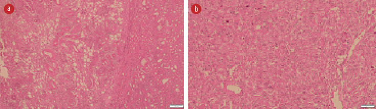
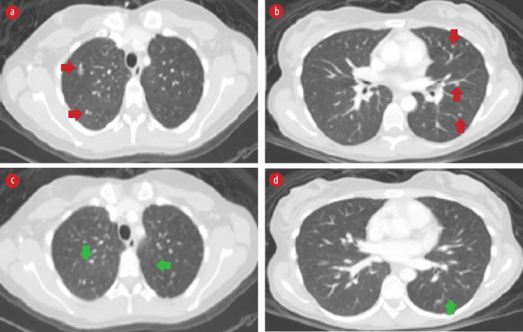
Epithelioid angiomyolipoma (EAML) is an uncommon renal neoplasm with malignant potential. It is classified under the group of perivascular epithelioid cell tumors and can be sporadic or as part of the tuberous sclerosis complex. On imaging, unlike classical AML that contains fat, EAML has a very low percentage of fat which can mimic the imaging findings of renal cell carcinoma. We reported a 31-year-old female who had a history of renal failure and bilateral renal masses. Magnetic resonance imaging of the abdomen revealed bilateral large renal masses replacing renal parenchyma with features suggestive of bilateral renal AML. The patient underwent left nephrectomy, and histopathology examination findings were consistent with the diagnosis of EAML.

Keywords: Angiomyolipoma; Carcinoma, Renal Cell; Magnetic Resonance Imaging; Oman; Tomography, X-Ray Computed.


Figures

Figure 1
Figure 1
(a) Axial and (b) coronal T2-weighted images show bilateral hyperintense renal masses replacing renal parenchyma (right renal mass: green star, left renal mass: red star). (c) Axial T1 in-phase and (d) T1 out-phase show a large area of drop in signal intensity of right renal mass (green star). Small focal of signal drop (red arrow) seen in the left renal mass (red arrow).
Figure 2
Figure 2
(a) Hematoxylin and eosin (H&E) stain showing many abnormally formed thick blood vessels, some with radiating smooth muscle fibers in the adjacent stroma, and adipose tissue component. (b) H&E slide with sheets of large polygonal epithelioid cells with pleomorphic nuclei and plenty of eosinophilic cytoplasm.
Figure 3
Figure 3
High-resolution chest computed tomography scan axial views. (a and b) Bilateral pulmonary nodules (red arrows). (c and d) Bilateral pulmonary cysts (green arrows).

Similar articles

Cited by

References

    1. Malinowska I, Kwiatkowski DJ, Weiss S, Martignoni G, Netto G, Argani P. Perivascular epithelioid cell tumors (PEComas) harboring TFE3 gene rearrangements lack the TSC2 alterations characteristic of conventional PEComas: further evidence for a biological distinction. Am J Surg Pathol 2012. May;36(5):783-784. 10.1097/PAS.0b013e31824a8a37 - DOI - PMC - PubMed
    1. Pea M, Bonetti F, Martignoni G, Henske EP, Manfrin E, Colato C, et al. Apparent renal cell carcinomas in tuberous sclerosis are heterogeneous: the identification of malignant epithelioid angiomyolipoma. Am J Surg Pathol 1998. Feb;22(2):180-187. 10.1097/00000478-199802000-00005 - DOI - PubMed
    1. Froemming AT, Boland J, Cheville J, Takahashi N, Kawashima A. Renal epithelioid angiomyolipoma: imaging characteristics in nine cases with radiologic-pathologic correlation and review of the literature. AJR Am J Roentgenol 2013. Feb;200(2):W178-86. 10.2214/AJR.12.8776 - DOI - PubMed
    1. Tsili AC, Ntorkou A, Argyropoulou MI. Renal epithelioid angiomyolipoma associated with pulmonary lymphangioleiomyomatosis: imaging findings. J Clin Imaging Sci 2017. May;7:18. 10.4103/jcis.JCIS_14_17 - DOI - PMC - PubMed
    1. Tsai CC, Wu WJ, Li CC, Wang CJ, Wu CH, Wu CC. Epithelioid angiomyolipoma of the kidney mimicking renal cell carcinoma: a clinicopathologic analysis of cases and literature review. Kaohsiung J Med Sci 2009. Mar;25(3):133-140. 10.1016/S1607-551X(09)70052-X - DOI - PMC - PubMed

Publication types

LinkOut - more resources