

Sarcocystis infection in red deer (Cervus elaphus) with eosinophilic myositis/fasciitis in Switzerland and involvement of red foxes (Vulpes vulpes) and hunting dogs in the transmission
- PMID: 33083225
- PMCID: PMC7551655
- DOI: 10.1016/j.ijppaw.2020.09.005
Sarcocystis infection in red deer (Cervus elaphus) with eosinophilic myositis/fasciitis in Switzerland and involvement of red foxes (Vulpes vulpes) and hunting dogs in the transmission
Abstract
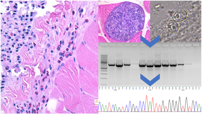
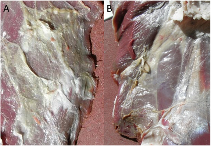
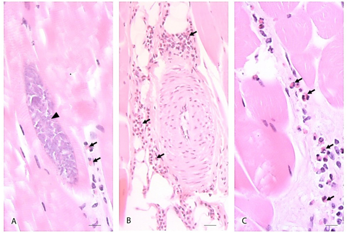
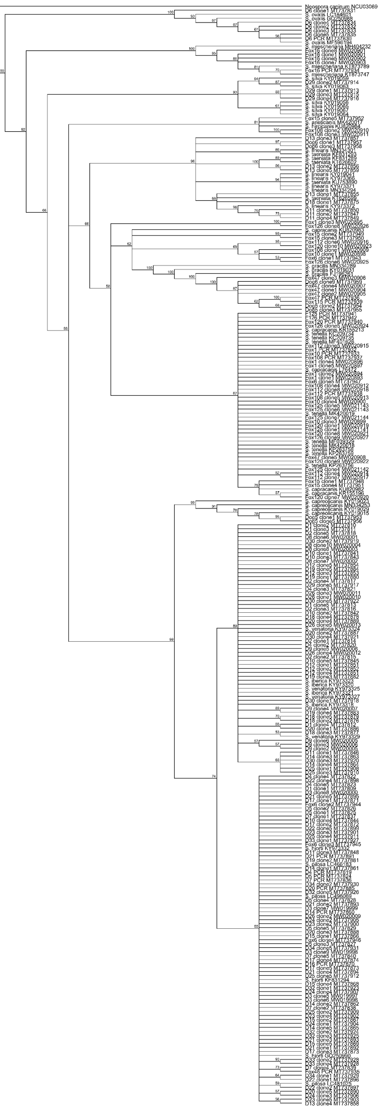
Red deer (Cervus elaphus) carcasses showing grey-greenish discolouration have been increasingly observed in the canton of Grisons, Switzerland. We investigated whether Sarcocystis infections were associated with this pathology, and whether wild and domestic canids were involved in their transmission. Meat from affected red deer (n = 26), faeces and intestines from red foxes (Vulpes vulpes) (n = 126), and faeces from hunting dogs (n = 12) from the region, were analysed. Eosinophilic myositis and/or fasciitis were diagnosed in 69% of the deer, and sarcocysts were observed in 89% of the animals. Molecular typing targeting a ~700bp variable region of the 18S rRNA gene revealed Sarcocystis hjorti in 73%, S. venatoria/S. iberica in 54%, S. linearis/S. taeniata in 12%, S. pilosa in 8% and S. ovalis in 4% of the deer samples. No inflammatory changes were observed in red deer carcasses with normal appearance (n = 8); however, sarcocysts were observed in one sample, and S. hjorti, S. venatoria/S. iberica or S. silva DNA was detected in five samples. Sarcocystis oocysts/sporocysts were observed in 11/106 faecal and 6/20 intestinal fox samples, and in 2/12 canine samples. Sarcocystis tenella (n = 8), S. hjorti (n = 2), S. gracilis (n = 2), and S. miescheriana (n = 1) were identified in foxes, and S. gracilis (n = 2), S. capreolicanis (n = 1) and S. linearis/S. taeniata (n = 1) in dogs. This study provides first molecular evidence of S. pilosa and S. silva infection in red deer and S. linearis/S. taeniata in dogs and represents the first record of S. ovalis transmitted by corvids in Central Europe. Although Sarcocystis species infecting red deer are not regarded as zoonotic, the affected carcasses can be declared as unfit for human consumption due to the extensive pathological changes.
Keywords: Dog (Canis familiaris); Molecular identification; Myositis/fasciitis; Red deer (Cervus elaphus); Red fox (Vulpes vulpes); Sarcocystis.
© 2020 The Authors. Published by Elsevier Ltd on behalf of Australian Society for Parasitology.
Conflict of interest statement
The authors declare that they have no known competing financial interests or personal relationships that could have appeared to influence the work reported in this paper.
Figures
References
-
- Abe N., Matsuo K., Moribe J., Takashima Y., Baba T., Gjerde B. Molecular differentiation of five Sarcocystis species in sika deer (Cervus nippon centralis) in Japan based on mitochondrial cytochrome c oxidase subunit I gene (cox1) sequences. Parasitol. Res. 2019;118(6):1975–1979. - PubMed
LinkOut - more resources
Full Text Sources
