Ascaris-mimicking common bile duct stone: A case report
- PMID: 33083410
- PMCID: PMC7559666
- DOI: 10.12998/wjcc.v8.i19.4499
Ascaris-mimicking common bile duct stone: A case report
Abstract
Background: In most cases, it is not difficult to differentiate common bile duct (CBD) stone from Ascaris infection because they are different disease entities and have different imaging findings. The two diseases usually demonstrate unique characteristic findings on computed tomography or magnetic resonance cholangiopancreatography. However, we report a rare case from our experience in which a CBD stone mimicked and was misdiagnosed as Ascaris.
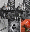
Case summary: A 72-year-old male presented with elevated serum liver enzymes. Computed tomography showed a hyper-attenuated, elongated lesion in the CBD lumen and associated biliary inflammation. Magnetic resonance cholangiopancreatography and endoscopic retrograde cholangiopancreatography revealed a linear filling defect in the bile duct. Moreover, elongated echogenic material with a central hypoechogenic area was seen on endoscopic ultrasound. Although the imaging findings caused us to suspect infection with the nematode Ascaris, the lesion was revealed to be a dark-brown-colored CBD stone through endoscopic extraction.
Conclusion: We report a rare case of a CBD stone that mimicked Ascaris. We also review the literature for side-by-side comparisons of the imaging features of CBD stones and ascariasis.
Keywords: Ascariasis; Ascaris; Case report; Common bile duct; Gallstones; Magnetic resonance cholangiopancreatography; Multidetector computed tomography.
©The Author(s) 2020. Published by Baishideng Publishing Group Inc. All rights reserved.
Conflict of interest statement
Conflict-of-interest statement: The authors state that they have no conflict of interest.
Figures


References
-
- Tazuma S. Gallstone disease: Epidemiology, pathogenesis, and classification of biliary stones (common bile duct and intrahepatic) Best Pract Res Clin Gastroenterol. 2006;20:1075–1083. - PubMed
-
- Yeh BM, Liu PS, Soto JA, Corvera CA, Hussain HK. MR imaging and CT of the biliary tract. Radiographics. 2009;29:1669–1688. - PubMed
-
- Lim JH, Kim SY, Park CM. Parasitic diseases of the biliary tract. AJR Am J Roentgenol. 2007;188:1596–1603. - PubMed
-
- Miller FH, Hwang CM, Gabriel H, Goodhartz LA, Omar AJ, Parsons WG., 3rd Contrast-enhanced helical CT of choledocholithiasis. AJR Am J Roentgenol. 2003;181:125–130. - PubMed
Publication types
LinkOut - more resources
Full Text Sources

