

Leaving no stone unturned: Allosteric targeting of SARS-CoV-2 spike protein at putative druggable sites disrupts human angiotensin-converting enzyme interactions at the receptor binding domain
- PMID: 33083517
- PMCID: PMC7561517
- DOI: 10.1016/j.imu.2020.100451
Leaving no stone unturned: Allosteric targeting of SARS-CoV-2 spike protein at putative druggable sites disrupts human angiotensin-converting enzyme interactions at the receptor binding domain
Abstract
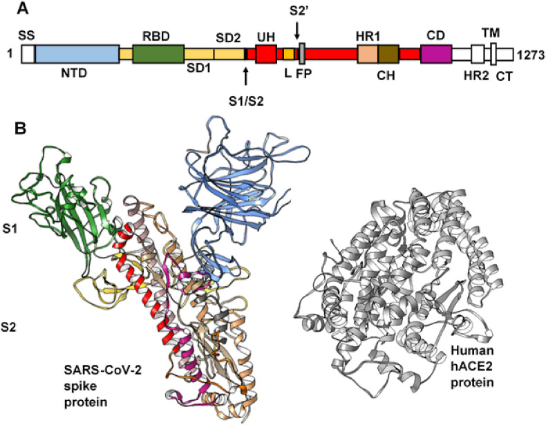
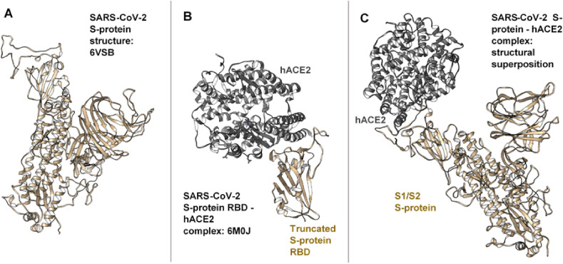
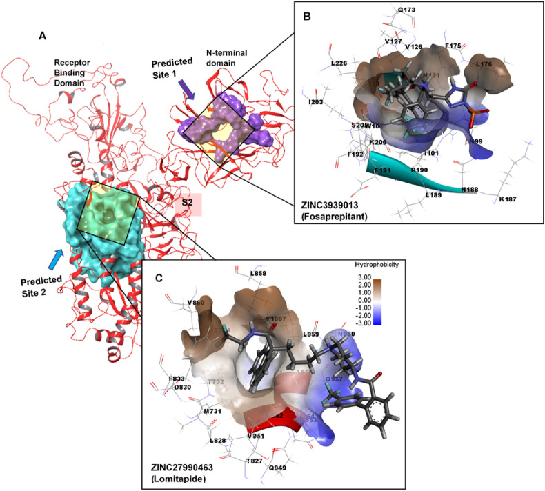
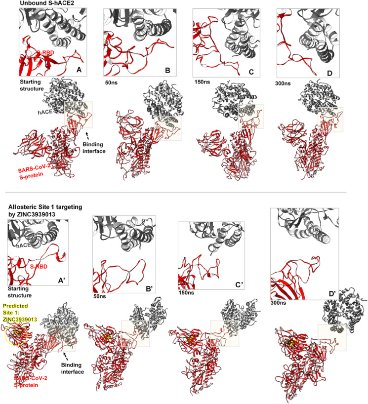
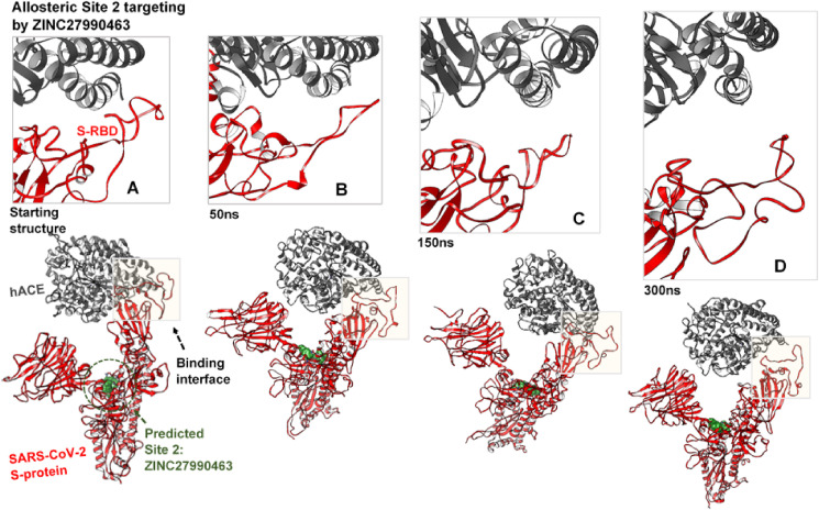
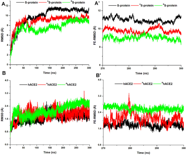
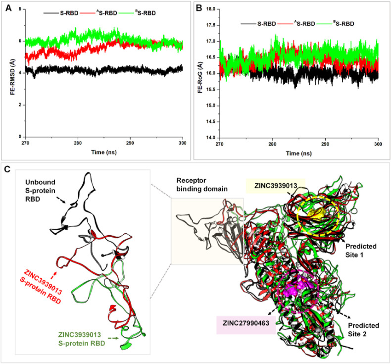
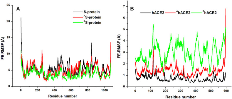
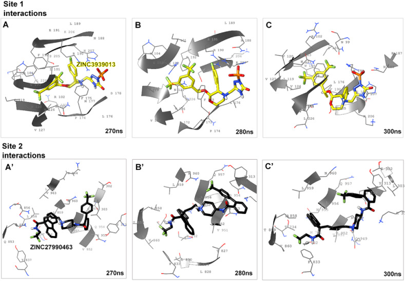
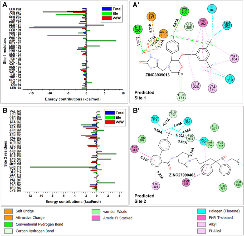
The systematic entry of SARS-CoV-2 into host cells, as mediated by its Spike (S) protein, is highly essential for pathogenicity in humans. Hence, targeting the viral entry mechanisms remains a major strategy for COVID-19 treatment. Although recent efforts have focused on the direct inhibition of S-protein receptor-binding domain (RBD) interactions with human angiotensin-converting enzyme 2 (hACE2), allosteric targeting remains an unexplored possibility. Therefore, in this study, for the first time, we employed an integrative meta-analytical approach to investigate the allosteric inhibitory mechanisms of SARS-CoV-2 S-protein and its association with hACE2. Findings revealed two druggable sites (Sites 1 and 2) located at the N-terminal domain (NTD) and S2 regions of the protein. Two high-affinity binders; ZINC3939013 (Fosaprepitant - Site 1) and ZINC27990463 (Lomitapide - Site 2) were discovered via site-directed high-throughput screening against a library of ~1500 FDA approved drugs. Interestingly, we observed that allosteric binding of both compounds perturbed the prefusion S-protein conformations, which in turn, resulted in unprecedented hACE2 displacement from the RBD. Estimated ΔG binds for both compounds were highly favorable due to high-affinity interactions at the target sites. In addition, Site 1 residues; R190, H207, K206 and K187, I101, R102, I119, F192, L226, V126 and W104 were identified for their crucial involvement in the binding and stability of ZINC3939013. Likewise, energy contributions of Q957, N953, Q954, L303, Y313, Q314, L858, V952, N953, and A956 corroborated their importance to ZINC27990463 binding at the predicted Site 2. We believe these findings would pave way for the structure-based discovery of allosteric SARS-CoV-2 S-protein inhibitors for COVID-19 treatment.
Keywords: Allosteric targeting; High-affinity binding; Receptor binding domain; SARS-CoV-2; Spike protein; Virtual high-throughput screening.
© 2020 The Author(s).
Conflict of interest statement
The authors declare that they have no known competing financial interests or personal relationships that could have appeared to influence the work reported in this paper.
Figures
Similar articles
-
A multiple-step in silico screening protocol to identify allosteric inhibitors of Spike-hACE2 binding.Phys Chem Chem Phys. 2022 Feb 16;24(7):4305-4316. doi: 10.1039/d1cp04736a. Phys Chem Chem Phys. 2022. PMID: 35107459
-
Role of tannic acid against SARS-cov-2 cell entry by targeting the interface region between S-protein-RBD and human ACE2.Front Pharmacol. 2022 Aug 8;13:940628. doi: 10.3389/fphar.2022.940628. eCollection 2022. Front Pharmacol. 2022. PMID: 36003511 Free PMC article.
-
Inhibition of S-protein RBD and hACE2 Interaction for Control of SARSCoV- 2 Infection (COVID-19).Mini Rev Med Chem. 2021;21(6):689-703. doi: 10.2174/1389557520666201117111259. Mini Rev Med Chem. 2021. PMID: 33208074 Review.
-
Improved Binding Affinity of Omicron's Spike Protein for the Human Angiotensin-Converting Enzyme 2 Receptor Is the Key behind Its Increased Virulence.Int J Mol Sci. 2022 Mar 21;23(6):3409. doi: 10.3390/ijms23063409. Int J Mol Sci. 2022. PMID: 35328828 Free PMC article.
-
Interactions of angiotensin-converting enzyme-2 (ACE2) and SARS-CoV-2 spike receptor-binding domain (RBD): a structural perspective.Mol Biol Rep. 2023 Mar;50(3):2713-2721. doi: 10.1007/s11033-022-08193-4. Epub 2022 Dec 23. Mol Biol Rep. 2023. PMID: 36562937 Free PMC article. Review.
Cited by
-
Distant residues modulate conformational opening in SARS-CoV-2 spike protein.Proc Natl Acad Sci U S A. 2021 Oct 26;118(43):e2100943118. doi: 10.1073/pnas.2100943118. Epub 2021 Oct 6. Proc Natl Acad Sci U S A. 2021. PMID: 34615730 Free PMC article.
-
Contributions of single-particle cryoelectron microscopy toward fighting COVID-19.Trends Biochem Sci. 2022 Feb;47(2):117-123. doi: 10.1016/j.tibs.2021.10.005. Epub 2021 Oct 30. Trends Biochem Sci. 2022. PMID: 34799235 Free PMC article. Review.
-
A Repurposed Drug Screen Identifies Compounds That Inhibit the Binding of the COVID-19 Spike Protein to ACE2.Front Pharmacol. 2021 Jun 14;12:685308. doi: 10.3389/fphar.2021.685308. eCollection 2021. Front Pharmacol. 2021. PMID: 34194331 Free PMC article.
-
Identifying promising druggable binding sites and their flexibility to target the receptor-binding domain of SARS-CoV-2 spike protein.Comput Struct Biotechnol J. 2023;21:2339-2351. doi: 10.1016/j.csbj.2023.03.029. Epub 2023 Mar 18. Comput Struct Biotechnol J. 2023. PMID: 36998674 Free PMC article.
-
The impact of structural bioinformatics tools and resources on SARS-CoV-2 research and therapeutic strategies.Brief Bioinform. 2021 Mar 22;22(2):742-768. doi: 10.1093/bib/bbaa362. Brief Bioinform. 2021. PMID: 33348379 Free PMC article. Review.
References
-
- Worldometer . Worldometer; 2020. Coronavirus cases.
LinkOut - more resources
Full Text Sources
Miscellaneous