

Hindlimb unloading causes regional loading-dependent changes in osteocyte inflammatory cytokines that are modulated by exogenous irisin treatment
- PMID: 33083525
- PMCID: PMC7542171
- DOI: 10.1038/s41526-020-00118-4
Hindlimb unloading causes regional loading-dependent changes in osteocyte inflammatory cytokines that are modulated by exogenous irisin treatment
Abstract
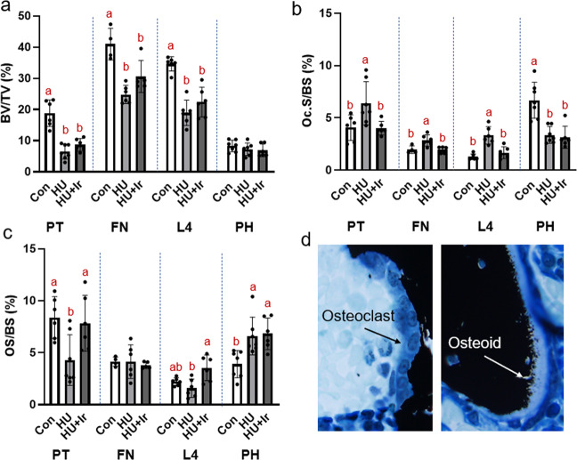
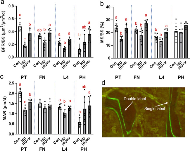
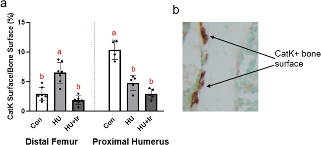
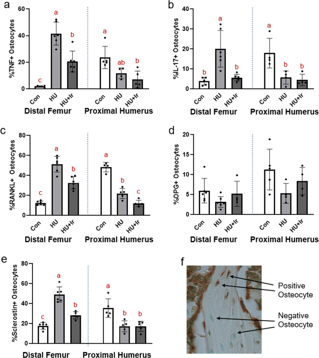
Disuse-induced bone loss is characterized by alterations in bone turnover. Accruing evidence suggests that osteocytes respond to inflammation and express and/or release pro-inflammatory cytokines; however, it remains largely unknown whether osteocyte inflammatory proteins are influenced by disuse. The goals of this project were (1) to assess osteocyte pro-inflammatory cytokines in the unloaded hindlimb and loaded forelimb of hindlimb unloaded rats, (2) to examine the impact of exogenous irisin during hindlimb unloading (HU). Male Sprague Dawley rats (8 weeks old, n = 6/group) were divided into ambulatory control, HU, and HU with irisin (HU + Ir, 3×/week). Lower cancellous bone volume, higher osteoclast surfaces (OcS), and lower bone formation rate (BFR) were present at the hindlimb and 4th lumbar vertebrae in the HU group while the proximal humerus of HU rats exhibited no differences in bone volume, but higher BFR and lower OcS vs. Con. Osteocyte tumor necrosis factor-α (TNF-α), interleukin-17 (IL-17), RANKL, and sclerostin were elevated in the cancellous bone of the distal femur of HU rats vs. Con, but lower at the proximal humerus in HU rats vs. Con. Exogenous irisin treatment increased BFR, and lowered OcS and osteocyte TNF-α, IL-17, RANKL, and sclerostin in the unloaded hindlimb of HU + Ir rats while having minimal changes in the humerus. In conclusion, there are site-specific and loading-specific alterations in osteocyte pro-inflammatory cytokines and bone turnover with the HU model of disuse bone loss, indicating a potential mechanosensory impact of osteocyte TNF-α and IL-17. Additionally, exogenous irisin significantly reduced the pro-inflammatory status of the unloaded hindlimb.
Keywords: Cytokines; Physiology.
© The Author(s) 2020.
Conflict of interest statement
Competing interestsC.E.M., S.A.N., and S.A.B. are inventors on a patent application through Texas A&M University on the use of irisin as an anti-inflammatory treatment.
Figures
Similar articles
-
Differential responses of mechanosensitive osteocyte proteins in fore- and hindlimbs of hindlimb-unloaded rats.Bone. 2017 Dec;105:26-34. doi: 10.1016/j.bone.2017.08.002. Epub 2017 Aug 3. Bone. 2017. PMID: 28782619
-
Osteocytes reflect a pro-inflammatory state following spinal cord injury in a rodent model.Bone. 2019 Mar;120:465-475. doi: 10.1016/j.bone.2018.12.007. Epub 2018 Dec 11. Bone. 2019. PMID: 30550849
-
Inflammatory Bowel Disease in a Rodent Model Alters Osteocyte Protein Levels Controlling Bone Turnover.J Bone Miner Res. 2017 Apr;32(4):802-813. doi: 10.1002/jbmr.3027. Epub 2017 Feb 28. J Bone Miner Res. 2017. PMID: 27796050
-
Pro-inflammatory Cytokines: Cellular and Molecular Drug Targets for Glucocorticoid-induced-osteoporosis via Osteocyte.Curr Drug Targets. 2019;20(1):1-15. doi: 10.2174/1389450119666180405094046. Curr Drug Targets. 2019. PMID: 29618305 Review.
-
The Role of Osteocytes in Inflammatory Bone Loss.Front Endocrinol (Lausanne). 2019 May 14;10:285. doi: 10.3389/fendo.2019.00285. eCollection 2019. Front Endocrinol (Lausanne). 2019. PMID: 31139147 Free PMC article. Review.
Cited by
-
Protective role of irisin on bone in osteoporosis: a systematic review of rodent studies.Osteoporos Int. 2025 Apr 7. doi: 10.1007/s00198-025-07470-9. Online ahead of print. Osteoporos Int. 2025. PMID: 40192854 Review.
-
Irisin at the crossroads of inter-organ communications: Challenge and implications.Front Endocrinol (Lausanne). 2022 Oct 4;13:989135. doi: 10.3389/fendo.2022.989135. eCollection 2022. Front Endocrinol (Lausanne). 2022. PMID: 36267573 Free PMC article. Review.
-
The effects of microgravity on bone structure and function.NPJ Microgravity. 2022 Apr 5;8(1):9. doi: 10.1038/s41526-022-00194-8. NPJ Microgravity. 2022. PMID: 35383182 Free PMC article. Review.
-
Irisin enhances chondrogenic differentiation of human mesenchymal stem cells via Rap1/PI3K/AKT axis.Stem Cell Res Ther. 2022 Aug 3;13(1):392. doi: 10.1186/s13287-022-03092-8. Stem Cell Res Ther. 2022. PMID: 35922833 Free PMC article.
-
Osteocytes function as biomechanical signaling hubs bridging mechanical stress sensing and systemic adaptation.Front Physiol. 2025 Jul 15;16:1629273. doi: 10.3389/fphys.2025.1629273. eCollection 2025. Front Physiol. 2025. PMID: 40735673 Free PMC article. Review.
References
-
- LeBlanc AD, Spector ER, Evans HJ, Sibonga JD. Skeletal responses to space flight and the bed rest analog: a review. J. Musculoskelet. Neuronal Interact. 2007;7:33–47. - PubMed
-
- LeBlanc AD, et al. Bone mineral and lean tissue loss after long duration space flight. J. Muscoskelet. Neuronal Interact. 2000;1:157–160. - PubMed
-
- Vico L, et al. Effects of long-term microgravity exposure on cancellous and cortical weight-bearing bones of cosmonauts. Lancet. 2000;355:1607–1611. - PubMed
-
- Bloomfield SA, Allen MR, Hogan HA, Delp MD. Site- and compartment-specific changes in bone with hindlimb unloading in mature adult rats. Bone. 2001;31:149–157. - PubMed
-
- Globus RK, Bikle DD, Morey-Holton E. The temporal response of bone to unloading. Endocrinology. 1986;118:733–742. - PubMed
LinkOut - more resources
Full Text Sources
