Case Reports
. 2020 Sep 29:20:100949.
doi: 10.1016/j.ajoc.2020.100949. eCollection 2020 Dec.

Acute idiopathic blind spot enlargement syndrome following influenza vaccination

Affiliations
Case Reports

Acute idiopathic blind spot enlargement syndrome following influenza vaccination

Xavier Quinones et al. Am J Ophthalmol Case Rep. .

Abstract

Purpose: To report a case of acute idiopathic blind spot enlargement syndrome (AIBSES) following influenza vaccination.

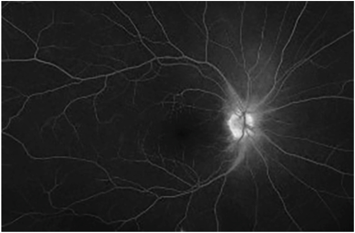
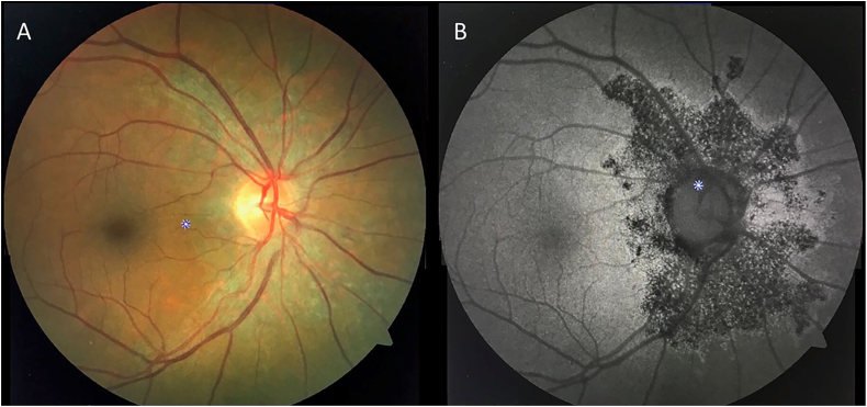
Observations: A 57-year old woman presented with a one-month history of photopsia, temporal visual field disturbance on the right eye, 11 days following the administration of the influenza virus vaccine. Visual acuity was 20/30, and color vision remained normal. Examination revealed mild venular dilation at the edge of the right optic disk and was otherwise unremarkable. Visual field testing revealed enlargement of the right physiological blind spot. Medical workup, including brain CT scan, brain MRI, RPR, Treponema pallidum antibodies, Chest X-Ray, ANA, and PPD testing, was found within normal limits. Ancillary testing was compatible with an assessment of AIBSES secondary to influenza virus vaccination.

Conclusions and importance: Although direct causation may not be absolutely established by a single report, our case suggests that the influenza virus vaccine may serve as an immunological trigger for some cases of AIBSES. Thoughtful vaccination history is of the utmost importance when evaluating patients with AIBSES, as it may help elucidate the underlying precipitating factor. To our knowledge, this is the first reported case of AIBSES following influenza virus vaccination.

Keywords: Acute blind spot enlargement syndrome; Influenza virus; Vaccination.


Conflict of interest statement

No funding or grant support was provided for this report. The following authors have no financial disclosures: XQ, JO, CS, AO, JR. All authors attest that they meet the current ICMJE criteria for Authorship.

Figures

Fig. 1
Fig. 1
Right eye visual field (Humphrey, Central 30-2 Threshold Test, Stimulus III, White, SITA-FAST). A. Upon presentation, there is an enlargement of the physiological blind spot. Mean deviation: −12.14 dB, P < 0.5% B. One-month post-presentation shows a mild improvement of the visual field defect and the mean deviation. Mean deviation: −7.25 dB, P < 0.5% C. At 1-year follow-up, there is the persistence of the abnormal scotoma. Mean deviation: −5.96 dB, P < 1%.
Fig. 2
Fig. 2
Fluorescein angiogram of the right fundus, late views (10 min post dye injection), revealing disk and perivascular hyperfluorescence with leakage in the peripapillary region.
Fig. 3
Fig. 3
Multifocal ERG of the right eye, 3 months after the onset of symptoms, revealing decreased wavelet amplitude in the macular regions corresponding to the existing scotoma. A. Field View B. Traces distribution. C. Response density tomography.
Fig. 4
Fig. 4
High Definition OCT, encompassing the right optic nerve and macula, four months after the initial presentation. The images reveal disruption of the ellipsoid zone and loss of regularity of the photoreceptor inner segment/outer segment (IS/OS) line in the peripapillary region (box).
Fig. 5
Fig. 5
Right eye fundus images obtained 5 months after presentation. A. Color photographs reveal mild peripapillary retinal pigment epithelium changes. B. Fundus autofluorescence images disclose a well-demarcated area of hypoautofluorescence, which is surrounded by a zone of hyperautofluorescence, which extends further temporally up to the nasal perifoveal region. (For interpretation of the references to color in this figure legend, the reader is referred to the Web version of this article.)

Similar articles

Cited by

References

    1. Fletcher W.A., Imes R.K., Goodman D., Hoyt W.F. Acute idiopathic blind spot enlargement: a big blind spot syndrome without optic disc edema. Arch Ophthalmol. 1988;106(1):44–49. doi: 10.1001/archopht.1988.01060130050026. - DOI - PubMed
    1. Holz F.G., Kim R.Y., Schwartz S.D. Acute zonal occult outer retinopathy (AZOOR) associated with multifocal choroidopathy. Eye. 1994;8(1):77–83. doi: 10.1038/eye.1994.15. - DOI - PubMed
    1. Volpe N.J., Rizzo J.F., Lessell S. Acute idiopathic blind spot enlargement syndrome: a review of 27 new cases. Arch Ophthalmol. 2001;119(1):59–63. http://www.ncbi.nlm.nih.gov/pubmed/11146727 (Chicago, Ill 1960) Accessed June 18, 2019. - PubMed
    1. Gass J.D.M., Hamed L.M. Acute macular neuroretinopathy and multiple evanescent white dot syndrome occurring in the same patients. Arch Ophthalmol. 1989;107(2):189–193. doi: 10.1001/archopht.1989.01070010195021. - DOI - PubMed
    1. Bouchenaki N., Cimino L., Auer C., Tran V.T., Herbort C.P. Vol 219. Klin Monbl Augenheilkd; 2002. Assessment and classification of choroidal vasculitis in posterior uveitis using indocyanine green angiography; pp. 243–249. (Klinische Monatsblatter Fur Augenheilkunde). - DOI - PubMed

Publication types

LinkOut - more resources