. 2020 Sep 6;23(10):101537.
doi: 10.1016/j.isci.2020.101537. eCollection 2020 Oct 23.

Metal- and UV- Catalyzed Oxidation Results in Trapped Amyloid-β Intermediates Revealing that Self-Assembly Is Required for Aβ-Induced Cytotoxicity

Affiliations

Metal- and UV- Catalyzed Oxidation Results in Trapped Amyloid-β Intermediates Revealing that Self-Assembly Is Required for Aβ-Induced Cytotoxicity

Mahmoud B Maina et al. iScience. .

Abstract

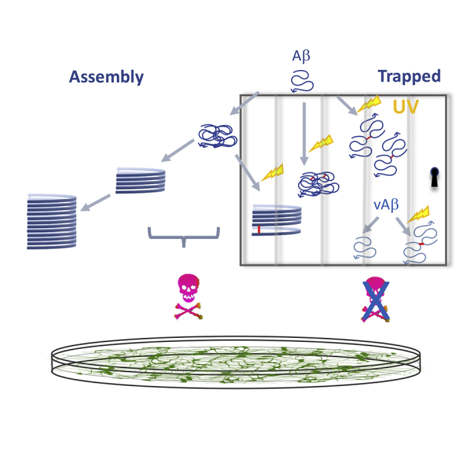
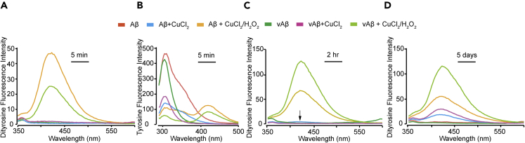
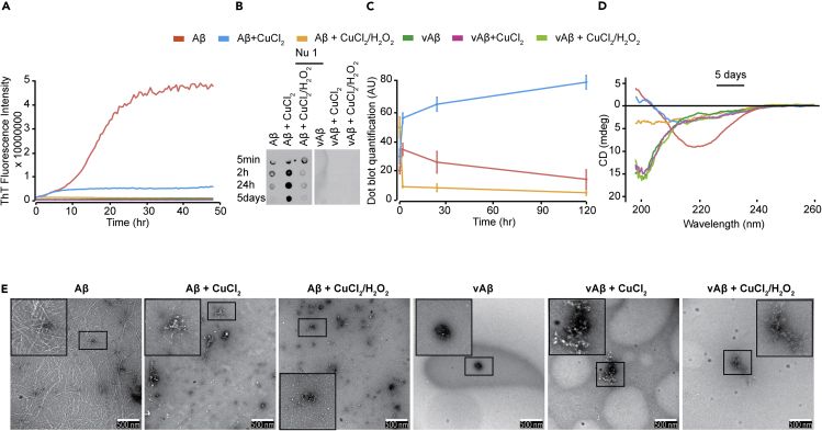
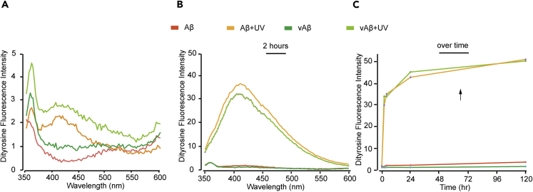
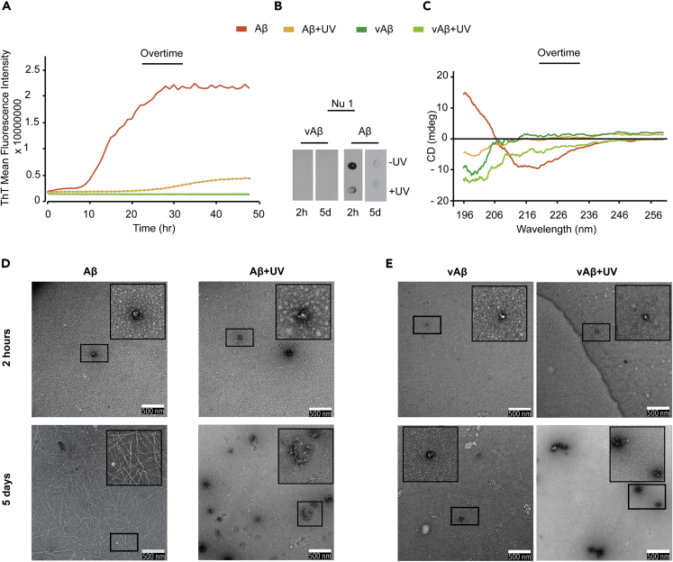
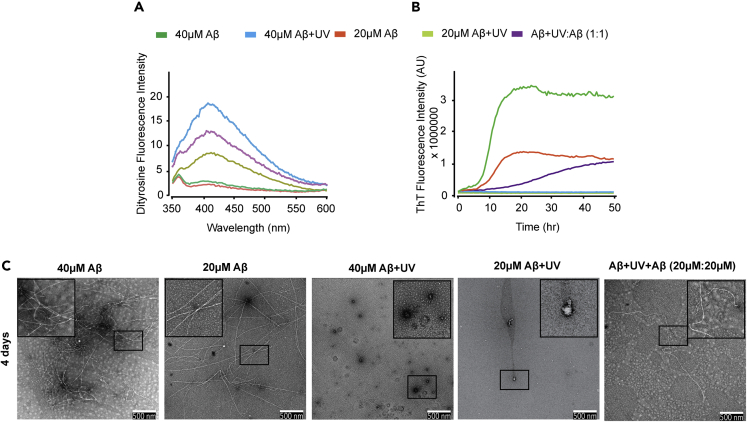
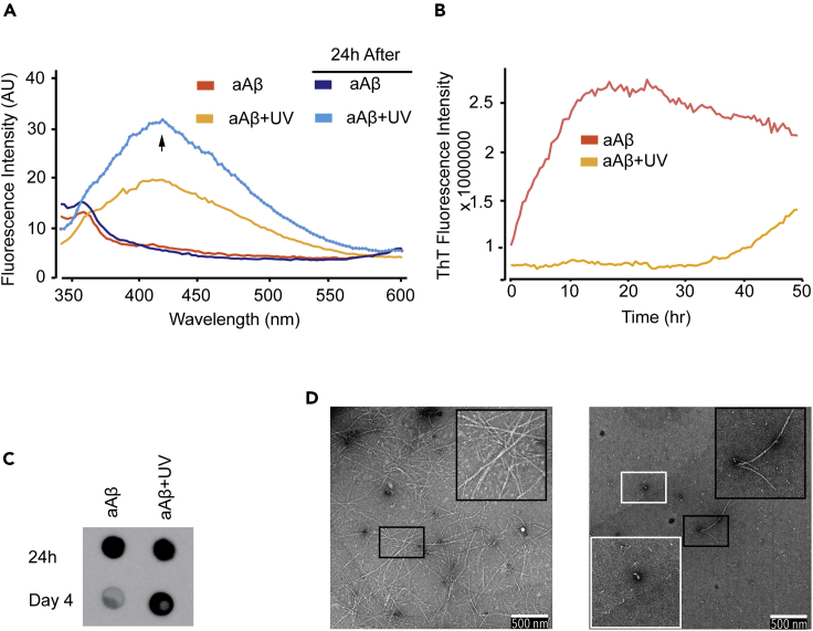
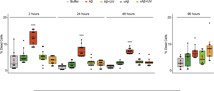
Dityrosine (DiY), via the cross-linking of tyrosine residues, is a marker of protein oxidation, which increases with aging. Amyloid-β (Aβ) forms DiY in vitro and DiY-cross-linked Aβ is found in the brains of patients with Alzheimer disease. Metal- or UV- catalyzed oxidation of Aβ42 results in an increase in DiY cross-links. Using DiY as a marker of oxidation, we compare the self-assembly propensity and DiY cross-link formation for a non-assembly competent variant of Aβ42 (vAβ) with wild-type Aβ42. Oxidation results in the formation of trapped wild-type Aβ assemblies with increased DiY cross-links that are unable to elongate further. Assembly-incompetent vAβ and trapped Aβ assemblies are non-toxic to neuroblastoma cells at all stages of self-assembly, in contrast to oligomeric, non-cross-linked Aβ. These findings point to a mechanism of toxicity that necessitates dynamic self-assembly whereby trapped Aβ assemblies and assembly-incompetent variant Aβ are unable to result in cell death.

Keywords: Biochemical Mechanism; Molecular Neuroscience; Neurotoxicology.


Conflict of interest statement

The authors declare there are no competing interests.

Figures

None
Graphical abstract
Figure 1
Figure 1
DiY Formation in Aβ and vAβ via Metal-Catalyzed Oxidation (A–D) Freshly prepared Aβ and vAβ (50 μM) were incubated at 37°C, without CuCl2, with CuCl2 (400 μM), or CuCl2 in combination with H2O2. Fluorescence spectra were collected 5 min post-incubation using fluorescent excitation wavelength of 320 nm and emission collected between 340 and 600 nm, with DiY peak signal observed between 400 and 420 nm (A). Fluorescence spectra were also collected at 5 min using an excitation wavelength of 280 nm and emission collected between 290 and 500, with peak tyrosine signal observed at 305 nm (B). Fluorescence spectra were collected again at 2 h (C) and then 5 days (D) to follow DiY formation over time. A minimum of three independent experiments was repeated to ensure the reproducibility of the findings.
Figure 2
Figure 2
Metal-Catalyzed Oxidation Results in the Formation of Stabilized Aβ Assemblies (A–E) Th-T fluorescence was monitored for the freshly prepared Aβ and vAβ (50 μM) incubated at 37°C, without CuCl2, with CuCl2 (400 μM), or CuCl2 in combination with H2O2 (A). Dot blotting using NU-1 antibody identified Aβ oligomers in the Aβ samples, but not in vAβ reactions (B). Quantification of dot blotting signal over time reveals that the oligomers in the CuCl2-oxidized Aβ remain stable for 5 days, unlike in the other reactions in which very low NU-1 affinity signal was observed (C). CD at 5 days showed a high β-sheet content in the unoxidized Aβ sample, with reduced signal for CuCl2 Aβ, and CuCl2/H2O2Aβ. All vAβ samples showed spectra consistent with random coil conformation (D). TEM imaging at 5 days revealed a network of fibers in the unoxidized Aβ, whereas the CuCl2 Aβ showed clumped assemblies with very little fiber density and the CuCl2/H2O2 sample revealed amorphous-like assemblies. Unoxidized vAβ showed no assemblies, whereas both oxidized vAβ reactions showed amorphous-like aggregates (E). A minimum of three independent experiments was repeated to ensure the reproducibility of the findings. Scale bars, 500 nm. Error bars are expressed as ±SEM.
Figure 3
Figure 3
DiY Formation in Aβ and vAβ via UV Photo-oxidation (A–C) Freshly prepared Aβ and vAβ (50 μM) were incubated under UV. Fluorescence spectra were collected 5 min post-incubation using fluorescent excitation wavelength of 320 nm and emission collected between 340 and 600 nm, with DiY peak signal observed between 400 and 420 nm after 5 min of incubation (A), which increased following 2 h of incubation (B). Fluorescence intensity at 410 nm against time showed that incubation of the 2 h UV-exposed Aβ and vAβ samples in the dark resulted in further increase in DiY formation in the absence of the UV (C). The Aβ and vAβ samples that were not exposed to UV showed no DiY signal. A minimum of three independent experiments was repeated to ensure the reproducibility of the findings.
Figure 4
Figure 4
UV-Induced DiY Cross-Linking of Early Aβ Assemblies Correlates with Formation of Stabilized Assemblies (A) Th-T fluorescence spectrum shows the expected increase in fluorescence for assembling Aβ, but Aβ+UV Th-T fluorescence was significantly reduced. vAβ incubated in the absence or presence UV showed no Th-T fluorescence. (B) Dot blotting using NU-1 antibody shows binding suggesting fewer oligomers in the oxidized Aβ+UV than in the unoxidized Aβ sample. No binding of NU-1 was observed for Aβ+UV 5 days post-UV exposure, but a small signal was detected in the Aβ-UV sample. (C) CD at 5 days showed a high β-sheet content in the Aβ sample, whereas the oxidized Aβ+UV showed a loss of signal but indicated some random coil. Oxidized and unoxidized vAβ samples showed random coil conformation. (D) TEM after 2 h and 5 days showed that the unoxidized Aβ at 2 h formed oligomers, which transformed into a network of fibers at 5 days. The oxidized Aβ+UV samples formed small assemblies at 2 h, some of which developed into amorphous-like assemblies at 5 days. (E) vAβ does not assemble into amyloid fibrils, but vAβ+UV forms some amorphous aggregates after 5 days. A minimum of three independent experiments was repeated to ensure the reproducibility of the findings. Scale bars, 500 nm.
Figure 5
Figure 5
Co-incubation with Photo-oxidized, DiY-Cross-Linked Aβ Assemblies Slows the Aggregation of Freshly Prepared Aβ (A) DiY fluorescence was measured for freshly prepared Aβ (40 and 20 μM) and Aβ+UV:Aβ (20 μM: 20 μM) mixture immediately after co-incubation of freshly prepared Aβ with DiY-cross-linked Aβ. DiY signal was not detected in unoxidized freshly prepared 40 and 20 μM Aβ but was induced in 2 h UV-oxidized (Aβ+UV) samples and in the mixture. (B) Th-T fluorescence showed that the unoxidized 40 and 20 μM Aβ assemble at different rates, which was significantly delayed for Aβ+UV:Aβ-UV mixture and completely absent in the oxidized 40 and 20 μM Aβ samples. (C) TEM imaging at 4 days revealed the presence of fibrils in both unoxidized 40 and 20 μM Aβ, which was significantly reduced in the Aβ+UV:Aβ-UV mixture and absent in the oxidized 40 and 20 μM Aβ samples. A minimum of three independent experiments was repeated to ensure the reproducibility of the findings. Scale bars, 500 nm.
Figure 6
Figure 6
Photo-oxidation of Pre-formed Aβ (aAβ) (24 h) Assemblies Slows Further Assembly of Aβ Oligomers/Protofibrils and Prolongs the Half-Life of the Oligomers (A) DiY signal was detected in the oxidized aAβ+UV sample, which continued over time. Unoxidized aAβ samples showed no DiY signal. (B) Th-T fluorescence showed that the unoxidized aAβ continues to assemble, whereas the oxidized aAβ+UV became significantly inhibited from further elongation up to 40 h. (C) Dot blotting using NU-1 antibody reveals the presence of oligomers in the unoxidized and oxidized aAβ at the starting time point. However, the oligomers are still strongly detected in the oxidized aAβ+UV at 4 days, unlike in the unoxidized samples. (D) TEM imaging at 4 days revealed the presence of fibrils in the unoxidized aAβ sample, whereas the oxidized aAβ+UV showed oligomers and a reduced number of fibrils. A minimum of three independent experiments was repeated to ensure the reproducibility of the findings. Scale bars, 500 nm
Figure 7
Figure 7
Oxidized, DiY-Containing Aβ Assemblies Are Not Toxic to Cells Differentiated SH-SY5Y neuroblastoma cells were incubated with UV oxidized or unoxidized vAβ or Aβ for 3 days, following which the percentage of dead cells was quantified using ReadyProbes Live/Dead Assay. The Aβ and vAβ samples were freshly prepared and UV oxidized for 2 h (Aβ+UV/vAβ+UV), or UV oxidized for 2 h followed by a further incubation on bench and in the dark for 24, 48, or 96 h, before being administered to cells. vAβ and Aβ samples not exposed to UV were used as reference. Only the unoxidized/assembling wild-type Aβ induced significant cell death. Experiments were repeated five times. ∗∗∗p < 0.001. p ≤  0.05 (∗), <0.01 (∗∗), <0.0001 (∗∗∗∗) and >0.05 was not significant. Error bars are expressed as ±SEM.

Similar articles

Cited by

References

    1. Al-Hilaly Y.K., Biasetti L., Blakeman B.J.F., Pollack S.J., Zibaee S., Abdul-Sada A., Thorpe J.R., Xue W.-F., Serpell L.C. The involvement of dityrosine crosslinking in α-synuclein assembly and deposition in Lewy Bodies in Parkinson’s disease. Sci. Rep. 2016;6:39171. - PMC - PubMed
    1. Al-Hilaly Y.K., Williams T.L., Stewart-Parker M., Ford L., Skaria E., Cole M., Bucher W.G., Morris K.L., Sada A.A., Thorpe J.R., Serpell L.C. A central role for dityrosine crosslinking of Amyloid-β in Alzheimer’s disease. Acta Neuropathol. Commun. 2013;1:83. - PMC - PubMed
    1. Ali F.E., Leung A., Cherny R.A., Mavros C., Barnham K.J., Separovic F., Barrow C.J. Dimerisation of N-acetyl-L-tyrosine ethyl ester and Abeta peptides via formation of dityrosine. Free Radic. Res. 2006;40:1–9. - PubMed
    1. Ali F.E., Separovic F., Barrow C.J., Cherny R.A., Fraser F., Bush A.I., Masters C.L., Barnham K.J. Methionine regulates copper/hydrogen peroxide oxidation products of Aβ. J. Pept. Sci. 2005;11:353–360. - PubMed
    1. Atwood C.S., Perry G., Zeng H., Kato Y., Jones W.D., Ling K.Q., Huang X., Moir R.D., Wang D., Sayre L.M. Copper mediates dityrosine cross-linking of Alzheimer's amyloid-beta. Biochemistry. 2004;43:560–568. - PubMed

LinkOut - more resources