[Preprint]. 2020 Oct 15:2020.10.13.20211664.
doi: 10.1101/2020.10.13.20211664.

IgM autoantibodies recognizing ACE2 are associated with severe COVID-19

Affiliations

IgM autoantibodies recognizing ACE2 are associated with severe COVID-19

Livia Casciola-Rosen et al. medRxiv. .

Update in

  • IgM anti-ACE2 autoantibodies in severe COVID-19 activate complement and perturb vascular endothelial function.
    Casciola-Rosen L, Thiemann DR, Andrade F, Trejo-Zambrano MI, Leonard EK, Spangler JB, Skinner NE, Bailey J, Yegnasubramanian S, Wang R, Vaghasia AM, Gupta A, Cox AL, Ray SC, Linville RM, Guo Z, Searson PC, Machamer CE, Desiderio S, Sauer LM, Laeyendecker O, Garibaldi BT, Gao L, Damarla M, Hassoun PM, Hooper JE, Mecoli CA, Christopher-Stine L, Gutierrez-Alamillo L, Yang Q, Hines D, Clarke WA, Rothman RE, Pekosz A, Fenstermacher KZ, Wang Z, Zeger SL, Rosen A. Casciola-Rosen L, et al. JCI Insight. 2022 May 9;7(9):e158362. doi: 10.1172/jci.insight.158362. JCI Insight. 2022. PMID: 35349483 Free PMC article.

Abstract

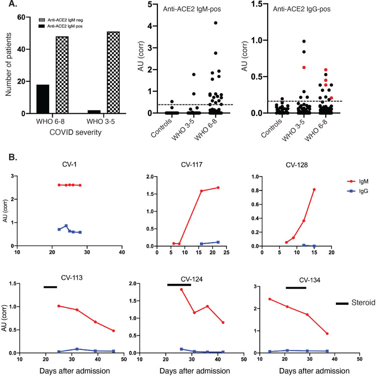
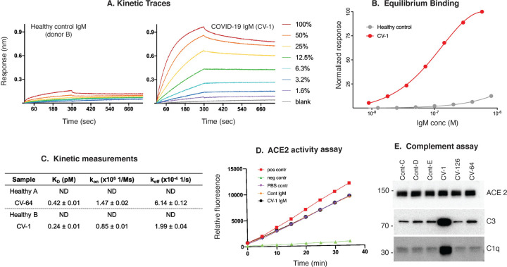
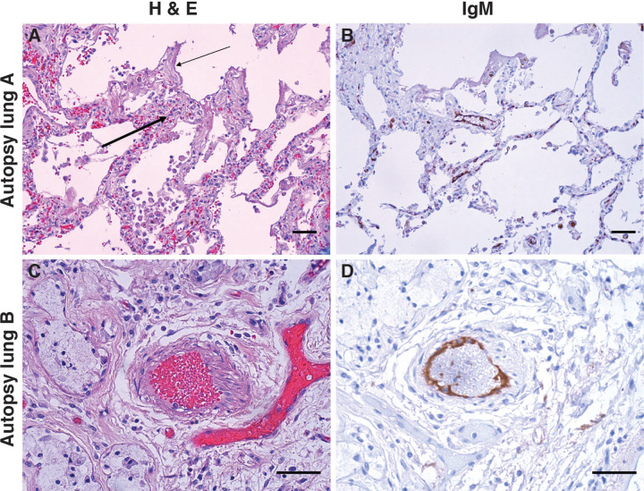
SARS-CoV-2 infection induces severe disease in a subpopulation of patients, but the underlying mechanisms remain unclear. We demonstrate robust IgM autoantibodies that recognize angiotensin converting enzyme-2 (ACE2) in 18/66 (27%) patients with severe COVID-19, which are rare (2/52; 3.8%) in hospitalized patients who are not ventilated. The antibodies do not undergo class-switching to IgG, suggesting a T-independent antibody response. Purified IgM from anti-ACE2 patients activates complement. Pathological analysis of lung obtained at autopsy shows endothelial cell staining for IgM in blood vessels in some patients. We propose that vascular endothelial ACE2 expression focuses the pathogenic effects of these autoantibodies on blood vessels, and contributes to the angiocentric pathology observed in some severe COVID-19 patients. These findings may have predictive and therapeutic implications.


Conflict of interest statement

Competing interests: Antony and Livia Casciola-Rosen are listed as inventors on a patent application filed by Johns Hopkins University that encompasses aspects of this publication. All other authors declare no competing interests.

Figures

Figure 1:
Figure 1:. Anti-ACE2 IgM antibodies are found in patients with COVID-19.
A: Antibodies were assayed by ELISA in the combined COVID cohort (N = 118 patients). Left panel: the number of patients with and without anti-ACE2 IgM antibodies is shown grouped by disease severity. 27.2% of severe patients were anti-ACE2 positive compared to 3.8% with moderate COVID (p = 0.0009; Fisher’s exact test). In the center and right panels, data from anti-ACE2 IgM and IgG ELISA assays, respectively, is presented as corrected OD 450 absorbance units. This data was obtained on all the COVID patients presented in the left panel, as well as from 30 healthy controls. Red dots in the IgG panel denote IgG-positive samples that also have anti-ACE2 IgM antibodies. The horizontal line on each plot represents the cutoff for assigning a positive antibody status. B: Longitudinal analysis of anti-ACE2 IgM antibodies. For all those anti-ACE2 IgM-positive patients with multiple banked sera available (16/18), anti-ACE2 IgM and IgG antibodies were quantitated over time. Red and blue lines on each plot denote anti-ACE2 IgM and IgG antibodies, respectively. Solid black bars represent steroid treatment periods. Additional examples are shown in Suppl Fig 3.
Figure 2:
Figure 2:. Clinical features of anti-ACE2 IgM-positive COVID-19 patients compared to those that do not have these antibodies.
A–E: Age, BMI, sex, temperature and CRP levels were compared between the anti-ACE2 IgM-positive and negative COVID patient groups. Red and blue colors denote anti-ACE2 IgM-antibody positive and negative status, respectively. Box plots show median, 25th and 75th percentiles, and whiskers min to max. Fig. 2D, E. IgM anti-ACE2 patients have higher average body temperature and CRP measurements beginning early after hospital admission. The IgM anti-ACE2-positive group had statistically significantly higher average temperatures and CRP levels over the first 10 days of hospitalization than the IgM-negative group (p = 0.0001 and 0.02, respectively). Analyses in both 2D and 2E use linear mixed-effects model Wald test with 4 degrees of freedom (see statistical methods. 2F: Anti-ACE2 IgM antibodies are detected in COVID-19 patients but not in other infectious and autoimmune disease controls.
Figure 3:
Figure 3:. Properties of anti-ACE2 IgM antibodies. (A–C): Kinetic analysis.
A: Kinetic traces of the binding interactions between immobilized human ACE2 and purified IgM, as determined by biolayer interferometry. Percentages represent twofold dilutions of IgM from patient CV-1 and Control B. B: Equilibrium binding titrations. Normalized responses at the indicated concentrations of purified IgM from the donors shown in (A) are plotted. C: Quantitation of the data obtained in A&B, and a separate patient and control shown in Supp. Fig 5A&B. D: Anti-ACE2 IgM antibodies do not inhibit ACE2 activity. ACE2 activity, in the presence or absence of IgM from patient CV-1 or Control B, was measured using a fluorescent substrate in a time course assay. The positive control was ACE2 alone, and the negative control was ACE2 plus ACE2 inhibitor (see Suppl Fig.5D for data obtained from another patient and control). E: Complement activation induced by IgM antibodies to ACE2. Dynabeads containing immune complexes of ACE2 and purified IgM from controls or anti-ACE2-positive COVID-19 (CV) patients were incubated with human complement. Deposition of C1q and C3 was visualized by immunoblotting. ACE2 is shown as a loading control. Markedly enhanced C1q binding in CV-1 observed in 3 separate experiments.
Figure 4:
Figure 4:. IgM deposition on endothelium in COVID-19 lung.
Lung paraffin sections from two autopsy patients (lung A, upper panels; lung B, lower panels) were stained with hematoxylin and eosin (A & C) or with an anti-IgM antibody (B & D). A: A section of the left upper lobe of the lung shows a widened interstitium with capillaries showing reactive endothelium (thick arrow). There are hyaline membranes lining alveolar spaces (thin arrow), consistent with the exudative phase of diffuse alveolar damage (acute lung injury). B: Anti-IgM immunohistochemical staining of the same tissue highlights capillary endothelium in that area. C: A small artery of a bronchiole stained with hematoxylin and eosin, with (D) endothelial staining for anti-IgM. Size bars represent 50 microns.

References

    1. Zhou P., Yang X.-L., Wang X.-G., Hu B., Zhang L., Zhang W., Si H.-R., Zhu Y., Li B., Huang C.-L., Chen H.-D., Chen J., Luo Y., Guo H., Jiang R.-D., Liu M.-Q., Chen Y., Shen X.-R., Wang X., Zheng X.-S., Zhao K., Chen Q.-J., Deng F., Liu L.-L., Yan B., Zhan F.-X., Wang Y.-Y., Xiao G.-F., Shi Z.-L., A pneumonia outbreak associated with a new coronavirus of probable bat origin. Nature. 579, 270–273 (2020). - PMC - PubMed
    1. Garibaldi B. T., Fiksel J., Muschelli J., Robinson M. L., Rouhizadeh M., Perin J., Schumock G., Nagy P., Gray J. H., Malapati H., Ghobadi-Krueger M., Niessen T. M., Kim B. S., Hill P. M., Ahmed M. S., Dobkin E. D., Blanding R., Abele J., Woods B., Harkness K., Thiemann D. R., Bowring M. G., Shah A. B., Wang M.-C., Bandeen-Roche K., Rosen A., Zeger S. L., Gupta A., Patient Trajectories Among Persons Hospitalized for COVID-19 : A Cohort Study. Annals of Internal Medicine (2020), doi: 10.7326/M20-3905 - DOI - PMC - PubMed
    1. Richardson S., Hirsch J. S., Narasimhan M., Crawford J. M., McGinn T., Davidson K. W., and the N. C.−19 R. Consortium, Barnaby D. P., Becker L. B., Chelico J. D., Cohen S. L., Cookingham J., Coppa K., Diefenbach M. A., Dominello A. J., Duer-Hefele J., Falzon L., Gitlin J., Hajizadeh N., Harvin T. G., Hirschwerk D. A., Kim E. J., Kozel Z. M., Marrast L. M., Mogavero J. N., Osorio G. A., Qiu M., Zanos T. P., Presenting Characteristics, Comorbidities, and Outcomes Among 5700 Patients Hospitalized With COVID-19 in the New York City Area. JAMA. 323, 2052–2059 (2020). - PMC - PubMed
    1. Kuri-Cervantes L., Pampena M. B., Meng W., Rosenfeld A. M., Ittner C. A. G., Weisman A. R., Agyekum R. S., Mathew D., Baxter A. E., Vella L. A., Kuthuru O., Apostolidis S. A., Bershaw L., Dougherty J., Greenplate A. R., Pattekar A., Kim J., Han N., Gouma S., Weirick M. E., Arevalo C. P., Bolton M. J., Goodwin E. C., Anderson E. M., Hensley S. E., Jones T. K., Mangalmurti N. S., Prak E. T. L., Wherry E. J., Meyer N. J., Betts M. R., Comprehensive mapping of immune perturbations associated with severe COVID-19. Science Immunology. 5 (2020), doi: 10.1126/sciimmunol.abd7114 - DOI - PMC - PubMed
    1. Lucas C., Wong P., Klein J., Castro T. B. R., Silva J., Sundaram M., Ellingson M. K., Mao T., Oh J. E., Israelow B., Takahashi T., Tokuyama M., Lu P., Venkataraman A., Park A., Mohanty S., Wang H., Wyllie A. L., Vogels C. B. F., Earnest R., Lapidus S., Ott I. M., Moore A. J., Muenker M. C., Fournier J. B., Campbell M., Odio C. D., Casanovas-Massana A., Herbst R., Shaw A. C., Medzhitov R., Schulz W. L., Grubaugh N. D., Cruz C. D., Farhadian S., Ko A. I., Omer S. B., Iwasaki A., Longitudinal analyses reveal immunological misfiring in severe COVID-19. Nature. 584, 463–469 (2020). - PMC - PubMed

Publication types