Review
. 2020 Oct 21;3(1):24.
doi: 10.1186/s42492-020-00061-x.

Recent advances in applications of multimodal ultrasound-guided photoacoustic imaging technology

Affiliations
Review

Recent advances in applications of multimodal ultrasound-guided photoacoustic imaging technology

Shanshan Wang et al. Vis Comput Ind Biomed Art. .

Abstract

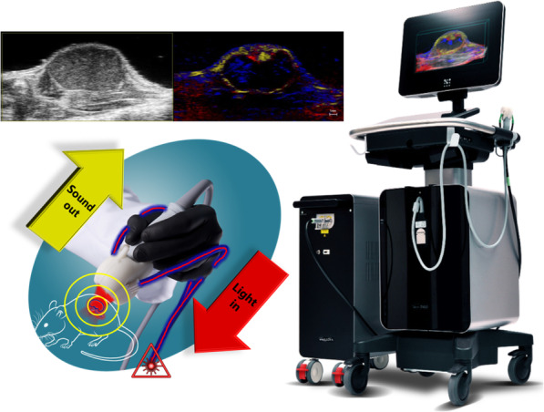
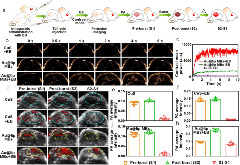
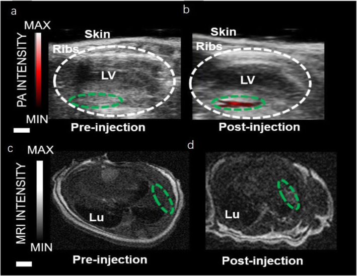
Photoacoustic imaging (PAI) is often performed simultaneously with ultrasound imaging and can provide functional and cellular information regarding the tissues in the anatomical markers of the imaging. This paper describes in detail the basic principles of photoacoustic/ultrasound (PA/US) imaging and its application in recent years. It includes near-infrared-region PA, photothermal, photodynamic, and multimode imaging techniques. Particular attention is given to the relationship between PAI and ultrasonic imaging; the latest high-frequency PA/US imaging of small animals, which involves not only B-mode, but also color Doppler mode, power Doppler mode, and nonlinear imaging mode; the ultrasonic model combined with PAI, including the formation of multimodal imaging; the preclinical imaging methods; and the most effective detection methods for clinical research for the future.

Keywords: Multi-mode imaging; Photoacoustic/ultrasound imaging; Photodynamic therapy; Photothermal therapy; The second near-infrared photoacoustic.


Conflict of interest statement

The authors declare that they have no competing interests.

Figures

Fig. 1
Fig. 1
Schematic of PA/US multimode imaging system
Fig. 2
Fig. 2
tHI caused prolonged deficits in cortical oxygenation. Doppler flow imaging and 3D reconstruction showed reduced perfusion in the ipsilateral hemisphere and the reversal of blood flow in the RICA in the Circle of Willis after RCCAO. Co-registered anatomical image (B-mode micro-US) and StO2 (StO2 in a red-white-blue color-map by PAI) showed bilateral difference in StO2 between the left (L, contralateral) and right (R, ipsilateral) cerebral cortex of an adult C57BL/6 mouse after RCCAO in normoxic (20.9% O2) or hypoxic (7.5% O2) conditions [16]
Fig. 3
Fig. 3
PAI of the injection and engraftment of PANP-labeled hESC-CMs in mouse hearts in vivo [17]
Fig. 4
Fig. 4
a PA spectra of 2 mg/mL SPNs2–3, PBS, and FBS; b PA signal of SPNs2–3 at various concentrations; c PA/US co-registered images without injection of SPNs2–3 (control) and with intratumoral injection of 50 μL, 500 μg/mL SPNs2–3 (dashed circles); d Fitted PA signal curves of SPNs2–3 solutions (1 mg/mL) at various depths in chicken breast tissue [22]
Fig. 5
Fig. 5
Preparation of dual-responsive AuNNP@SN38 Ve loaded with BLZ-945. The smaller AuNNP@PEG/PSN38VP then penetrated the deeper tumor regions and released the SN38 prodrug in the reductive environment, leading to tumor cell apoptosis. PA imaging in the NIR-II window further enabled drug release monitoring and guided therapy [23]
Fig. 6
Fig. 6
MRI and PAI performance in vivo [25]
Fig. 7
Fig. 7
PAI of fibronectin-positive MDAMB-231 tumors at 0, 1, 2, 6, 12 and 24 h postinjection of SQ1 nanoprobe or SQ1@DSPE (control) i.v [26].
Fig. 8
Fig. 8
Schematic algorithm to obtain PDT treatment prediction map from the US and PA images for identifying treatment responders from non-responders and for prediction of tumor regrowth [28]
Fig. 9
Fig. 9
ACSN was injected i.v. to detect 4 T1 tumor-bearing mice by PAI [29]
Fig. 10
Fig. 10
Background-free PAI to study tumor blood perfusion with Au@lip MBs [32]
Fig. 11
Fig. 11
PAI and MRI of Gd (III)-SMNP implanted into mouse hearts [34]

Similar articles

Cited by

References

    1. Zackrisson S, van de Ven SMWY, Gambhir SS. Light in and sound out: emerging translational strategies for photoacoustic imaging. Cancer Res. 2014;74(4):979–1004. doi: 10.1158/0008-5472.CAN-13-2387. - DOI - PMC - PubMed
    1. Needles A, Heinmiller A, Sun J, Theodoropoulos C, Bates D, Hirson D, et al. Development and initial application of a fully integrated photoacoustic micro-ultrasound system. IEEE Trans Ultrason Ferroelectr Freq Control. 2013;60(5):888–897. doi: 10.1109/TUFFC.2013.2646. - DOI - PubMed
    1. Nam SY, Chung E, Suggs LJ, Emelianov SY. Combined ultrasound and photoacoustic imaging to noninvasively assess burn injury and selectively monitor a regenerative tissue-engineered construct. Tissue Eng Part C Methods. 2015;21(6):557–566. doi: 10.1089/ten.tec.2014.0306. - DOI - PMC - PubMed
    1. Huang DD, Qiu Q, Lin WZ, Liu JY, Huang YL, Zhao QL. Recent advances in biomedical applications of dual-modality photoacoustic/ultrasound imaging technology. J Light Scatt. 2019;31(1):1–10.
    1. Zafar H, Breathnach A, Subhash HM, Leahy MJ. Linear-array-based photoacoustic imaging of human microcirculation with a range of high frequency transducer probes. J Biomed Opt. 2015;20(4):051021. doi: 10.1117/1.JBO.20.5.051021. - DOI - PubMed

LinkOut - more resources