

Notch signaling defects in NK cells in patients with cancer
- PMID: 33083905
- PMCID: PMC10991167
- DOI: 10.1007/s00262-020-02763-w
Notch signaling defects in NK cells in patients with cancer
Abstract
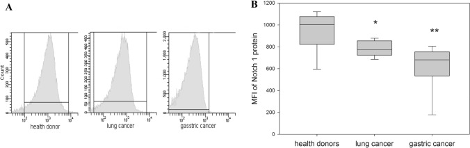
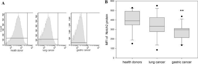
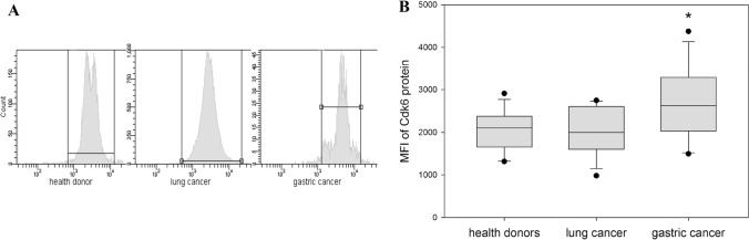
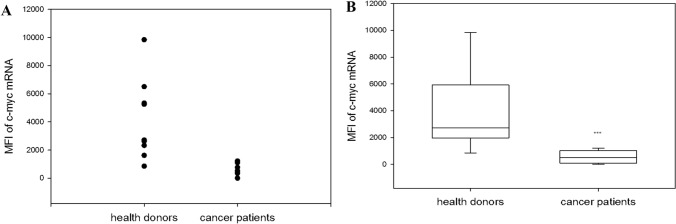
Altered expressions of proto-oncogenes have been reported during normal lymphocytes mitogenesis and in T and B lymphocytes in patients with autoimmune diseases. We have recently demonstrated a significantly decreased expression of c-kit and c-Myc in NK cells isolated from patients with cancer, which might be related to the functional deficiency of NK cells in the tumor environment. Here, focusing on the regulatory mechanisms of this new clinical phenomenon, we determined expression of c-Myc, Notch1, Notch2, p-53, Cdk6, Rb and phosphorylated Rb in NK cells isolated from the healthy donors and cancer patients. The results of our study revealed a significant down-regulation of expression of Notch receptors and up-regulation of Cdk6 expression in NK cells in cancer, while no significant changes in the expression of p53 and Rb proteins were seen. These data revealed novel signaling pathways altered in NK cells in the tumor environment and support further investigation of the origin of deregulated expression of proto-oncogenes in NK cells patients with different types of cancer.
Keywords: NK cells; Notch1; Notch2; T cells; c-myc.
Conflict of interest statement
The authors declare no potential conflicts of interest.
Figures
Similar articles
-
A microRNA-mediated regulatory loop modulates NOTCH and MYC oncogenic signals in B- and T-cell malignancies.Leukemia. 2015 Apr;29(4):968-76. doi: 10.1038/leu.2014.302. Epub 2014 Oct 14. Leukemia. 2015. PMID: 25311243 Free PMC article.
-
Alterations of oncogenes expression in NK cells in patients with cancer.Immun Inflamm Dis. 2017 Dec;5(4):493-502. doi: 10.1002/iid3.179. Epub 2017 Jul 10. Immun Inflamm Dis. 2017. PMID: 28695716 Free PMC article.
-
Differential Notch1 and Notch2 expression and frequent activation of Notch signaling in gastric cancers.Arch Pathol Lab Med. 2011 Apr;135(4):451-8. doi: 10.5858/2009-0665-OA.1. Arch Pathol Lab Med. 2011. PMID: 21466361
-
Effect of notch1,2,3 genes silicing on NF-κB signaling pathway of macrophages in patients with atherosclerosis.Biomed Pharmacother. 2016 Dec;84:666-673. doi: 10.1016/j.biopha.2016.09.078. Epub 2016 Sep 30. Biomed Pharmacother. 2016. PMID: 27697639
-
Unravelling disparate roles of NOTCH in bladder cancer.Nat Rev Urol. 2018 Jun;15(6):345-357. doi: 10.1038/s41585-018-0005-1. Nat Rev Urol. 2018. PMID: 29643502 Review.
Cited by
-
A comprehensive multi-omics analysis reveals molecular features associated with cancer via RNA cross-talks in the Notch signaling pathway.Comput Struct Biotechnol J. 2022 Jul 26;20:3972-3985. doi: 10.1016/j.csbj.2022.07.036. eCollection 2022. Comput Struct Biotechnol J. 2022. PMID: 35950189 Free PMC article.
-
Notch signaling pathway: architecture, disease, and therapeutics.Signal Transduct Target Ther. 2022 Mar 24;7(1):95. doi: 10.1038/s41392-022-00934-y. Signal Transduct Target Ther. 2022. PMID: 35332121 Free PMC article. Review.
-
The Notch signaling pathway: a potential target for cancer immunotherapy.J Hematol Oncol. 2023 May 2;16(1):45. doi: 10.1186/s13045-023-01439-z. J Hematol Oncol. 2023. PMID: 37131214 Free PMC article. Review.
References
-
- Hofmann JW, Zhao X, De Cecco M, Peterson AL, Pagliaroli L, Manivannan J, Hubbard GB, Ikeno Y, Zhang Y, Feng B, Li X, Serre T, Qi W, Van Remmen H, Miller RA, Bath KG, de Cabo R, Xu H, Neretti N, Sedivy JM. Reduced expression of MYC increases longevity and enhances healthspan. Cell. 2015;160:477–488. doi: 10.1016/j.cell.2014.12.016. - DOI - PMC - PubMed
MeSH terms
Substances
Grants and funding
LinkOut - more resources
Full Text Sources
Medical
Research Materials
Miscellaneous
