Comparative Study
. 2021 May 1;529(7):1703-1718.
doi: 10.1002/cne.25051. Epub 2020 Oct 20.

Ultrastructural localization of glutamate delta 1 (GluD1) receptor immunoreactivity in the mouse and monkey striatum

Affiliations
Comparative Study

Ultrastructural localization of glutamate delta 1 (GluD1) receptor immunoreactivity in the mouse and monkey striatum

Andrew H Hoover et al. J Comp Neurol. .

Abstract

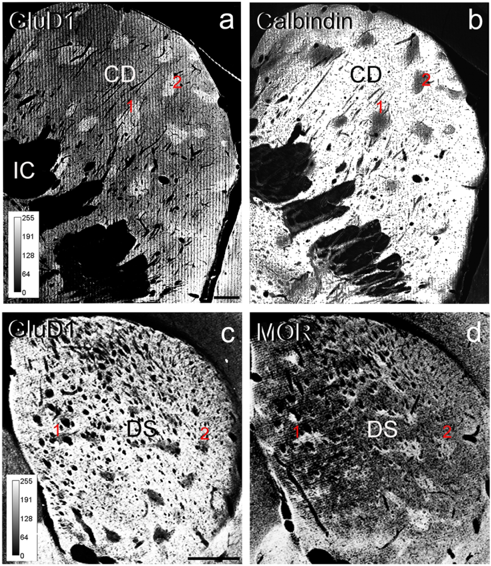
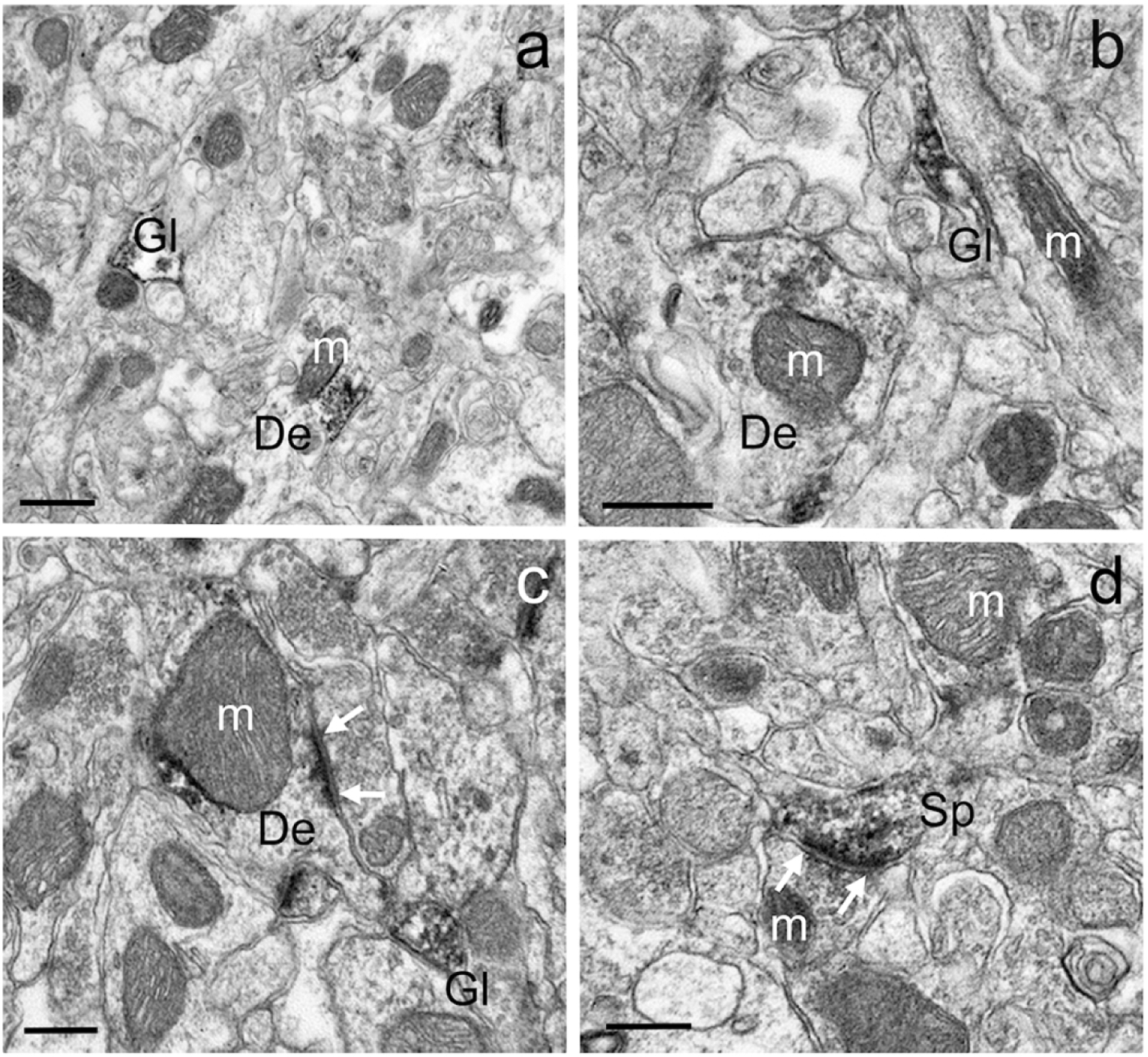
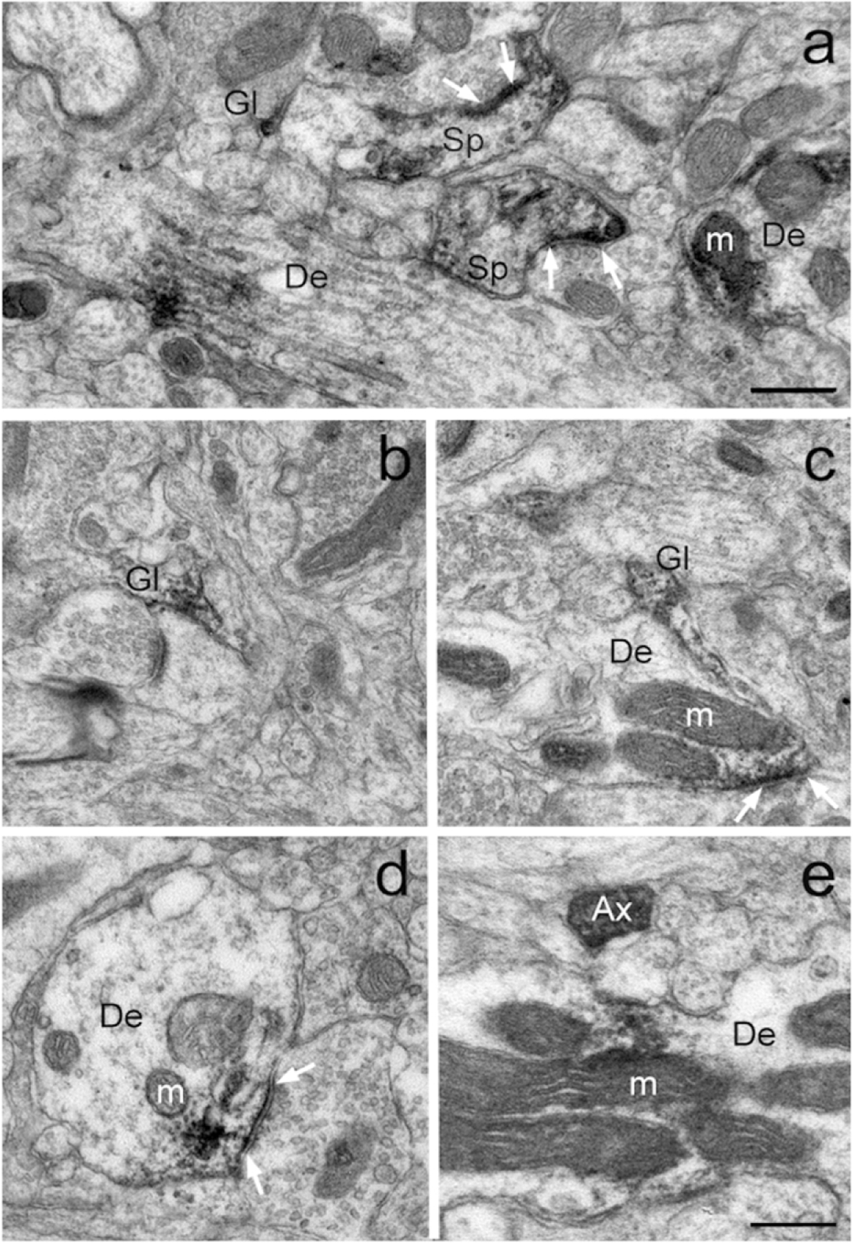
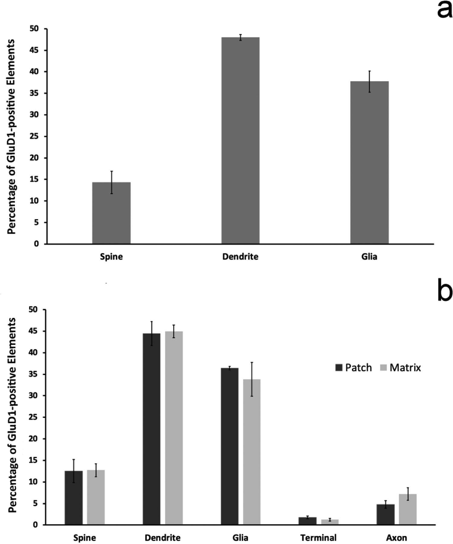
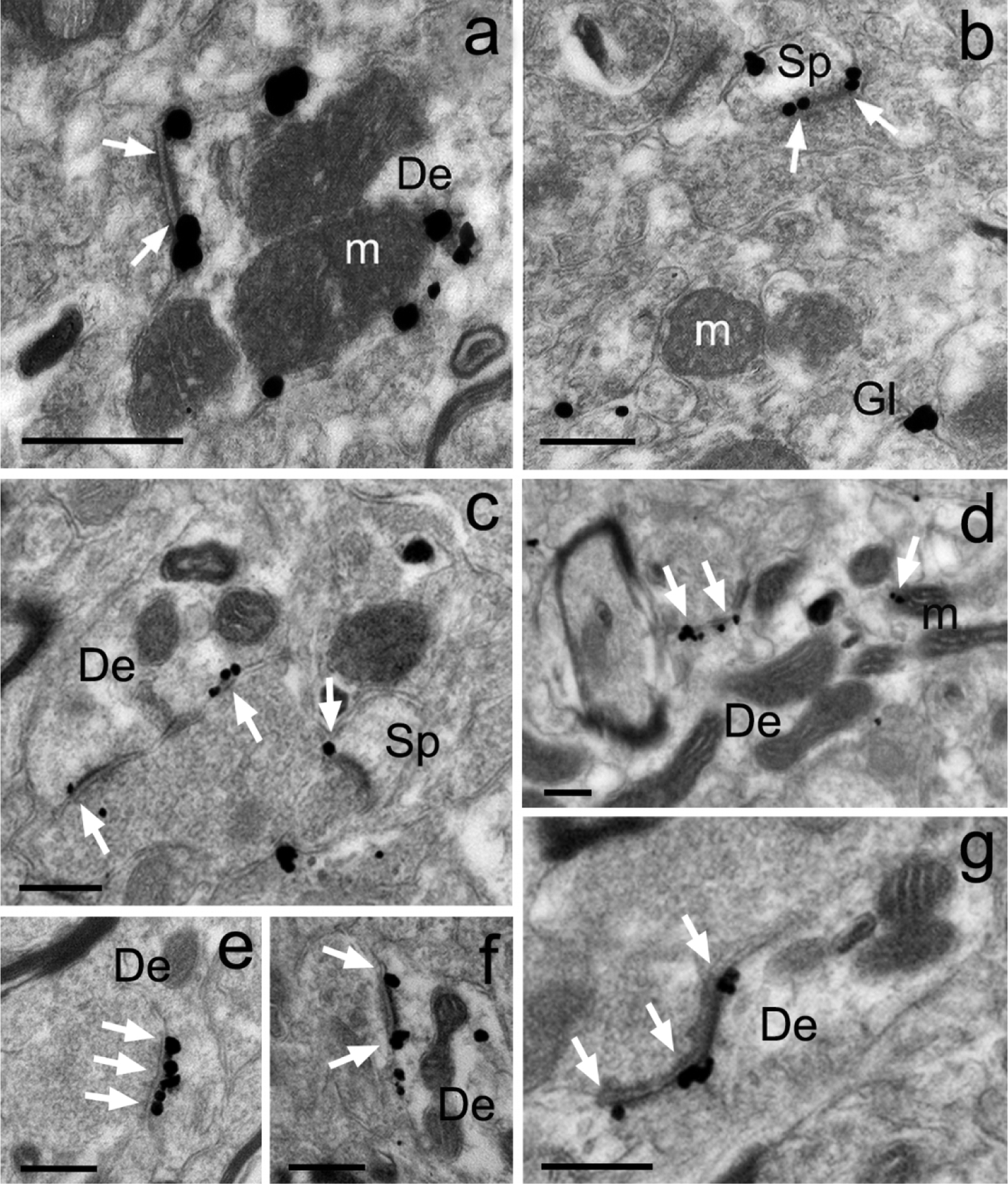
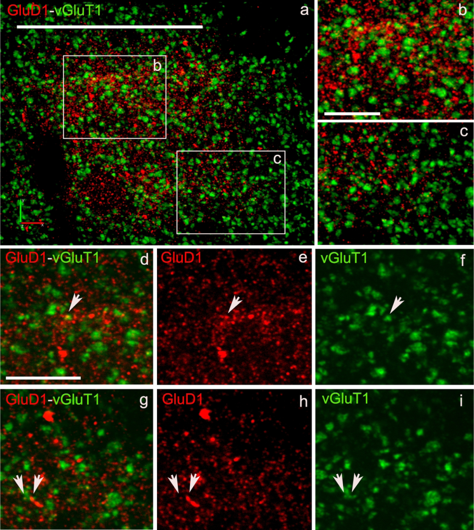
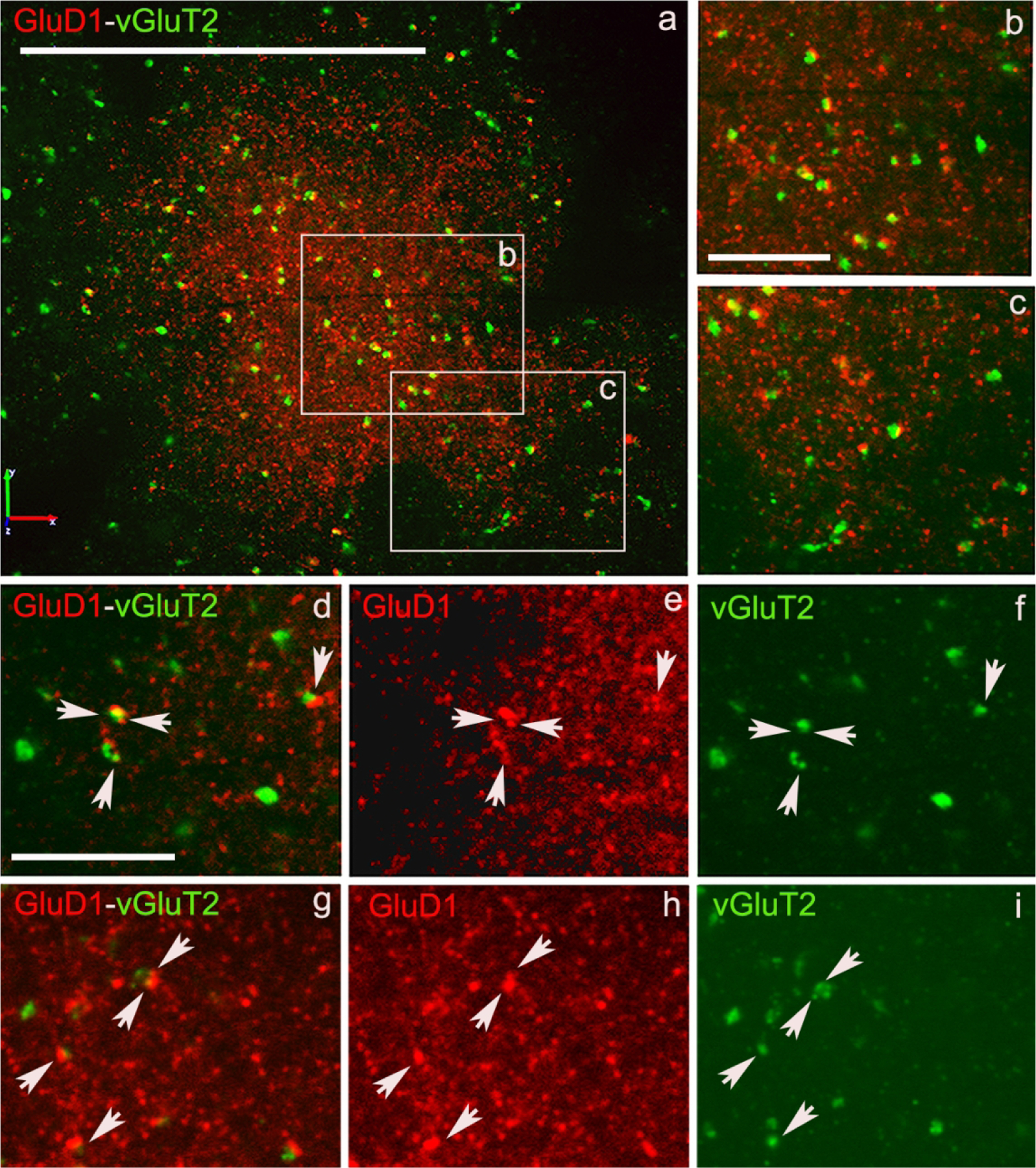
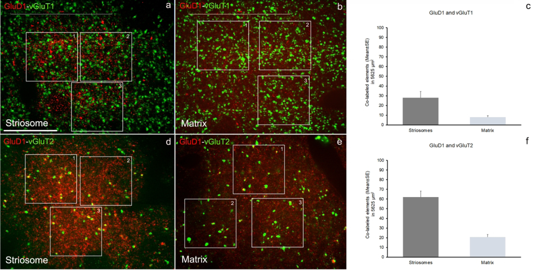
The glutamate receptor delta 1 (GluD1) is strongly expressed in the striatum. Knockout of GluD1 expression in striatal neurons elicits cognitive deficits and disrupts the thalamostriatal system in mice. To understand the potential role of GluD1 in the primate striatum, we compared the cellular and subcellular localization of striatal GluD1 immunoreactivity (GluD1-IR) in mice and monkeys. In both species, striatal GluD1-IR displayed a patchy pattern of distribution in register with the striosome/matrix compartmentation, but in an opposite fashion. While GluD1 was more heavily expressed in the striosomes than the matrix in the monkey caudate nucleus, the opposite was found in the mouse striatum. At the electron microscopic level, GluD1-IR was preferentially expressed in dendritic shafts (47.9 ± 1.2%), followed by glia (37.7 ± 2.5%), and dendritic spines (14.3 ± 2.6%) in the matrix of the mouse striatum. This pattern was not statistically different from the labeling in the striosome and matrix compartments of the monkey caudate nucleus, with the exception of a small amount of GluD1-positive unmyelinated axons and axon terminals in the primate striatum. Immunogold staining revealed synaptic and perisynaptic GluD1 labeling at putative axo-dendritic and axo-spinous glutamatergic synapses, and intracellular labeling on the surface of mitochondria. Confocal microscopy showed that GluD1 is preferentially colocalized with thalamic over cortical terminals in both the striosome and matrix compartments. These data provide the anatomical substrate for a deeper understanding of GluD1 regulation of striatal glutamatergic synapses, but also suggest possible extrasynaptic, glial, and mitochondrial GluD1 functions.

Keywords: RRIDs-AB_2571757; AB_2314065; AB_2716850; AB_2571759; AB_2301751; AB_2315569; cerebellin; matrix; parafascicular; patch; striosome; thalamostriatal.


Conflict of interest statement

Conflict of Interest: The authors declare nonfinancial competing interests or other conflicts of interests for this article.

Figures

Figure 1:
Figure 1:. Striosome/Matrix Compartmentation of GluD1 in the monkey and mouse striatum.
Light micrographs of GluD1 immunoperoxidase staining in the pre-commissural caudate nucleus of a monkey (a) and the pre-commissural dorsal striatum of a mouse (c). (b) and (d) illustrate adjacent sections immunostained for calbindin D28k (b) or MOR (d) to differentiate the striosomes from the matrix compartments of the monkey and mouse striatum, respectively. Note the close correspondence between dense patches of GluD1 immunostaining in a with calbindin-immunonegative striosomes in b. In mouse, areas of low GluD1 labeling in c correspond to MOR-enriched striosomes in d. In both sets of sections, red numbers 1 and 2 indicate corresponding striosomes. The colors in all panels have been inverted, and the levels of immunoreactivity correspond to the intensity scales shown in panel (a). Abbreviations: CD: caudate nucleus, DS: dorsal striatum, IC: internal capsule. Scale bar in a (applies to b) = 1 mm. Scale bar in c (applies to d) = 1 mm.
Figure 2:
Figure 2:. GluD1-immunoreactive elements in the mouse striatum.
Examples of different GluD1-immunoreactive dendrites (a-c), spines (d) and glia (a,b) in the mouse dorsal striatum. In c and d, white arrows indicate asymmetric axo-dendritic (c) and axo-spinous (d) synapses. Note some GluD1 peroxidase staining associated with the surface of mitochondria in a-c. Abbreviations: Sp: spine, De: dendrite, Gl: glia and m: mitochondria. Scale bar in a = 0.50μm. Scale bar in b = 0.35μm. Scale bar in c = 0.25μm. Scale bar in d = 0.25μm.
Figure 3:
Figure 3:. GluD1-immunoreactive elements in the monkey striatum.
Examples of GluD1-positive neuronal and glial structures in the monkey striatum. White arrows indicate asymmetric synapses with dense aggregates of GluD1 imunolabeling in their close vicinity. Abbreviations: Sp: spine, De: dendrite, Gl: glia, m: mitochondria and Ax: unmyelinated axon. Scale bar in a (applies to b-e) = 0.30μm.
Figure 4:
Figure 4:. Relative Distribution of GluD1-immunostained elements.
(a) shows the mean relative percentages (+/− SEM) of GluD1-positive neuronal and glial structures in the matrix compartment of the mouse dorsal striatum (N=3 animals; n=157 images). (b) illustrates the relative percentage of GluD1-immunostained elements in the striosome vs matrix compartments of the monkey caudate nucleus (N=3 animals; n=324 images). There is no statistically significant difference in the relative percentages of the different neuronal and glial elements between the striosome and matrix compartments (two sample t-test; dendrites p=0.870; glia p=0.548; spines p=0.953; axons p=0.243; terminals p=0.268). There is also no statistically significant difference between the percentages of GluD1-immunoreactive elements of the mouse vs the monkey matrix compartment (two-sample t-test; dendrites p=0.140; glia p=0.451; spines p=0.633).
Figure 5:
Figure 5:. Immunogold localization of GluD1 in the mouse and monkey striatum.
Examples of GluD1-immunoreactive elements in the mouse (a,b) and monkey (c-g) pre-commissural striatum as revealed with the pre-embedding immunogold technique. (a-b) Peri-synaptic gold labeling at an asymmetric axo-dendritic (a) and axo-spinous (b) synapse. Intracellular gold particles closely apposed to the external membrane of a mitochondria are also depicted in panel “a”. (c-g) Examples of peri-synaptic (c,f,g) and synaptic (e,g) GluD1 immunolabeling at asymmetric synapses in the monkey caudate nucleus. White arrows indicate GluD1-labeled asymmetric synapses. Abbreviations: Sp: spine, De: dendrite, Gl: glia and m: mitochondria. Scale bars = 0.25μm in all panels.
Figure 6:
Figure 6:. Confocal images of GluD1/vGluT1 in the monkey striatum.
(a) Low magnification image of a putative striosome (enriched in GluD1 immunoreactivity) in the caudate nucleus of monkey striatum double immunostained for GluD1 (red) and vGluT1 (Figure 6) or vGluT2 (Figure 7) (green). (b,c) High magnification images of boxed areas marked in panel a. (d,g) Colocalization of GluD1 and vGluT1/vGluT2 in double immunostained “terminal-like” structures (puncta). The same puncta structures (arrows in d-i) are identified in single immunofluorescence images for GluD1 (e,h) and vGluT1 or vGluT2 (f,i). Scale bar in a = 50μm in b (applies to c) and d (applies to e-i) = 10μm.
Figure 7:
Figure 7:. Confocal images of GluD1/vGluT2 in the monkey striatum.
(a) Low magnification image of a putative striosome (enriched in GluD1 immunoreactivity) in the caudate nucleus of monkey striatum double immunostained for GluD1 (red) and vGluT1 (Figure 6) or vGluT2 (Figure 7) (green). (b,c) High magnification images of boxed areas marked in panel a. (d,g) Colocalization of GluD1 and vGluT1/vGluT2 in double immunostained “terminal-like” structures (puncta). The same puncta structures (arrows in d-i) are identified in single immunofluorescence images for GluD1 (e,h) and vGluT1 or vGluT2 (f,i). Scale bar in a = 50μm in b (applies to c) and d (applies to e-i) = 10μm.
Figure 8:
Figure 8:. Quantitative analysis of double immunostained confocal images.
Quantification of co-labeled puncta structures in double immunostained sections have been done in striatal areas (caudate) with high GluD1-immunofluorescence (striosome-like) (a, d) and areas with low GluD1-immunofluorescence (matrix-like) (b,e). The quantitative results for co-labeled elements for GluD1/vGluT1 (c) and GluD1/vGluT2 (d) in striosome- and matrix-like areas have been obtained from three areas/image (boxed areas) and three images/striosome- and /matrix-like areas for each double immunostaining, GluD1/vGluT1 or GluD1/vGluT2. The data are shown as the mean value (±SE) of co-labeled elements in the total area analyzed (5625 μm2). The detailed quantitative data for co-labeled puncta elements and single labeled structures are shown in the Table 3. Scale bar in a = 25μm.

Similar articles

Cited by

References

    1. Antel JP, Lin YH, Cui Q-L, Pernin F, Kennedy TE, Ludwin SK, & Healy LM (2019). Immunology of oligodendrocyte precursor cells in vivo and in vitro. Journal of Neuroimmunology, 331, 28–35. 10.1016/j.jneuroim.2018.03.006 - DOI - PubMed
    1. Benamer N, Marti F, Lujan R, Hepp R, Aubier TG, Dupin A. a. M., Frébourg G, Pons S, Maskos U, Faure P, Hay YA, Lambolez B, & Tricoire L (2018). GluD1, linked to schizophrenia, controls the burst firing of dopamine neurons. Molecular Psychiatry, 23(3), 691–700. 10.1038/mp.2017.137 - DOI - PMC - PubMed
    1. Bergles DE, Jabs R, & Steinhäuser C (2010). Neuron-glia synapses in the brain. Brain Research Reviews, 63(1–2), 130–137. 10.1016/j.brainresrev.2009.12.003 - DOI - PMC - PubMed
    1. Bergles DE, Roberts JDB, Somogyi P, & Jahr CE (2000). Glutamatergic synapses on oligodendrocyte precursor cells in the hippocampus. Nature, 405(6783), 187–191. 10.1038/35012083 - DOI - PubMed
    1. Berridge G, Menassa DA, Moloney T, Waters PJ, Welding I, Thomsen S, Zuberi S, Fischer R, Aricescu AR, Pike M, Dale RC, Kessler B, Vincent A, Lim M, Irani SR, & Lang B (2018). Glutamate receptor δ2 serum antibodies in pediatric opsoclonus myoclonus ataxia syndrome. Neurology, 91(8), e714–e723. 10.1212/WNL.0000000000006035 - DOI - PMC - PubMed

Publication types

Substances