. 2020 Nov 12;63(21):12799-12813.
doi: 10.1021/acs.jmedchem.0c01178. Epub 2020 Oct 21.

Structural Insights into the Recognition of Mono- and Diacetylated Histones by the ATAD2B Bromodomain

Affiliations

Structural Insights into the Recognition of Mono- and Diacetylated Histones by the ATAD2B Bromodomain

Jonathan T Lloyd et al. J Med Chem. .

Abstract

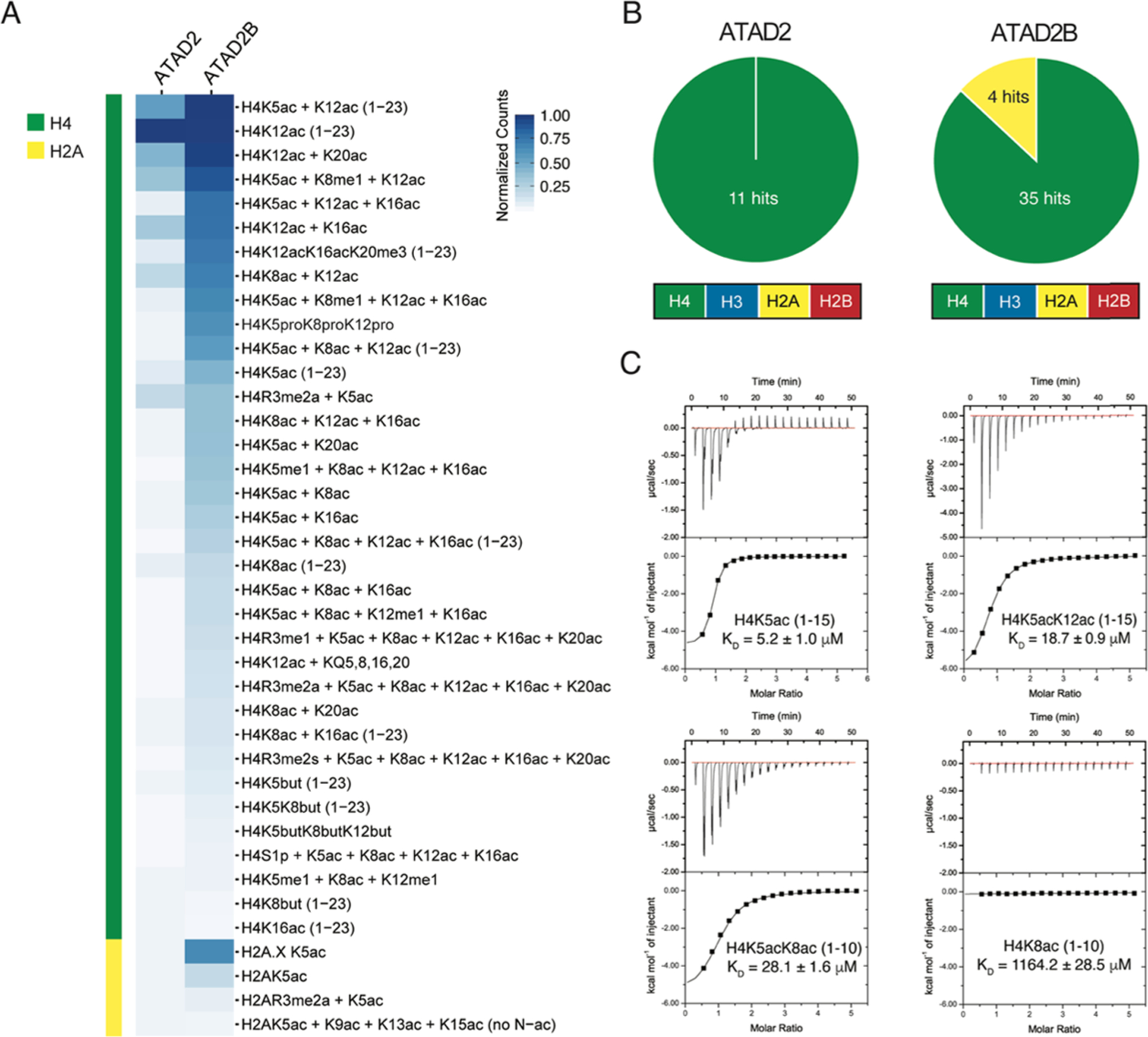
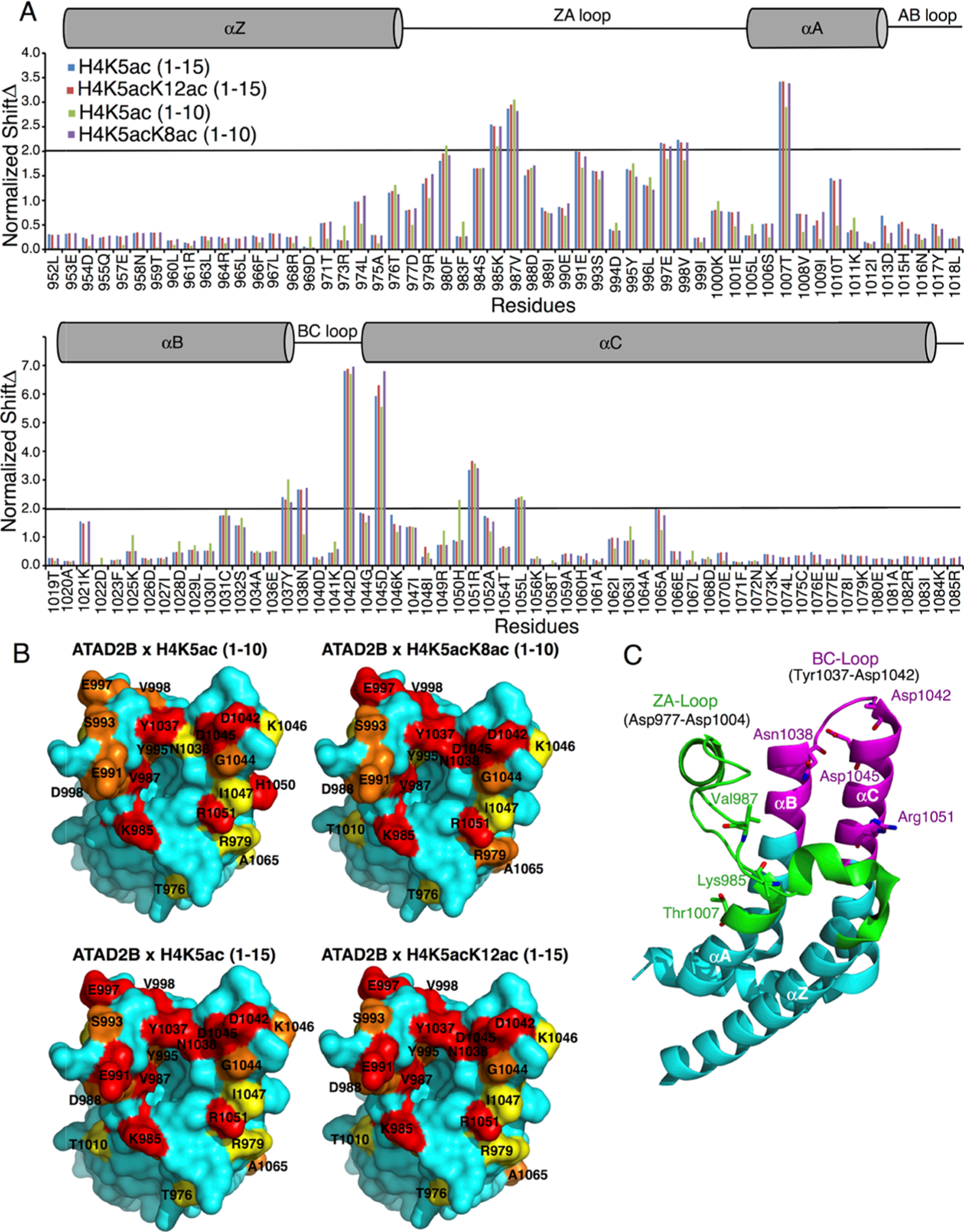
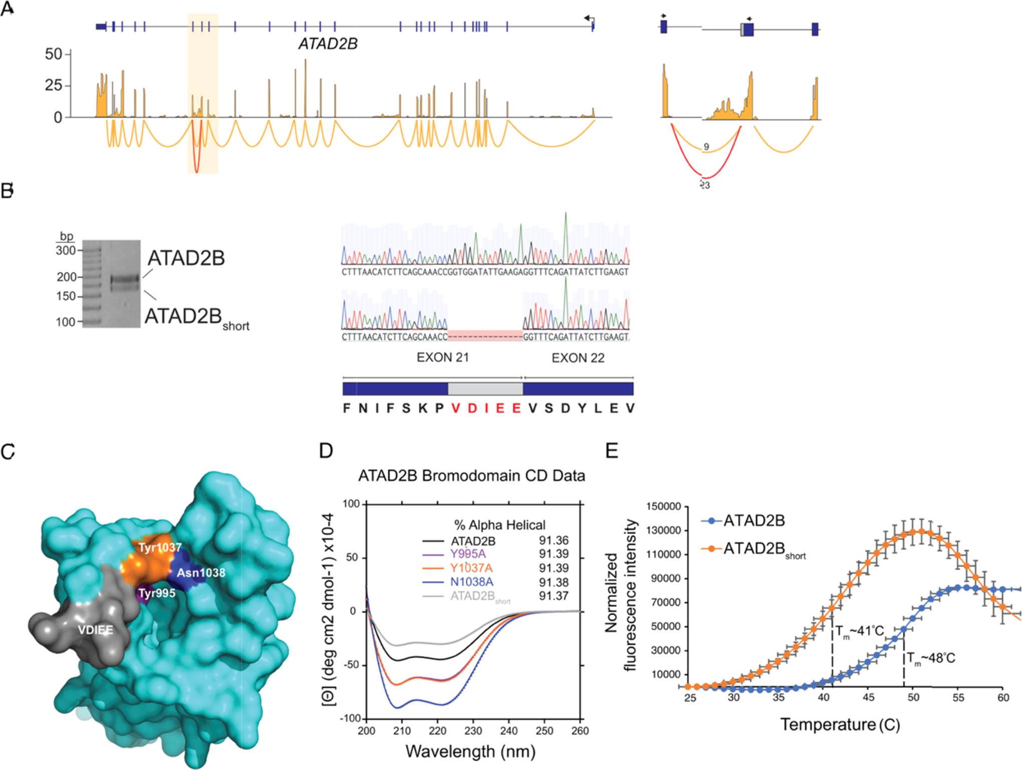
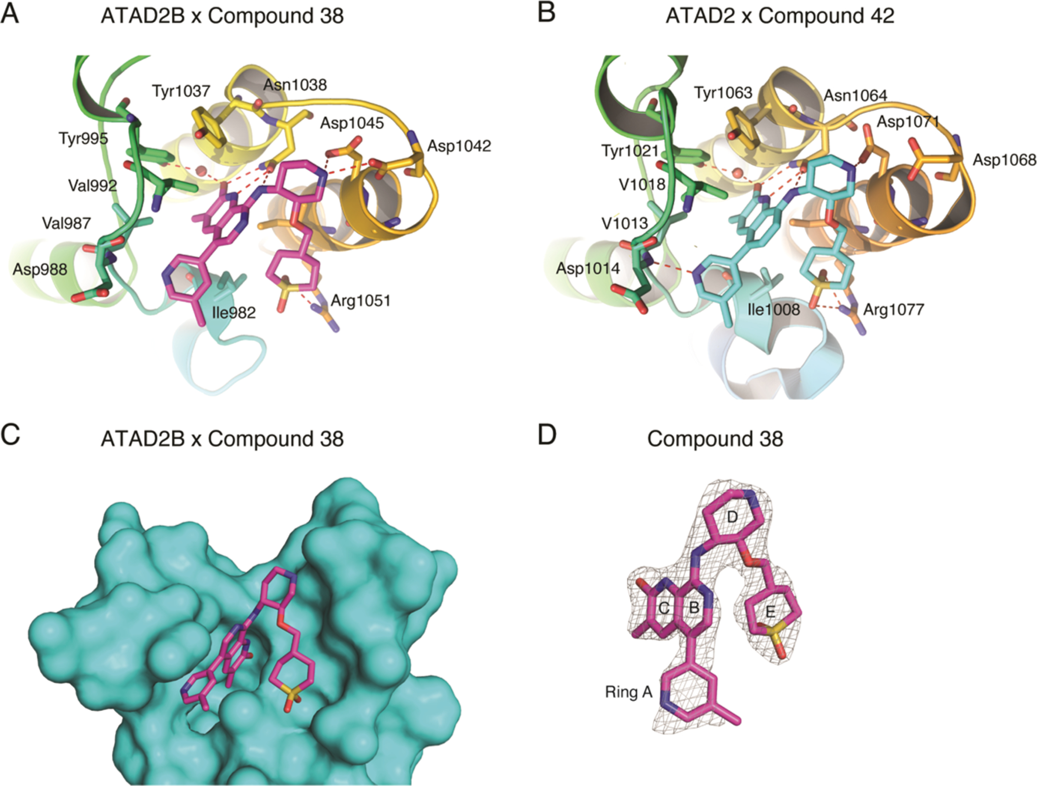
Bromodomains exhibit preferences for specific patterns of post-translational modifications on core and variant histone proteins. We examined the ligand specificity of the ATAD2B bromodomain and compared it to its closely related paralogue in ATAD2. We show that the ATAD2B bromodomain recognizes mono- and diacetyllysine modifications on histones H4 and H2A. A structure-function approach was used to identify key residues in the acetyllysine-binding pocket that dictate the molecular recognition process, and we examined the binding of an ATAD2 bromodomain inhibitor by ATAD2B. Our analysis demonstrated that critical contacts required for bromodomain inhibitor coordination are conserved between the ATAD2/B bromodomains, with many residues playing a dual role in acetyllysine recognition. We further characterized an alternative splice variant of ATAD2B that results in a loss of function. Our results outline the structural and functional features of the ATAD2B bromodomain and identify a novel mechanism regulating the interaction of the ATAD2B protein with chromatin.


Conflict of interest statement

The authors declare the following competing financial interest(s): EpiCypher (M.R.M. and M.-C.K.) is a commercial developer of the dCypher peptide-binding platform used in this study.

Figures

Figure 1.
Figure 1.
ATAD2B bromodomain recognizes acetylated histones. (A) Heat map represents the relative binding of GST-tagged ATAD2 and ATAD2B bromodomains to histone peptides using AlphaScreen technology (i.e., dCypher phase B: see the Methods section). α counts (n = 2) were normalized for each protein to the highest fluorescent intensity signal for each respective assay, and the relative binding strength is indicated by the color gradient. (B) Pie charts for ATAD2/B bromodomains indicate the number of peptides classified as positive binders (α counts having signals >5000, which is at least twice that of the relevant unmodified control peptide) (full dCypher peptide screen data in Tables S1 and S2, and Resources Table A). (C) Exothermic ITC enthalpy plots for the binding of the ATAD2B bromodomain to mono- and diacetylated histone ligands. The calculated binding constants are indicated.
Figure 2.
Figure 2.
Mapping of the mono- and diacetylated histone ligand binding interfaces on the ATAD2B bromodomain. (A) Histogram shows the normalized 1H–15N chemical shift changes observed in the backbone amides of the ATAD2B bromodomain upon the addition of peptides H4K5ac (1–10) (green), H4K5acK8ac (1–10) (purple), H4K5ac (1–15) (blue), and H4K5acK12ac (1–15) (red) in a 1:5 (protein/peptide) molar ratio. The secondary structure elements for the bromodomain are represented above the histogram. A cylinder indicates an α-helical structure, and a straight line depicts loop regions. (B) Chemical shift perturbations mapped onto the solvent-accessible surface representation of the apo ATAD2B bromodomain (PDB ID: 3LXJ) with the addition of H4K5ac (1–10), H4K5acK8ac (1–10), H4K5ac (1–15), and H4K5acK12ac (1–15). CSPs that are 0.5, 1, and 2 standard deviations from the average shift changes are colored in yellow, orange, and red, respectively. (C) Cartoon representation of the ATAD2B bromodomain with the ZA loop region (residues 977–1004) colored in green and the extended region around the BC loop (residues 1031–1055) involved in chemical shift perturbations in magenta. The seven amino acid residues exhibiting the largest chemical shift changes are labeled in stick representation.
Figure 3.
Figure 3.
Analysis of targeted mutations and noncanonical splice variants in the ATAD2B bromodomain. (A) RNA-seq and sashimi plots showing expression and splice junctions across the ATAD2B gene. Normalized expression values are on the y-axis. The highlighted region indicates exons 20–22 that encode the ATAD2B bromodomain. The junctions for these exons are shown on the right where the number of reads spanning each junction is shown, and the noncanonical splice variant is in red. (B) Reverse transcription-polymerase chain reaction (RT-PCR) and Sanger sequencing showing both ATAD2B and ATAD2Bshort isoforms. The amino acids excluded from ATAD2Bshort as a result of alternative 5′ splice donor sites are highlighted in red. (C) Surface representation of the apo ATAD2B bromodomain (PDB ID: 3LXJ) showing the location of specific point mutations introduced to the binding pocket by site-directed mutagenesis and the location of the residues excluded from the ATAD2Bshort splice variant. (D) Circular dichroism spectra in the far-UV region of the ATAD2B, ATAD2B mutant, and ATAD2Bshort bromodomains. The percent α-helical content for each protein is listed in the inset. (E) Thermal stability assay showing the ATAD2B bromodomain unfolding curve (blue) with a Tm = 48 °C, while the ATAD2Bshort bromodomain (orange) demonstrated a significantly lower Tm = 41 °C.
Figure 4.
Figure 4.
Coordination of bromodomain inhibitor molecules by the ATAD2 and ATAD2B bromodomains. (A) ATAD2B bromodomain in complex with compound 38 (PDB ID: 6VEO). (B) ATAD2 bromodomain in complex with compound 42. Hydrogen bonds are indicated by a red dashed line. (C) Simulated annealing composite omits map (gray) representing the bound compound 38 (green) in complex with the ATAD2B bromodomain (cyan) contoured at 1σ. (D) Isolated image of the simulated annealing composite omits map (gray) around compound 38 (green) contoured at 1σ. The simulated annealing composite omits maps shown in (C) and (D) were calculated prior to building the ligand into the structure. Figures were generated with the PyMOL Molecular Graphics System, version 2.3, Schrödinger, LLC.

References

    1. Hebbes TR; Thorne AW; Crane-Robinson C A direct link between core histone acetylation and transcriptionally active chromatin. EMBO J 1988, 7, 1395–1402. - PMC - PubMed
    1. Luger K; Richmond TJ The histone tails of the nucleosome. Curr. Opin. Genet. Dev 1998, 8, 140–146. - PubMed
    1. Norton VG; Imai BS; Yau P; Bradbury EM Histone acetylation reduces nucleosome core particle linking number change. Cell 1989, 57, 449–457. - PubMed
    1. Li Y; Li Z; Dong L; Tang M; Zhang P; Zhang C; Cao Z; Zhu Q; Chen Y; Wang H; Wang T; Lv D; Wang L; Zhao Y; Yang Y; Wang H; Zhang H; Roeder RG; Zhu WG Histone H1 acetylation at lysine 85 regulates chromatin condensation and genome stability upon DNA damage. Nucleic Acids Res 2018, 46, 7716–7730. - PMC - PubMed
    1. Banerjee DR; Deckard CE 3rd; Zeng Y; Sczepanski JT Acetylation of the histone H3 tail domain regulates base excision repair on higher-order chromatin structures. Sci. Rep 2019, 9, No. 15972. - PMC - PubMed

Publication types

MeSH terms

LinkOut - more resources