. 2020 Dec;9(1):2455-2464.
doi: 10.1080/22221751.2020.1840311.

Yin-Yang 1 and HBx protein activate HBV transcription by mediating the spatial interaction of cccDNA minichromosome with cellular chromosome 19p13.11

Affiliations

Yin-Yang 1 and HBx protein activate HBV transcription by mediating the spatial interaction of cccDNA minichromosome with cellular chromosome 19p13.11

Congle Shen et al. Emerg Microbes Infect. 2020 Dec.

Abstract

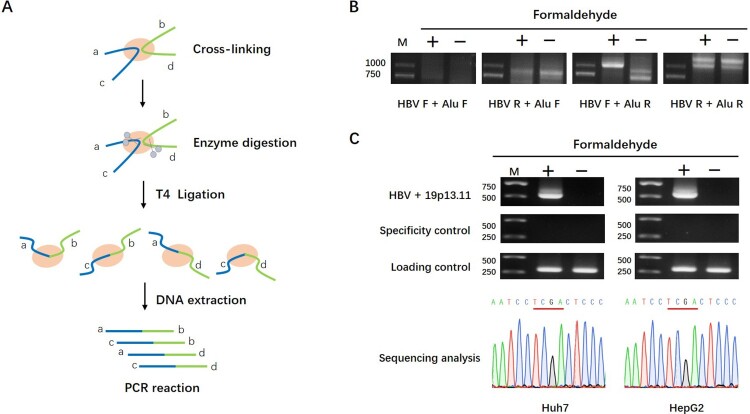
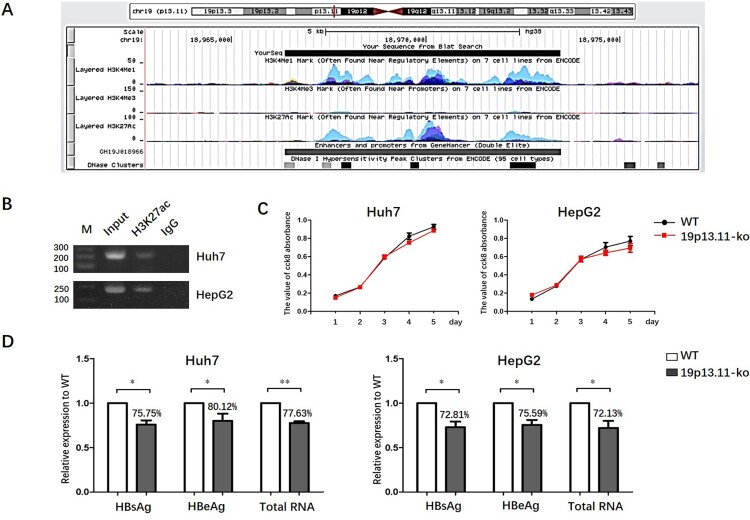
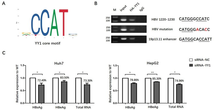
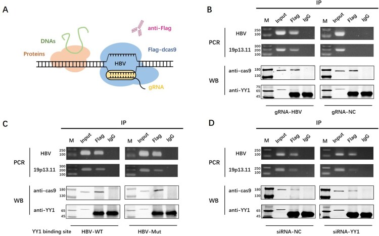
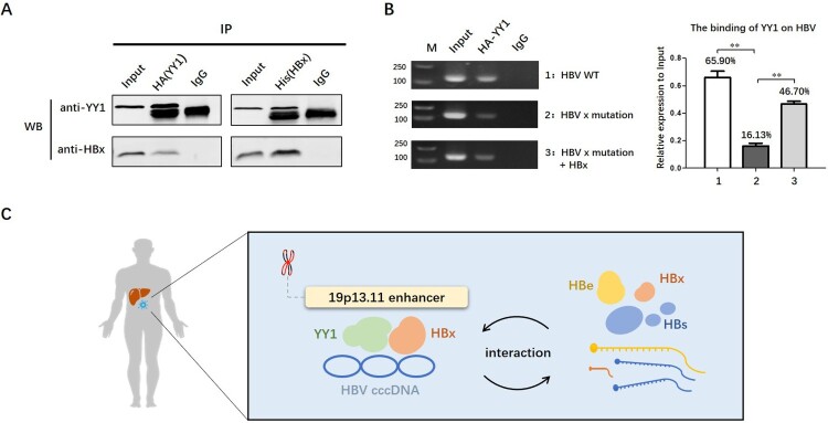
HBV cccDNA stably exists in the nuclei of infected cells as an episomal munichromosome which is responsible for viral persistence and failure of current antiviral treatments. However, the regulatory mechanism of cccDNA transcription by viral and host cellular factors is not well understood. In this study, we investigated whether cccDNA could be recruited into a specific region of the nucleus via specific interaction with a cellular chromatin to regulate its transcription activity. To investigate this hypothesis, we used chromosome conformation capture (3C) technology to search for the potential interaction of cccDNA and cellular chromatin through rcccDNA transfection in hepatoma cells and found that cccDNA is specifically associated with human chromosome 19p13.11 region, which contains a highly active enhancer element. We also confirmed that cellular transcription factor Yin-Yang 1 (YY1) and viral protein HBx mediated the spatial regulation of HBV cccDNA transcription by 19p13.11 enhancer. Thus, These findings indicate that YY1 and HBx mediate the recruitment of HBV cccDNA minichromosomes to 19p13.11 region for transcription activation, and YY1 may present as a novel therapeutic target against HBV infection.

Keywords: HBV cccDNA; HBV transcription regulation; YY1; chromosome 19p13.11; spatial interaction.


Conflict of interest statement

No potential conflict of interest was reported by the author(s).

Figures

Figure 1.
Figure 1.
Physical proximity of 19p13.11 region and HBV cccDNA detected by 3C assays. A: Schematic diagram of 3C procedure. B: HBV-Alu ligation fragments detected by 3C assays. The PCR products were obtained from cross-linked Huh7 cells with specific pairs of primers of HBV and Alu sequence: HBV F + Alu F, HBV R + Alu F, HBV F + Alu R, HBV R + Alu R. Among them, a unique PCR product was shown by using HBV F + Alu R primers. C: 19p13.11-HBV ligation fragments detected by 3C assays. The PCR products were obtained from cross-linked Huh7 and HepG2 cells respectively with a specific pair of primers of 19p13.11 and HBV sequence: 19p13.11 R + HBV F (540bp) (upper). The sequencing of these fragments identified 19p13.11 – HBV piecing sequences mediated by TaqI enzyme site (TCGA) (lower). HBV + ACTB (350bp) was used as a specificity control, and ACTB gene (288 bp) was used as a loading control.
Figure 2.
Figure 2.
19p13.11 enhancer could regulate the transcriptional activity of HBV cccDNA. A: Analysis of 19p13.11 enhancer in UCSC database. Scale: the length of the enhancer, 7106bp. Chr19: the location of the enhancer, from 18966372 to 18973477 in hg38. Layered H3K4me1, H3K4me3, H3K27ac: the intensity of H3K4me1, H3K4me3, H3K27ac modification of the enhancer in seven cell lines distinguished by different colours including GM12878, H1-hESC, HSMM, HUVEC, K562, NHEK and NHLF cells. The high intensity of H3K4me1 and H3K27ac were mostly shown in HSMM, HUVEC, K562, NHEK and NHLF cells. GH19J018966: the ID card of the regulatory element in GeneCards database and colours are used to distinguish promoters and enhancers. Red: promoters, Grey: enhancers. DNase clusters: clusters of DNaseI hypersensitivity derived from assays in 95 cell types. Regulatory regions in general, and promoters in particular, tend to be DNase-sensitive. B: Chip-PCR shows obvious H3K27ac binding in 19p13.11 site in Huh7 and HepG2 liver cancer cells. The intended PCR product is 228bp. C: The value of CCK8 absorbance of wild type or 19p13.11-ko Huh7 and HepG2 cells from first to fifth day. Three independent experiments were performed for each assay. D: The relative expression of HBsAg and HBeAg in supernatant detected by ELISA and HBV total RNAs intracellular detected by q-PCR of wild type or 19p13.11-ko Huh7 and HepG2 cells co-transfecting prcccDNA and pCMV-Cre plasmids three days later. Three independent experiments were performed for each assay. * P<0.05. ** P<0.01.
Figure 3.
Figure 3.
YY1 could combine with 19p13.11 enhancer as well as HBV genome and regulate the transcriptional activity of HBV cccDNA. A: core motif of YY1 binding sites from JASPAR database. B: ChIP-PCR by anti-HA antibody shows obvious YY1 binding in potential 19p13.11 site (189bp) and HBV wild or mutated genome (130bp) in Huh7 cell line. Nucleotides underlined present core motif of YY1 binding and nucleotides marked red present mutation. C: The relative expression of HBsAg and HBeAg in supernatant detected by ELISA and HBV total RNAs intracellular detected by q-PCR of siRNA-mock or siRNA-YY1 as well as prcccDNA and pCMVCre plasmids transfected Huh7 and HepG2 cells 48 h later. Three independent experiments were performed for each assay. * P<0.05. ** P<0.01.
Figure 4.
Figure 4.
YY1 is the structural regulator of 19p13.11 enhancer and HBV cccDNA. A: Schematic diagram of enChIP technique. B: dCas9 protein (170kDa) – HBV DNA (130bp) – YY1 protein (65kDa) – 19p13.11 DNA (189bp) complex was detected by gRNA-HBV, and the abundance of YY1 protein and 19p13.11 DNA were both reduced when the binding site of YY1 was mutated in HBV (C) or the expression of YY1 was knock down (D) in Huh7 cells. The most obvious print in WB-YY1 was the heavy chain of antibody (55kDa).
Figure 5.
Figure 5.
HBx could interact with YY1 and promote the expression of YY1. A: co-IP experiments show that HBx protein (17kd) could be detected by anti-HA antibody and YY1 protein (65kd) could be detected by anti-His antibody. B: Chip-PCR experiments show the binding of YY1 on HBV cccDNA in Huh7 cells transfecting wild HBV plasmid, HBx-mutated plasmid with or without His-HBx plasmid (130bp). * P<0.05. ** P<0.01. C: Schematic diagram of spatial interaction between 19p13.11 enhancer and HBV cccDNA.

Similar articles

Cited by

References

    1. Wei XL, Luo HY, Li CF, et al. . Hepatitis B virus infection is associated with younger median age at diagnosis and death in cancers. Int J Cancer. 2017;141(1):152–159. - PubMed
    1. World Health Organization . Hepatitis B 2018. Available from: http://www.who.int/news-room/fact-sheets/detail/hepatitis-b.
    1. Gane EJ. Future anti-HBV strategies. Liver Int 2017;37:40–44. - PubMed
    1. McNaughton AL, Arienzo VD, Ansariet MA, et al. . Insights from deep sequencing of the HBV genome-unique, tiny, and misunderstood. Gastroenterology. 2019;156(2):384–399. - PMC - PubMed
    1. Wang J, Huang H, Liu Y, et al. . HBV genome and life cycle. Adv Exp Med Biol. 2020;1179:17–37. - PubMed

MeSH terms