Review
. 2020 Dec 7;49(23):8749-8773.
doi: 10.1039/d0cs00600a. Epub 2020 Oct 21.

Covalent labeling of nucleic acids

Affiliations
Review

Covalent labeling of nucleic acids

Nils Klöcker et al. Chem Soc Rev. .

Abstract

Labeling of nucleic acids is required for many studies aiming to elucidate their functions and dynamics in vitro and in cells. Out of the numerous labeling concepts that have been devised, covalent labeling provides the most stable linkage, an unrivaled choice of small and highly fluorescent labels and - thanks to recent advances in click chemistry - an incredible versatility. Depending on the approach, site-, sequence- and cell-specificity can be achieved. DNA and RNA labeling are rapidly developing fields that bring together multiple areas of research: on the one hand, synthetic and biophysical chemists develop new fluorescent labels and isomorphic nucleobases as well as faster and more selective bioorthogonal reactions. On the other hand, the number of enzymes that can be harnessed for post-synthetic and site-specific labeling of nucleic acids has increased significantly. Together with protein engineering and genetic manipulation of cells, intracellular and cell-specific labeling has become possible. In this review, we provide a structured overview of covalent labeling approaches for nucleic acids and highlight notable developments, in particular recent examples. The majority of this review will focus on fluorescent labeling; however, the principles can often be readily applied to other labels. We will start with entirely chemical approaches, followed by chemo-enzymatic strategies and ribozymes, and finish with metabolic labeling of nucleic acids. Each section is subdivided into direct (or one-step) and two-step labeling approaches and will start with DNA before treating RNA.


Conflict of interest statement

Conflicts of interest

There are no conflicts to declare.

Figures

Figure 1
Figure 1
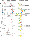
Schematic overview of labeling approaches and their application in cells. The illustrated methods include chemical and chemo-enzymatic one-step labeling, chemical and chemo-enzymatic two-step labeling and metabolic labeling.
Figure 2
Figure 2
Schematic illustration of select click reactions commonly used for nucleic acid labeling. A) Copper(I)-catalyzed azide-alkyne cycloaddition (CuAAC). B) Reaction scheme of the strain-promoted azide-alkyne cycloaddition (SPAAC). C) Reaction scheme of the inverse-electron demand Diels-Alder cycloaddition (IEDDA).
Figure 3
Figure 3
Strategies for labeling nucleic acids based on solid-phase synthesis. A) Co-synthetic incorporation of expanded nucleobases B) Post-synthetic fluorescent labeling, achieved by a two-step approach: co-synthetic incorporation of a functional group and subsequent labeling via click chemistry. Similar approaches are usually used for DNA (R = H) and RNA (R = O-TOM) labeling.
Figure 4
Figure 4
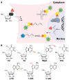
Structures of fluorescent nucleobase analogs sorted according to their emission wavelengths. A) Structure of 2-aminopurine. B) Examples of expanded nucleobase analogs that are fluorescent. C) Examples of expanded nucleobase analogs that are fluorescent and two-photon excitable. 1: 7-amino-1-ribose-quinazoline-2,4(1H,3H)-dione, 2: 5-(furan-2-yl)-2′-deoxyuridine, 3: X=S: 5-(thiophen-2-yl)-2′-deoxyuridine, X=Se: SeU 4: 5-(thiophen-2-yl)-6-aza-uridine. R denotes the (deoxy)ribose.
Figure 5
Figure 5
Functionality transfer for RNA labeling. A) Proposed reaction mechanism. The 1-aryl-2-methylydene-1,3-diketone building block from the S-functionalized 6-thio-9-methyl-guanine is transferred onto the exocyclic amino function of cytidine or guanosine depending on the pH in the area were the guide-DNA hybridizes with the target RNA. R1 is usually a methyl or phenyl group R2 is the fluorophore (pyrene, ATTO550, Cy3). B) Scheme illustrating RNA labeling guided by a DNA oligonucleotide containing modified 6-thioguanosine (Gs).
Figure 6
Figure 6
Polymerase-based fluorescent labeling strategies for nucleic acids. A) Direct enzymatic incorporation of fluorescent nucleotides. B) Two-step labeling approach. Enzymatic incorporation of a functional group for subsequent fluorescent labeling via click chemistry. Templates for polymerases are omitted.
Figure 7
Figure 7
Structure of fluorescent (d)NTPs. A) Structures of rigid phenylethyne linkers of increasing lengths lead to a bathochromic shift increasing with the length of the linker. B) Structure of the fluorescent dNTP/NTP analogs dCBdpTP (BODIPY), dCTBdpTP (thiophene-linked tetramethylbodipy), dCFLTP (fluorene), dCTrpTP (tryptophan), 4CIN-TP (4-cyanoindole-2′- deoxyribonucleoside-5′-triphosphate) and thGTP (thieno). R denotes 5′-triphosphorylated (deoxy)ribose.
Figure 8
Figure 8
Direct post-synthetic enzymatic labeling strategies for nucleic acids. A) M.TaqI was used to transfer a Cy3 bearing aziridine cofactor onto the exocyclic amino function of adenine in the 5′-TCGA-3′ sequence of the two plasmids, pUC19 and pBR322. Mammalian CHO-K1 cells were transfected with these labeled plasmids, which could then be detected by fluorescence microscopy. B) TGT from E. coli can transfer fluorescent PreQ1 analogs onto a small 17 nt hairpin motif and label RNA in fixed cells. C) Hen1 2′-O-methyltransferase utilizing a Cy3-bearing AdoMet analog for labeling ssRNA at the 3′-end. Together with a Cy5-labeled complementary ssDNA for the labeled ssRNA was detected by FRET.
Figure 9
Figure 9
Modified (d)NTPs for chemo-enzymatic two-step labeling of nucleic acids. A) dNTPs for chemo-enzymatic two-step labeling of DNA. B) NTPs for for chemo-enzymatic two-step labeling of RNA. C) NTPs with unnatural base-pairs for chemo-enzymatic labeling of RNA using the dTPT3-dNaM system. R denotes 5′-triphosphorylated (deoxy)ribose.
Figure 10
Figure 10
Schematic overview of post-synthetic enzyme-mediated two-step covalent labeling of nucleic acids and its applications. A) Beta-glucosyltransferase (β-GT)-directed tagging of 5-hydroxymethylcytosine (5-hmC) in DNA. B) Labeling of DNA and different RNA species using AdoMet analogs and various MTases. C) tRNAIle- agmatine synthetase (Tias)-directed tagging of RNA. D) tRNA guanine transglycosylase (TGT)-mediated labeling.
Figure 11
Figure 11
Chemical structures of functionalities transferred to nucleic acids using β-GT, MTase, Tias or TGT and the corresponding cofactors.
Figure 12
Figure 12
Ribozyme-assisted labeling of nucleic acids. A) Twin ribozyme-mediated insertion of short-labeled RNA fragment by strand exchange. B) Ribozyme-directed reaction between a labeled electrophile and nucleophilic groups in RNA. C) Polymerase ribozyme-based strategy to attach functionalized or labeled nucleotides at the 3′-end. D) DNAzyme-assisted labeling of 2′-OH of internal adenosine residues using functionalized GTP.
Figure 13
Figure 13
Chemically introduced reporters (besides fluorophores) and their application. A) Introduction of spin labels in RNA and DNA using click chemistry. B) Attachment of quantum dots to functionalized DNA via NHS-esterification.
Figure 14
Figure 14
Chemo-enzymatically introduced reporters (besides fluorophores) and their applications. A) Incorporation of photocrosslinker via MTase. B) DNA biotinylation based on β-GT modification. C) Biosensor for detection of 5-hmC by modifying immobilized DNA (magnetic beads are illustrated as black circle). D) RNA MTase-directed modification and subsequent clicking with biotin.
Figure 15
Figure 15
A) Concept of metabolic DNA labeling. Deoxynucleosides with small modifications (red triangle) are taken up by cells and enter the salvage pathway to dNTPs. After polymerase-based incorporation into DNA, click chemistry is used for (fluorescent) labeling. B) Deoxynucleosides successfully used for metabolic labeling. Abbreviations: EdU: 5-ethynyl-2′-deoxyuridine, F-ara-EdU: 2′-deoxy-2′-fluoro-5-ethynyluridine, dF-EdU: 2′-deoxy-2′, 2′-difluoro-5-ethinyluridine, EdC: 2′-deoxy-5-ethynylcytidine, EdA: 7-deaza-7-ethynyl-2′-deoxyadenosine; AzC: 1-(2-azido-2′-deoxy-β-D-arabinofuranosyl)cytosine, AmdU: 5-(azidomethyl)-2′-deoxyuridine; VdU: 5-vinyl-2′-deoxyuridine; VTdT: 5-(vinylthio-ethoxycarbamoyl-methyl)-2′-deoxythymidine.
Figure 16
Figure 16
A) Concept of metabolic RNA labeling. Nucleosides with small modifications are taken up by cells and enter the salvage pathway to NTPs. After incorporation into different types of RNA during transcription or by PAPS, click chemistry is used for (fluorescent) labeling. B). Nucleosides successfully used for RNA metabolic labeling. Abbreviations: EU: 5-ethynyluridine, EA: 2-ethynyladenosine, N6pA: N -propargyl-adenosine, 5VU: 5-vinyluridine, 2VA: 2-vinyladenosine, 7-dVA: 7-deaza-7-vinyl-adenosine, 2′-azA: 2′-azido-2′-deoxy-adenosine. Arrows indicate uptake of nucleosides and export of RNA.
Figure 17
Figure 17
Approaches for cell-specific RNA labeling. A) Cells overexpressing UPRT, which is lacking in mammals can convert 5EC to EU-monophosphate. WT cells can use 5EU to a limited extent, causing background. B-C) Cells overexpressing UCK2 or an improved variant can process 5AmU and 2’AzUd to the respective monophosphates. The monophosphates are processed to the respective triphosphates and incorporated into RNA, which can be labeled. In mammalian wildtype cells, low (A, B) or no (C) background labeling is detected.
Figure 18
Figure 18
Approaches to increase uptake of deoxynucleoside phosphates. A) Membrane-permeable phosphotriester derivatives successfully used as pro-labels for DNA metabolic labeling. B) Schematic illustration of the synthetic nucleoside-triphosphate transporter (SNTT). A labeled dNTP binds to the transporter that enters the cell via a cell-penetrating peptide. Cellular ATP binds to the transporter, facilitating release of the labeled dNTP and subsequent DNA metabolic labeling.
Scheme 1
Scheme 1
Overview of select applications of labeled nucleic acids in different areas.

References

    1. Mardis ER. Nat Protoc. 2017;12:213–218. - PubMed
    1. Steffen FD, Khier M, Kowerko D, Cunha RA, Borner R, Sigel RKO. Nat Commun. 2020;11 104. - PMC - PubMed
    1. Chen J, Tsai A, O’Leary SE, Petrov A, Puglisi JD. Curr Opin Struct Biol. 2012;22:804–814. - PMC - PubMed
    1. Ray S, Widom JR, Walter NG. Chem Rev. 2018;118:4120–4155. - PMC - PubMed
    1. Vera M, Biswas J, Senecal A, Singer RH, Park HY. Annu Rev Genet. 2016;50:267–291. - PMC - PubMed