

Abdominal symptoms as initial manifestation of COVID-19: a case series
- PMID: 33084795
- PMCID: PMC7546681
- DOI: 10.31744/einstein_journal/2020RC5831
Abdominal symptoms as initial manifestation of COVID-19: a case series
Abstract
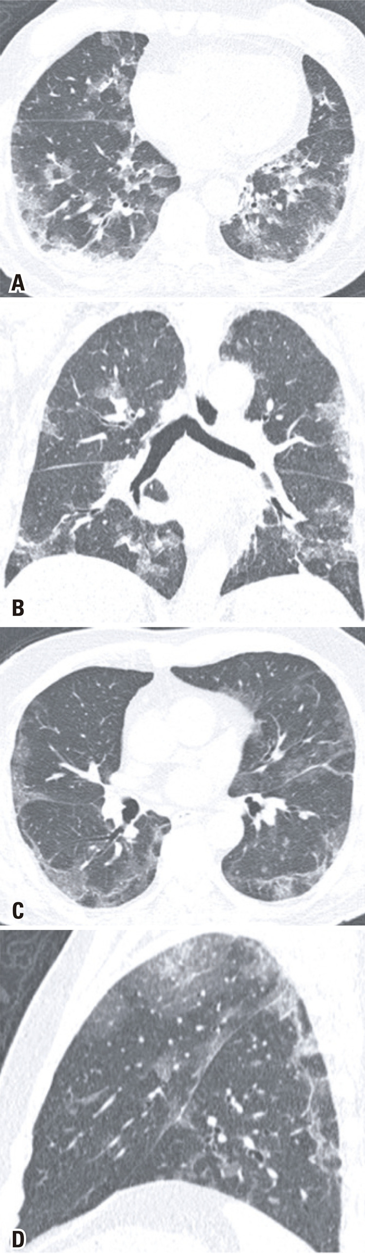
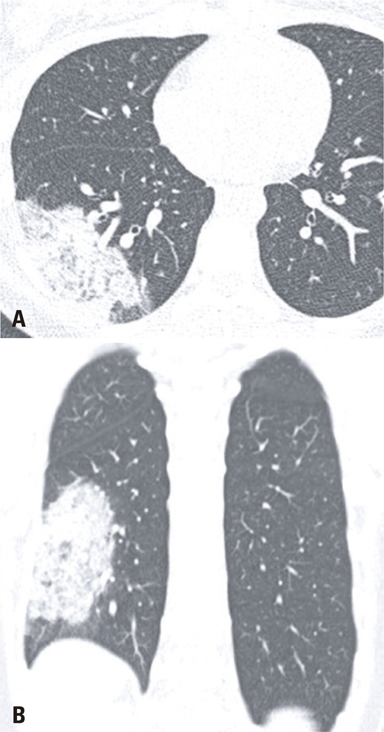
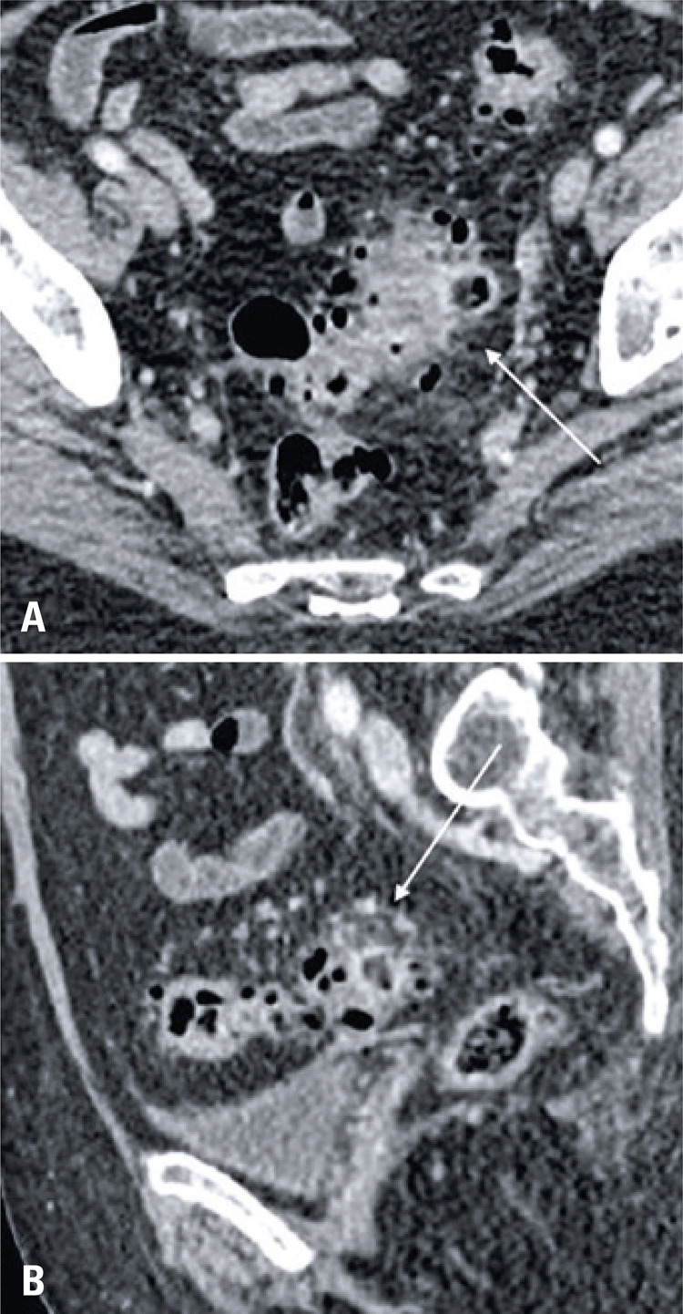
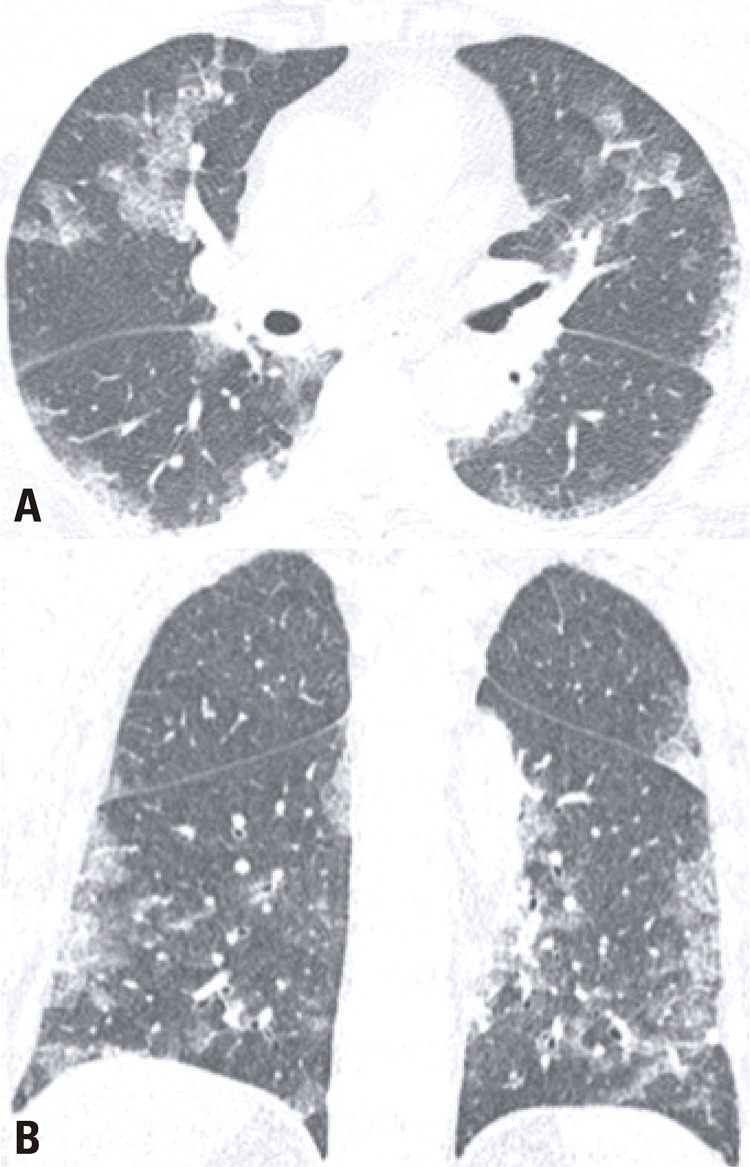
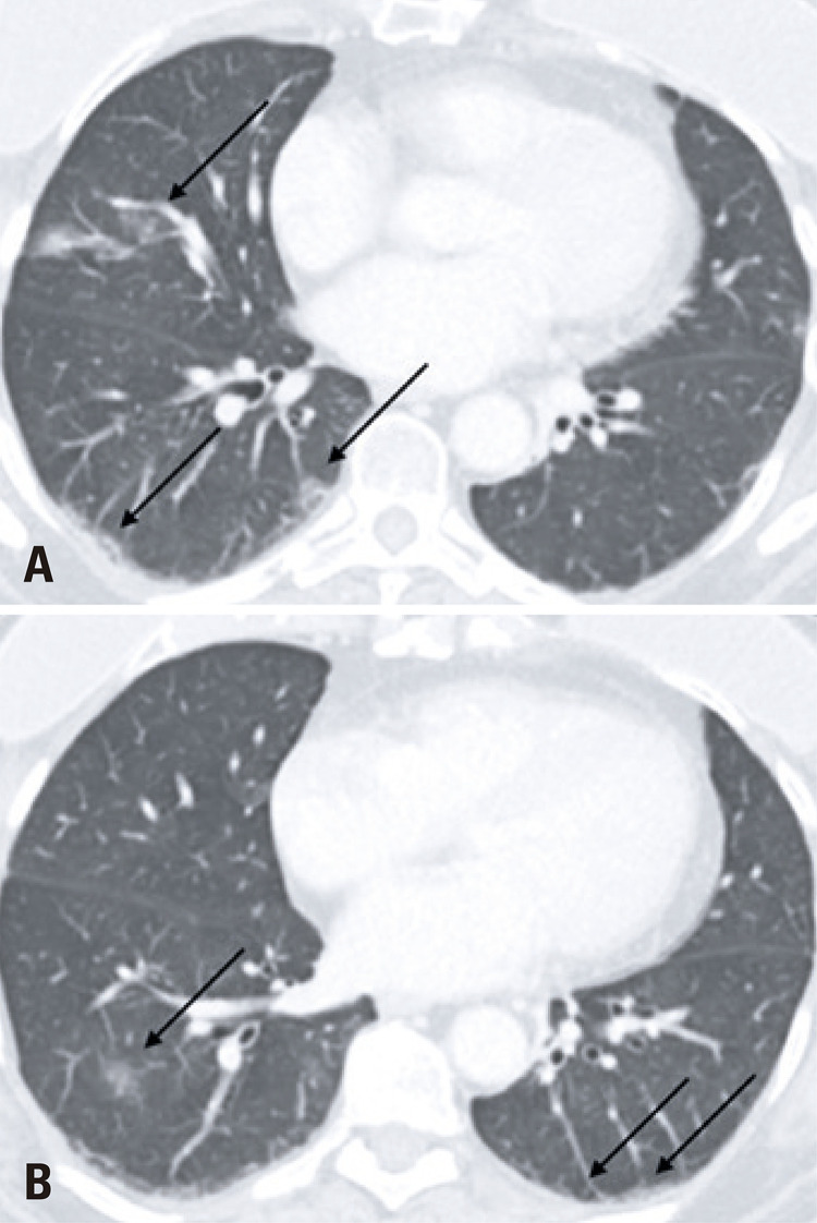
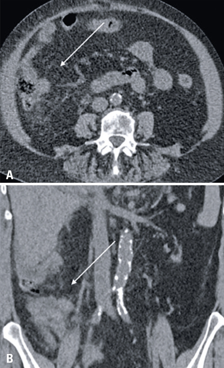
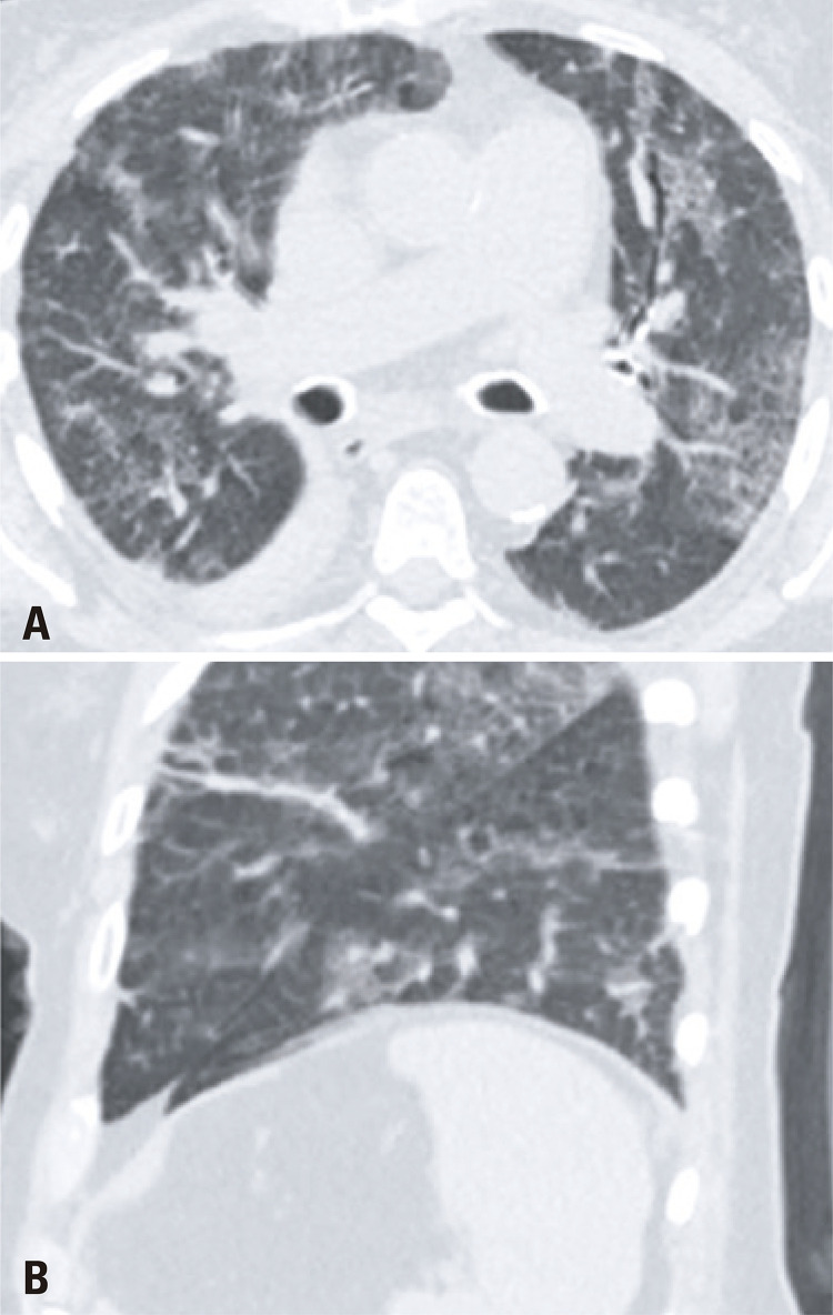
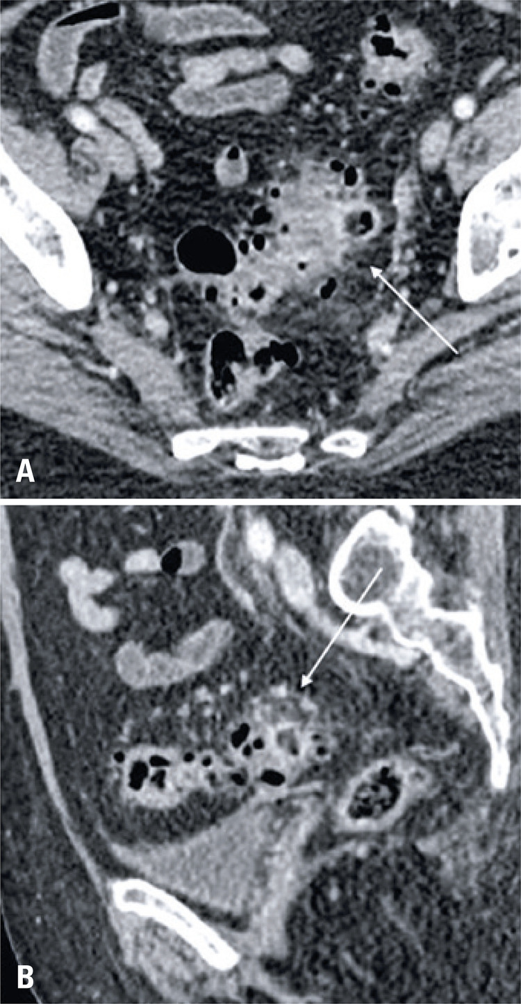
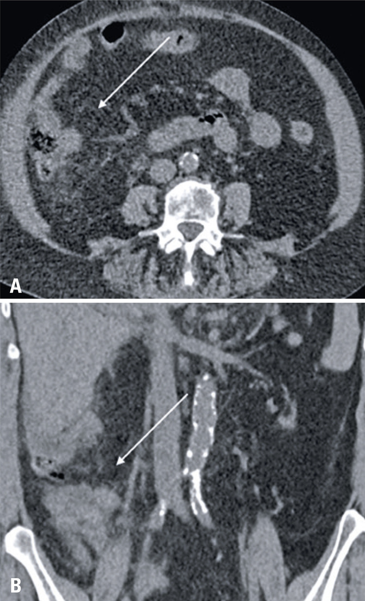
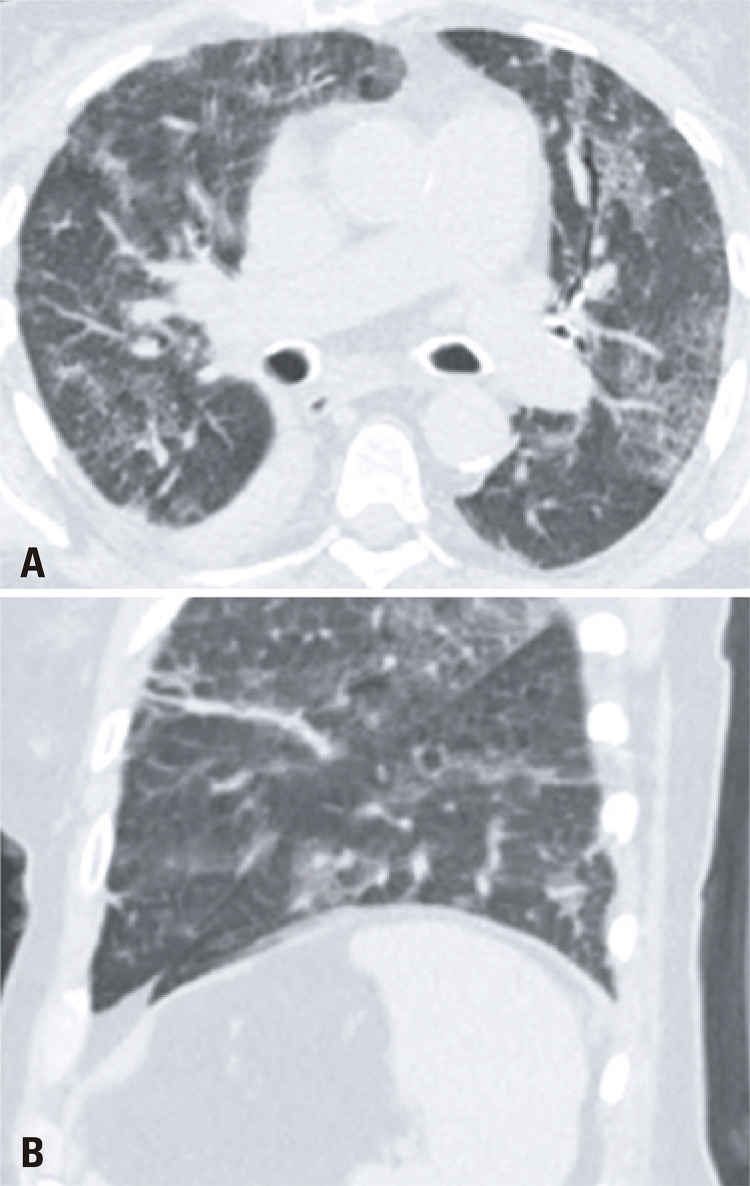
The COVID-19 became a pandemic in early 2020. It was found, at first, that the main manifestations of this new virus occur through respiratory and constitutional symptoms. Therefore, chest tomography was elected as the best imaging test to assess the extent of pulmonary involvement and as a good prognostic predictor for the disease. However, as new studies were produced, the gastrointestinal involvement of COVID-19 becomes more evident, with reports from patients who manifested mainly or only gastrointestinal symptoms in the course of the disease. Thus, in some cases, the initial investigation is carried out at the emergency department with an abdominal computed tomography. We report a case series of ten patients who came to the emergency department of our institution with a chief gastrointestinal complaint, and were initially submitted to an abdominal computed tomography as the first investigation. Although most of the patients did not have significant changes in the abdominal images, most reported patients had pulmonary findings visualized at the lung bases, which were later designated as typical COVID-19 pulmonary findings on chest computed tomography. Only one patient had atypical COVID-19 lung changes on chest computed tomography. All patients had a positive real-time polymerase chain reaction for COVID-19. It is imperative to alert radiologists, especially abdominal radiologists, with the possibility of COVID-19 isolated gastrointestinal symptoms. Besides, it must become a habit to radiologists to assess the pulmonary basis on abdominal scans, a site commonly affected by the new coronavirus.
RESUMO: A COVID-19 foi declarada uma pandemia no início de 2020. Constatou-se, inicialmente, que as principais manifestações desse novo vírus ocorrem por meio de sintomas respiratórios e constitucionais. A tomografia do tórax foi eleita o exame de imagem para avaliar a extensão do comprometimento pulmonar e como um fator preditivo do prognóstico para a doença. No entanto, à medida que novos estudos são produzidos, o envolvimento gastrointestinal da COVID-19 torna-se mais evidente, com relatos de pacientes que manifestaram principalmente ou apenas sintomas gastrointestinais no decorrer da doença. Em alguns casos, a investigação inicial é realizada no pronto-socorro, com tomografia computadorizada do abdome. Relatamos uma série de casos de dez pacientes que compareceram ao serviço de emergência da instituição com uma queixa principal gastrointestinal e foram submetidos inicialmente a uma tomografia computadorizada de abdome como primeira investigação. Embora a maioria dos pacientes não tenha apresentado alterações significativas nas imagens abdominais, eles apresentaram achados pulmonares visualizados nas bases pulmonares, que depois foram caracterizadas como achados pulmonares típicos de COVID-19 nas tomografias de tórax subsequentes. Apenas um paciente apresentou achados atípicos para COVID-19 na tomografia. Todos os pacientes tiveram reação em cadeia da polimerase em tempo real positiva para o novo coronavírus. É muito importante alertar os radiologistas, principalmente os radiologistas abdominais, da possibilidade de sintomas gastrointestinais isolados no contexto da COVID-19. Além disso, deve ser um hábito para todos os radiologistas avaliar as bases pulmonares nas tomografias de abdome, local comumente afetado pela COVID-19.
Figures


Similar articles
-
Quantitative analysis in COVID-19: report of an initial experience.Einstein (Sao Paulo). 2020 Oct 7;18:eAI5842. doi: 10.31744/einstein_journal/2020AI5842. eCollection 2020. Einstein (Sao Paulo). 2020. PMID: 33053015 Free PMC article. No abstract available.
-
CT imaging of the COVID-19.J Formos Med Assoc. 2020 May;119(5):990-992. doi: 10.1016/j.jfma.2020.04.006. Epub 2020 Apr 16. J Formos Med Assoc. 2020. PMID: 32307320 Free PMC article.
-
Lung Base Findings of Coronavirus Disease (COVID-19) on Abdominal CT in Patients With Predominant Gastrointestinal Symptoms.AJR Am J Roentgenol. 2020 Sep;215(3):607-609. doi: 10.2214/AJR.20.23232. Epub 2020 Apr 17. AJR Am J Roentgenol. 2020. PMID: 32301631
-
Clinical characteristics and diagnostic challenges of pediatric COVID-19: A systematic review and meta-analysis.J Formos Med Assoc. 2020 May;119(5):982-989. doi: 10.1016/j.jfma.2020.04.007. Epub 2020 Apr 16. J Formos Med Assoc. 2020. PMID: 32307322 Free PMC article.
-
Computed Tomography Features of Coronavirus Disease 2019 (COVID-19): A Review for Radiologists.J Thorac Imaging. 2020 Jul;35(4):211-218. doi: 10.1097/RTI.0000000000000527. J Thorac Imaging. 2020. PMID: 32427651 Review.
Cited by
-
Access to Information, and Concerns, Myths and Truths about Food Safety during the COVID-19 Pandemic: An Overview of the Portuguese Population.Foods. 2023 Jul 24;12(14):2802. doi: 10.3390/foods12142802. Foods. 2023. PMID: 37509894 Free PMC article.
-
Atypical presentation of COVID-19 with abdominal pain and no respiratory symptoms: a case series.Singapore Med J. 2023 Nov;64(11):687-689. doi: 10.11622/smedj.2021196. Singapore Med J. 2023. PMID: 34749489 Free PMC article. No abstract available.
-
Coronavirus disease-2019 and the intestinal tract: An overview.World J Gastroenterol. 2021 Apr 7;27(13):1255-1266. doi: 10.3748/wjg.v27.i13.1255. World J Gastroenterol. 2021. PMID: 33833480 Free PMC article. Review.
-
Gastrointestinal manifestations of COVID-19 in a single center in the Eastern Province of Saudi Arabia.Saudi J Gastroenterol. 2022 May-Jun;28(3):218-224. doi: 10.4103/sjg.sjg_547_21. Saudi J Gastroenterol. 2022. PMID: 35042321 Free PMC article.
-
Extrapulmonary onset manifestations of COVID-19.Clinics (Sao Paulo). 2021 Jul 5;76:e2900. doi: 10.6061/clinics/2021/e2900. eCollection 2021. Clinics (Sao Paulo). 2021. PMID: 34231709 Free PMC article. Review.
References
-
- 1. World Health Organization (WHO) . WHO announces COVID-19 outbreak a pandemic [ Internet]. Geneva : WHO ; 2020 [ cited 12 May 2020 ]. Available from: https://www.euro.who.int/en/health-topics/health-emergencies/coronavirus...
- World Health Organization (WHO) WHO announces COVID-19 outbreak a pandemic. Geneva: WHO; 2020. [cited 12 May 2020]. https://www.euro.who.int/en/health-topics/health-emergencies/coronavirus... Internet].
-
- 2. Zu ZY , Jiang MD , Xu PP , Chen W , Ni QQ , Lu GM , et al . Coronavirus disease 2019 (COVID-19): a perspective from China . Radiology . 2020 ; 296 ( 2 ): E15 - E25 . Review . - PMC - PubMed
- Zu ZY, Jiang MD, Xu PP, Chen W, Ni QQ, Lu GM, et al. Coronavirus disease 2019 (COVID-19): a perspective from China. Radiology. 2020;296(2):E15–E25. Review. - PMC - PubMed
-
- 3. Wan Y , Shang J , Sun S , Tai W , Chen J , Geng Q , et al . Molecular mechanism for antibody-dependent enhancement of coronavirus entry . J Virol . 2020 ; 94 ( 5 ).pii: e02015 - 19 . - PMC - PubMed
- Wan Y, Shang J, Sun S, Tai W, Chen J, Geng Q, et al. Molecular mechanism for antibody-dependent enhancement of coronavirus entry. J Virol. 2020;94(5):e02015–e02019. - PMC - PubMed
-
- 4. Liang W , Feng Z , Rao S , Xiao C , Xue X , Lin Z , et al . Diarrhoea may be underestimated: a missing link in 2019 novel coronavirus . Gut . 2020 ; 69 ( 6 ): 1141 - 3 . - PubMed
- Liang W, Feng Z, Rao S, Xiao C, Xue X, Lin Z, et al. Diarrhoea may be underestimated: a missing link in 2019 novel coronavirus. Gut. 2020;69(6):1141–1143. - PubMed
-
- 5. Jin X , Lian JS , Hu JH , Gao J , Zheng L , Zhang YM , et al . Epidemiological, clinical and virological characteristics of 74 cases of coronavirus-infected disease 2019 (COVID-19) with gastrointestinal symptoms . Gut . 2020 ; 69 ( 6 ): 1002 - 9 . - PMC - PubMed
- Jin X, Lian JS, Hu JH, Gao J, Zheng L, Zhang YM, et al. Epidemiological, clinical and virological characteristics of 74 cases of coronavirus-infected disease 2019 (COVID-19) with gastrointestinal symptoms. Gut. 2020;69(6):1002–1009. - PMC - PubMed
