. 2021 Jan 6;29(21):3504-3515.
doi: 10.1093/hmg/ddaa234.

The prevalent I686T human variant and loss-of-function mutations in the cardiomyocyte-specific kinase gene TNNI3K cause adverse contractility and concentric remodeling in mice

Affiliations

The prevalent I686T human variant and loss-of-function mutations in the cardiomyocyte-specific kinase gene TNNI3K cause adverse contractility and concentric remodeling in mice

Peiheng Gan et al. Hum Mol Genet. .

Abstract

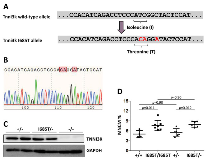
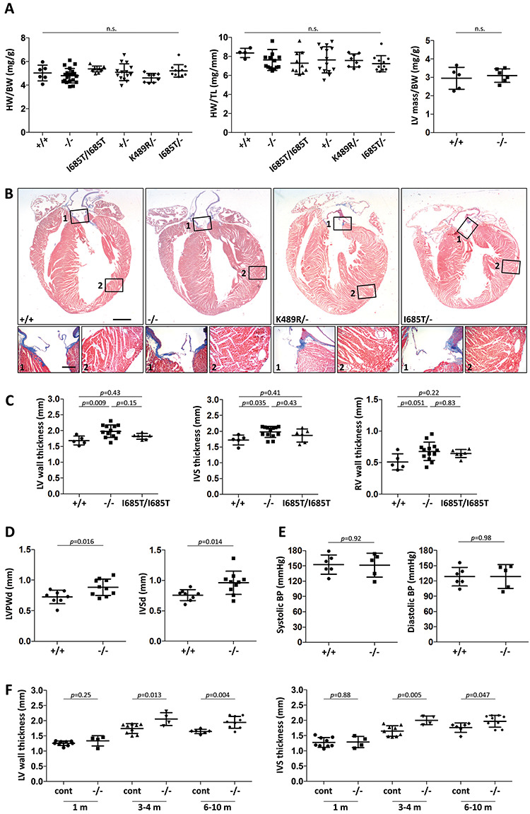
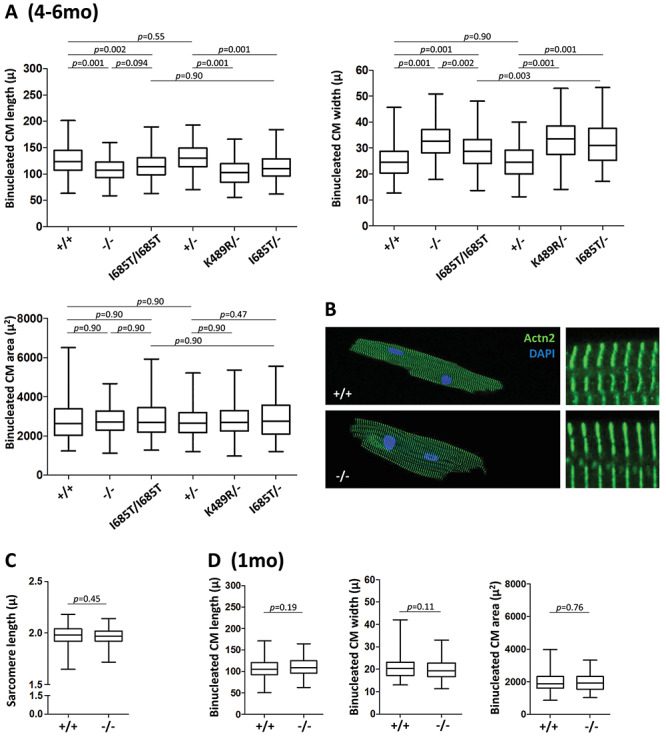
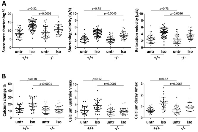
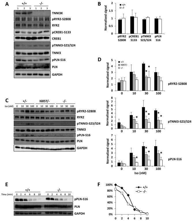
TNNI3K expression worsens disease progression in several mouse heart pathology models. TNNI3K expression also reduces the number of diploid cardiomyocytes, which may be detrimental to adult heart regeneration. However, the gene is evolutionarily conserved, suggesting a beneficial function that has remained obscure. Here, we show that C57BL/6J-inbred Tnni3k mutant mice develop concentric remodeling, characterized by ventricular wall thickening and substantial reduction of cardiomyocyte aspect ratio. This pathology occurs in mice carrying a Tnni3k null allele, a K489R point mutation rendering the protein kinase-dead, or an allele corresponding to human I686T, the most common human non-synonymous TNNI3K variant, which is hypomorphic for kinase activity. Mutant mice develop these conditions in the absence of fibrosis or hypertension, implying a primary cardiomyocyte etiology. In culture, mutant cardiomyocytes were impaired in contractility and calcium dynamics and in protein kinase A signaling in response to isoproterenol, indicating diminished contractile reserve. These results demonstrate a beneficial function of TNNI3K in the adult heart that might explain its evolutionary conservation and imply that human TNNI3K variants, in particular the widespread I686T allele, may convey elevated risk for altered heart geometry and hypertrophy.


Figures

Figure 1
Figure 1
Creation and analysis of the Tnni3k I685T allele. (A) The relevant portion of the wild-type C57BL/6J Tnni3k exon 21 sequence is shown above, and below in red are the base changes made. This manipulation changed an isoleucine (ATC) codon to a threonine (ACA) codon, and introduced an EcoNI restriction site (CCTNNNNNAGG) into the sequence to facilitate genotyping. The additional base change made in the third position of the following glycine codon does not change the encoded amino acid and was introduced to suppress CRISPR/Cas9 retargeting. (B) Sequence trace of this portion of the gene in an I685T/I685T homozygous mouse. (C) Western blot showing that Tnni3k protein is expressed at normal levels in hearts of I685T/− mice. Each lane is lysate from a different mouse of the genotypes indicated above. (D) Ventricular mononuclear cardiomyocyte percentage from single cell preparations; each dot is the measurement of a different adult mouse of the indicated genotype.
Figure 2
Figure 2
Phenotypic evaluation of Tnni3k mutant mouse hearts. (A) Heart weight to body weight ratio, heart weight to tibia length and left ventricle mass to body weight ratio measurements for hearts of the indicated genotypes; all mice were 3–10 months old, and each dot represents a unique mouse. All pair-wise comparisons were statistically non-significant (n.s.). (B) Trichrome-stained long-axis histology sections of representative hearts from 7 month old mice fixed in diastole; the magnified regions (numbered) show positive collagen staining (blue) in the aortic valve (number 1 boxes) but little if any staining in the myocardium (number 2 boxes). All corresponding images are at the same magnification. Scale bar in larger images, 1 mm; scale bar for smaller images, 200 μ. (C) Measurements of left ventricular (LV) wall, interventricular septum (IVS), and right ventricle (RV) wall thicknesses in transverse sections taken at a cross-sectional plane through the papillary muscles. LV wall thickness was measured between the two papillary muscles, and IVS and RV thicknesses directly opposite. Representative images of sections are shown in Supplementary Material, Figure S1A. Analysis of additional genotypes (+/−, K489R/−, I685T/−) is shown in Supplementary Material, Figure S1B. (D) Measurement of LV posterior wall (LVPW) and interventricular septal (IVS) diastolic diameters in live mice by echocardiography. (E) Tail-cuff measurement of systolic and diastolic blood pressure in non-anesthetized mice. (F) LV and IVS thickness in control (+/+ and +/− combined) and null (−/−) mice at different ages as measured in transverse sections.
Figure 3
Figure 3
Cardiomyocyte cellular remodeling. (A) Length, width and area measurements of individual binucleated cardiomyocytes from 4 to 6 month old mice of the indicated genotypes, shown as a bars and whiskers plot (mean, middle quartiles and full range of data). Numbers of cells measured are listed in Table 2. Measurements were aggregated from roughly equal numbers of cells from 4 to 6 mice of the indicated genotypes. The (minimal) variance between cell preparations from individual mice is shown in Supplementary Material, Figure S2A. (B) Visualization of sarcomere structure and organization in cardiomyocytes from 6 month old mice by alpha-actinin2 (Actn2) staining and confocal microscopy. The cells shown correspond to the average dimensions measured in panel A. (C) Measurement of sarcomere length, obtained from the same evaluation used to measure sarcomere shortening during contraction that is shown in Figure 4A. (D) Length, width and area measurements of individual binucleated cardiomyocytes from 1 month old mice of the indicated genotypes; see also Table 2. The same analysis expressed on a per mouse basis is shown in Supplementary Material, Figure S2B.
Figure 4
Figure 4
Cardiomyocyte contractility and calcium dynamics without and with isoproterenol treatment. (A) Cardiomyocyte contractility in paced cells. Sarcomere shortening percent at peak contraction, shortening velocity and relaxation velocity were measured for individual cardiomyocytes during the contraction cycle, in untreated (untr) and isoproterenol (Iso) treated cells. Cells were either untreated or treated, but not both. Single cell preparations from 11 +/+ mice and 9 −/− mice were used in this analysis. (B) Calcium dynamics in paced cells measured by Fura-2 signal. Percent change in cytoplasmic calcium fluorescence at peak amplitude, and maximum fluorescence upstroke and fluorescence decay velocities (units of fluorescence/sec) were measured for individual cardiomyocytes during the contraction cycle. Each dot indicates measurement for an individual cell from 8 +/+ mice and 8 −/− mice. See also Supplementary Material, Table S1 for statistical comparison of untreated versus treated cells by genotype.
Figure 5
Figure 5
Altered PKA activity in Tnni3k mutant cardiomyocytes. (A, B) Western blot using phospho-specific and total protein antibodies with lysates from 3 +/+ and 3 −/− mouse hearts. The blots shown in A were quantified and graphed in B. (C, D) Tnni3k mutant cardiomyocytes have impaired response to isoproterenol. Cells were isolated and plated, treated with the indicated concentration of isoproterenol (Iso) for 8 min, then harvested. One western blot is shown for results with cells from individual mice of the genotypes shown; the aggregated quantified data from three independent blots was used to assemble the charts shown in (D). The control cells in the blot shown were from a heterozygous mouse (+/−); and were from +/+ mice in the other two blots; all three were aggregated in the quantitation shown in (D) (labeled as +/+). Asterisk indicates statistically significant differences (P < 0.05), a complete compilation of all statistical comparisons for western blots is provided in Supplementary Material, Table S2. A parallel analysis that includes K489R/− cardiomyocytes is shown in Supplementary Material, Figure S3A–B. (E, F) Measurement of phospholamban S16 dephosphorylation. Cells were pretreated with 300 nM isoproterenol, then washed and cultured in media without isoproterenol and supplemented instead with propranolol. The times indicated refer to time after removal of isoproterenol. Quantified data are graphed in (F). This analysis was repeated three times with comparable results.

Similar articles

Cited by

References

    1. Zhao Y., Meng X.M., Wei Y.J., Zhao X.W., Liu D.Q., Cao H.Q., Liew C.C. and Ding J.F. (2003) Cloning and characterization of a novel cardiac-specific kinase that interacts specifically with cardiac troponin I. J. Mol. Med. (Berl), 81, 297–304. - PubMed
    1. Gan P., Patterson M., Velasquez A., Wang K., Tian D., Windle J.J., Tao G., Judge D.P., Makita T., Park T.J. et al. (2019) Tnni3k alleles influence ventricular mononuclear diploid cardiomyocyte frequency. PLoS Genet., 15, e1008354. - PMC - PubMed
    1. Wheeler F.C., Tang H., Marks O.A., Hadnott T.N., Chu P.L., Mao L., Rockman H.A. and Marchuk D.A. (2009) Tnni3k modifies disease progression in murine models of cardiomyopathy. PLoS Genet., 5, e1000647. - PMC - PubMed
    1. Patterson M., Barske L., Van Handel B., Rau C.D., Gan P., Sharma A., Parikh S., Denholtz M., Huang Y., Yamaguchi Y. et al. (2017) Frequency of mononuclear diploid cardiomyocytes underlies natural variation in heart regeneration. Nat. Genet., 49, 1346–1353. - PMC - PubMed
    1. Karczewski K.J., Francioli L.C., Tiao G., Cummings B.B., Alföldi J., Wang Q., Collins R.L., Laricchia K.M., Ganna A., Birnbaum D.P. et al. (2020) The mutational constraint spectrum quantified from variation in 141,456 humans. Nature, 581, 434–443. - PMC - PubMed

Publication types

Substances