Review
. 2020 Oct 30;134(20):2681-2706.
doi: 10.1042/CS20190893.

Regulation of cellular senescence by extracellular matrix during chronic fibrotic diseases

Affiliations
Review

Regulation of cellular senescence by extracellular matrix during chronic fibrotic diseases

Kaj E C Blokland et al. Clin Sci (Lond). .

Abstract

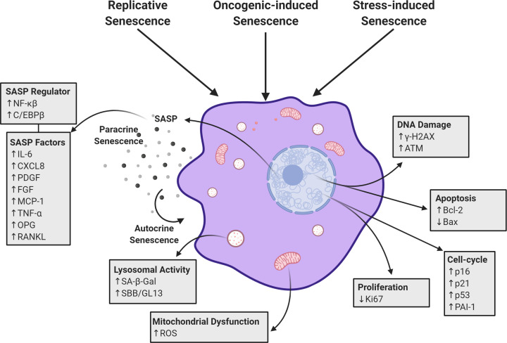
The extracellular matrix (ECM) is a complex network of macromolecules surrounding cells providing structural support and stability to tissues. The understanding of the ECM and the diverse roles it plays in development, homoeostasis and injury have greatly advanced in the last three decades. The ECM is crucial for maintaining tissue homoeostasis but also many pathological conditions arise from aberrant matrix remodelling during ageing. Ageing is characterised as functional decline of tissue over time ultimately leading to tissue dysfunction, and is a risk factor in many diseases including cardiovascular disease, diabetes, cancer, dementia, glaucoma, chronic obstructive pulmonary disease (COPD) and fibrosis. ECM changes are recognised as a major driver of aberrant cell responses. Mesenchymal cells in aged tissue show signs of growth arrest and resistance to apoptosis, which are indicative of cellular senescence. It was recently postulated that cellular senescence contributes to the pathogenesis of chronic fibrotic diseases in the heart, kidney, liver and lung. Senescent cells negatively impact tissue regeneration while creating a pro-inflammatory environment as part of the senescence-associated secretory phenotype (SASP) favouring disease progression. In this review, we explore and summarise the current knowledge around how aberrant ECM potentially influences the senescent phenotype in chronic fibrotic diseases. Lastly, we will explore the possibility for interventions in the ECM-senescence regulatory pathways for therapeutic potential in chronic fibrotic diseases.

Keywords: Antifibrotics; DAMPs; Senescence; Senolytics; extracellular matrix; fibrosis.


Conflict of interest statement

The authors declare that there are no competing interests associated with the manuscript.

Figures

Figure 1
Figure 1. Characteristics of senescent cells
Cells that undergo senescence display several changed features that can be used for identification. An increased secretory phenotype can be detected by measuring growth factors and cytokines. These factors contribute to the spreading of senescence to neighbouring cells or reinforce senescence via an autocrine process. Increased lysosomal activity can be detected using SA-β-Gal and SBB expression. Mitochondrial dysfunction is detectable by measuring ROS levels such as superoxide production. As cells undergo irreversible cell-cycle arrest, markers such as p16, p21 and p53 are increased while proliferation markers like Ki67 decrease. Resistance to apoptosis can be measured by changes in Bcl-2 and Bax expression. In addition, the DDR can be measured by immunofluorescence microscopy for increased formation of phosphorylated ATM and γ-H2AX in the nucleus. Abbreviation: SA-β-Gal, senescence-associated β-galactosidase.
Figure 2
Figure 2. ECM DAMP receptor activation leading to increased pro-inflammatory cytokine release
ECM DAMPs such as soluble decorin, tenascin-c and fibrinogen can activate an inflammatory response by binding to the TLR2 and TLR4 receptors. TLR activation leads to nuclear translocation of NF-κB and subsequent activation of pro-inflammatory cytokines that comprise the SASP such as IL-1β, IL-6, CXCL8, interferon (IFN)-γ and TNF-α.
Figure 3
Figure 3. ECM–cell interactions and their downstream targets potentially contribute to cellular senescence
The diagram highlights the different pathways that ECM proteins activate and the downstream targets that potentially contribute to cellular senescence in fibrosis. Fibronectin binds to integrin αvβ3 and activates the PI3k/Akt pathway, resulting in the inhibition of cellular senescence. Decorin binds to receptor tyrosine kinase and activates the RAS/RAF pathway, which eventually leads to the up-regulation of the cell-cycle inhibitors p16, p21 and phospo-p53. CCN1 binds to the α6β3 integrin which colocalises with HSPG to activate the downstream RAC1 pathway, resulting in increases in ROS production. Subsequently, increased levels of ROS leads to a DNA damage repair response that induces cell-cycle arrest via increases in p16, p21 and phospo-p53. Elastin fragments binds to EBP to interact with PPARγ, which lead to cell-cycle arrest via changes in the expression of p16. Tenascin-c binds to αvβ3 and induce the activation and nuclear translocation of NF-κB which leads to increases in apoptosis resistance as well as the up-regulation of growth factor and cytokine expression. Fibulin-1 may increase ROS production leading to cell-cycle arrest while limiting the Wnt/β-catenin pathway which protects against senescence. Periostin can directly activate the TGF-β pathway which leads to increase production of profibrotic factors. Laminin can directly activate the TGF-β pathway leading to profibrotic factor production and NF-κB activation which reinforce growth factor production and resistance to apoptosis.
Figure 4
Figure 4. Summary of ECM–cell interaction that contribute to cellular senescence
The binding of ECM DAMPs to TLR2/4 activates the production of pro-inflammatory cytokines that comprise the SASP of senescent cells in fibrosis. Binding of ECM proteins to their respective receptors (i.e. integrins or RTK) leads to the induction of cell-cycle arrest and an increase in apoptosis resistance. Additionally, up-regulation of profibrotic/pro-inflammatory cytokines reinforces the senescent phenotype in an autocrine dependent manner while also spreading senescence to neighbouring cells as part of a feedback loop.

Similar articles

Cited by

References

    1. Bonnans C., Chou J. and Werb Z. (2014) Remodelling the extracellular matrix in development and disease. Nat. Rev. Mol. Cell Biol. 15, 786–801 10.1038/nrm3904 - DOI - PMC - PubMed
    1. Jansen K.A., Atherton P. and Ballestrem C. (2017) Mechanotransduction at the cell-matrix interface. Semin. Cell Dev. Biol. 71, 75–83 10.1016/j.semcdb.2017.07.027 - DOI - PubMed
    1. Leiphart R.J. et al. . (2019) Mechanosensing at cellular interfaces. Langmuir 35, 7509–7519 10.1021/acs.langmuir.8b02841 - DOI - PubMed
    1. Sapir L. and Tzlil S. (2017) Talking over the extracellular matrix: how do cells communicate mechanically? Semin. Cell Dev. Biol. 71, 99–105 10.1016/j.semcdb.2017.06.010 - DOI - PubMed
    1. Tschumperlin D.J. et al. . (2018) Mechanosensing and fibrosis. J. Clin. Invest. 128, 74–84 10.1172/JCI93561 - DOI - PMC - PubMed

Publication types