. 2021 Feb;51(2):222-230.
doi: 10.1007/s00247-020-04826-7. Epub 2020 Oct 21.

Overview of chest involvement at computed tomography in children with coronavirus disease 2019 (COVID-19)

Affiliations

Overview of chest involvement at computed tomography in children with coronavirus disease 2019 (COVID-19)

Xuehua Peng et al. Pediatr Radiol. 2021 Feb.

Abstract

Background: Chest computed tomography (CT) findings in children with coronavirus disease 2019 (COVID-19) have been rarely reported in a comprehensive and systematic manner.

Objective: We investigated the chest CT findings in children with COVID-19, and explored the differences in these findings between symptomatic patients and asymptomatic patients.

Materials and methods: Demographic findings, clinical characteristics, duration of hospital stay and viral shedding, and chest CT findings in 201 children infected with severe acute respiratory syndrome coronavirus-2 (SARS-CoV-2) were retrospectively analyzed from January 15 to March 20, 2020, and divided into two groups: symptomatic group (n=136) and asymptomatic group (n=65). Chi-square test and Student's t-test were used for statistical analysis.

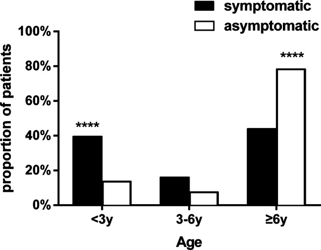
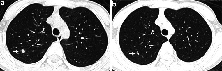
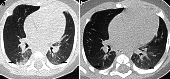
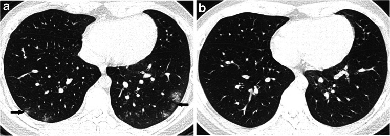
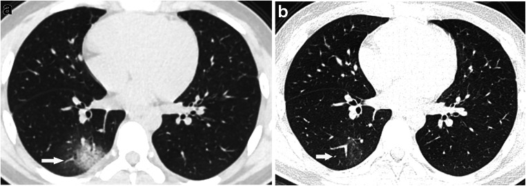
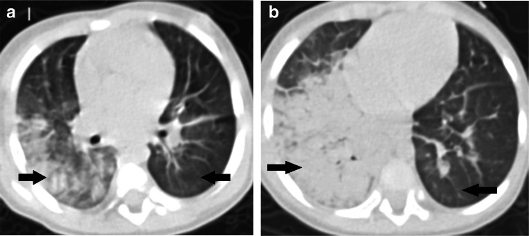
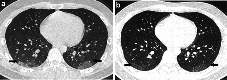
Results: Symptomatic patients were mainly young children ≤3 years old (54/63, 86%),while asymptomatic patients were mainly children ≥ 6 years old (51/111, 46%). Fever (41%) and cough (41%) were the most common symptoms. Overall, 119/201 (59%) patients had chest CT findings, and symptomatic patients accounted for 82% (98/119). The CT findings presented as bilateral multiple lesions (60/119, 50.4%), ground-glass opacities (83/119, 70%) and/or consolidation (44/119, 37%) with a peripheral and subpleural distribution (62/83, 75%). Fifteen of 87 (7.2%) patients with lung lesions showed complete lesion absorption, and 42/87 (48%) improved within a mean of 9.1 (standard deviation [SD] 3.2) days. The mean duration of viral shedding was 8.7 (SD 4.9) days. Pleural effusion was very rare. No lymphadenopathy was found in either group.

Conclusion: Symptoms associated with pulmonary involvement were most common in infants and young children. The lung lesions of most patients were absorbed and improved in about 9 days.

Keywords: COVID-19; Chest; Children; Computed tomography; Lungs; SARS-CoV-2.


Conflict of interest statement

None

Figures

Fig. 1
Fig. 1
The age distribution of COVID-19 pediatric patients. Comparison of proportion of population with different age distribution in the symptomatic group and the asymptomatic group. There were significant differences in the group younger than 3 years old and the group older than 6 years. **** P<0.0001
Fig. 2
Fig. 2
A 10-year-old boy with COVID-19, with fever and a history of exposure. a Axial thin-section non-contrast CT shows nodule shadow in the posterior segment of the right upper lobe (arrow). b Follow-up CT image shows the lesion is markedly reduced after 21 days (arrow)
Fig. 3
Fig. 3
A 2-month-old symptomatic girl with COVID-19 and a history of definite contact presented with paroxysmal cough and rhinorrhea. a Axial thin-section non-contrast CT shows ground-glass opacities and consolidation in the subpleural zone of both lower lobes (arrows). b A follow-up CT image shows the lesions are markedly absorbed after 1 month (arrows)
Fig. 4
Fig. 4
A 15-year-old symptomatic boy with COVID-19 and a history of definite contact presented with fever, cough, muscular soreness and fatigue. a Axial thin-section non-contrast CT shows scattered patchy ground-glass opacities in both lower lobes with a peripheral distribution (arrows). b A follow-up CT image shows normal lung after 1 month
Fig. 5
Fig. 5
A 15-year-old asymptomatic boy with COVID-19 and a history of definite contact. a Axial thin-section non-contrast CT shows a subpleural rounded area of consolidation surrounded by ground glass, consistent with the halo sign and situated at the right lower lobe (arrow). b A follow-up CT image shows light ground-glass opacities after 9 days (arrow)
Fig. 6
Fig. 6
A 1-year old critically ill boy with COVID-19 with diarrhea, fever and a history of exposure. a, b Axial thin-section non-contrast CT images show diffuse ground-glass opacities bilaterally, extensive consolidation at the right lung and reticulation bilaterally (arrows)
Fig. 7
Fig. 7
A 13-year-old symptomatic boy with COVID-19 with fever, cough and a history of exposure. a Axial thin-section non-contrast CT shows patchy peripheral ground-glass opacities in both lower lobes (arrows). b A follow-up CT image shows light ground-glass opacities after 5 days (arrows)

Similar articles

Cited by

References

    1. Ai T, Yang Z, Hou H, et al. Correlation of chest CT and RT-PCR testing in coronavirus disease 2019 (COVID-19) in China: a report of 1014 cases. Radiology. 2020;296:E32–E40. doi: 10.1148/radiol.2020200642. - DOI - PMC - PubMed
    1. Shi H, Han X, Jiang N, et al. Radiological findings from 81 patients with COVID-19 pneumonia in Wuhan, China: a descriptive study. Lancet Infect Dis. 2020;20:425–434. doi: 10.1016/S1473-3099(20)30086-4. - DOI - PMC - PubMed
    1. Ma H, Hu J, Tian J, et al. A single-center, retrospective study of COVID-19 features in children: a descriptive investigation. BMC Med. 2020;18:123. doi: 10.1186/s12916-020-01596-9. - DOI - PMC - PubMed
    1. Xia W, Shao J, Guo Y, et al. Clinical and CT features in pediatric patients with COVID-19 infection: different points from adults. Pediatr Pulmonol. 2020;55:1169–1174. doi: 10.1002/ppul.24718. - DOI - PMC - PubMed
    1. Hansell DM, Bankier AA, MacMahon H, et al. Fleischner society: glossary of terms for thoracic imaging. Radiology. 2008;246:697–722. doi: 10.1148/radiol.2462070712. - DOI - PubMed