Comparative Study
. 2020 Oct;3(5):e1257.
doi: 10.1002/cnr2.1257. Epub 2020 Jun 8.

Direct comparison of five different 3D extracellular matrix model systems for characterization of cancer cell migration

Affiliations
Comparative Study

Direct comparison of five different 3D extracellular matrix model systems for characterization of cancer cell migration

Yoshinari Shinsato et al. Cancer Rep (Hoboken). 2020 Oct.

Abstract

Background: Three-dimensional (3D) in vitro model systems can bridge the gap between regular two-dimensional cell culture and whole-animal studies. Analyses of cancer cell migration and invasion increasingly use differing 3D systems, which may produce conflicting findings.

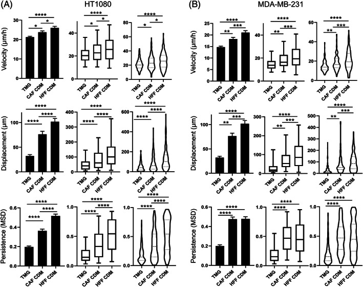
Aims: We directly compared different 3D extracellular matrix systems for studying cancer cell migration/invasion by analyzing cell morphologies and quantifying aspects of cell migration including speed and directional persistence using automated computer-based cell tracking.

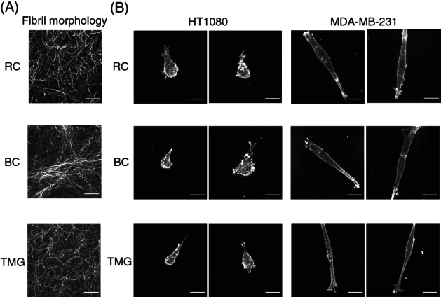
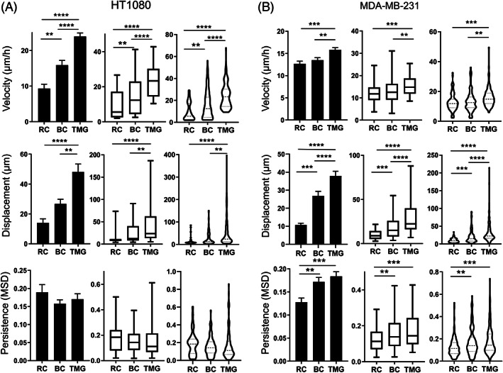
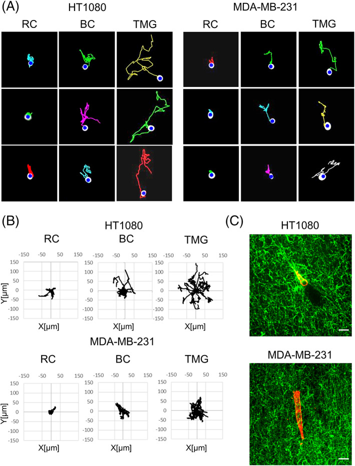
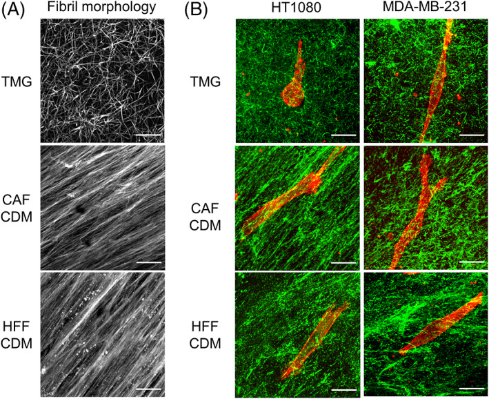
Methods and results: We performed direct comparisons of five different 3D extracellular matrix cell culture systems using both HT1080 fibrosarcoma and MDA-MB-231 breast carcinoma cell lines. The reconstituted 3D systems included two types of collagen hydrogel and tissue matrix gel (TMG) vs cell-derived matrices extracted from cultured primary human or cancer-associated fibroblasts. The fibrillar matrix architecture of these systems differed. 3D rat tail collagen and TMG matrices had short, randomly oriented collagen fibrils; bovine collagen had long, larger fibril bundles; and the cell-derived matrices were strongly oriented. HT1080 cells displayed rounded morphologies in all three reconstituted 3D matrices but became spindle shaped in the two cell-derived matrices. MDA-MB-231 cell morphologies were elongated in all matrices. Quantitative measures of cell migration parameters differed markedly between the different types of 3D matrix. Comparing the reconstituted matrices, cells migrated the most rapidly and furthest in TMG. Comparing TMG with cell-derived matrices, cells migrated more efficiently in the cell-derived matrices. The most notable differences were in directional persistence of migration, which was greatest in the two cell-derived matrices.

Conclusion: The morphologies of matrix fibrils and cell shape, and particularly the efficiency and directionality of cell migration, differed substantially depending on the type of 3D matrix system. We suggest that it is important to employ the 3D model system that most closely resembles the matrix environment being studied for analyses of cancer cell migration and invasion.

Keywords: cancer; cell migration; collagen; extracellular matrix; invasion; three-dimensional culture.


Conflict of interest statement

The authors have no conflict of interest to report.

Figures

FIGURE 1
FIGURE 1
Production of the different types of 3D extracellular matrix for comparisons of cell migration/invasion. All coverslips were silanized, and each 3D matrix was generated as described in detail under section 2
FIGURE 2
FIGURE 2
3D matrix fibril morphologies and cell morphologies of two cancer cell lines in 3D matrices reconstituted from rat tail collagen, bovine collagen, or tissue matrix gel. A, Maximum intensity projection confocal microscopy images of 10‐μm‐thick 3D matrices stained covalently using fluorescent NHS‐ester 647. B, Cellular F‐Actin labeled with rhodamine‐phalloidin. RC: Rat tail collagen matrix, BC: Bovine collagen matrix, TMG: Tissue matrix gel. Scale bars: 20 μm
FIGURE 3
FIGURE 3
Cell migration patterns in rat tail collagen matrix, bovine collagen matrix, or tissue matrix gel. Column bar graph, box‐and‐whisker plots (5th to 95th percentile), and violin plots with the migration parameters indicated along the Y axis for HT1080 sarcoma cells, A, and MDA‐MB‐231 carcinoma cells, B, in each of the reconstituted 3D matrices. Migration parameters were quantified by automated computer tracking for velocity, displacement, and persistence. N (number of independent experiments) > 3, n > 50 cells. Error bars: SEM. MSD: mean square displacement. **P < .01, ***P < .01, ****P < .0001. RC: rat tail collagen matrix, BC: bovine collagen matrix, TMG: tissue matrix gel
FIGURE 4
FIGURE 4
Cell migration tracks from time‐lapse video recordings of HT1080 or MDA‐MB‐231 cells in each of the three different reconstituted 3D matrices. A, Migration tracks for HT1080 cells and MDA‐MB‐231 cells in each of the reconstituted 3D matrices. B, Wind rose plots of representative HT1080 and MDA‐MB‐231 cell migration tracks in each of the three reconstituted 3D matrices. n = 20. C, Merged images of cells and collagen fibers in tissue matrix gel (TMG). Collagen in TMG was stained using anti‐pig‐collagen I + III antibody (green). HT1080 and MDA‐MB‐231 cells were stained with rhodamine‐phalloidin (red). RC: rat tail collagen matrix, BC: bovine collagen matrix, TMG: tissue matrix gel. Scale bars: 20 μm
FIGURE 5
FIGURE 5
3D matrix fibril and cell morphologies in tissue matrix gel and cell‐derived matrix from cancer‐associated fibroblasts vs human foreskin fibroblasts. A, TMG and CDM were stained using fluorescent NHS‐ester 647. B, Merged images of cells and collagen fibers in TMG compared to the two CDMs. The cells were stained with rhodamine‐phalloidin (red). Collagen in TMG was stained using anti‐pig‐collagen I + III antibody (green). Collagen in the CDMs was stained with anti‐collagen I antibody (green). HT1080 and MDA‐MB‐231 cells were stained using rhodamine‐phalloidin (red). TMG: tissue matrix gel, CDM: cell‐derived matrix, CAF: cancer associated fibroblast, HFF: human foreskin fibroblast. Scale bars: 20 μm
FIGURE 6
FIGURE 6
Cell migration patterns in tissue matrix gel compared to two types of cell‐derived matrix. Column bar graph, box‐and‐whisker plots (5th to 95th percentile), and violin plots with migration parameters of HT1080 sarcoma cells, A, and MDA‐MB‐231 carcinoma cells, B, in TMG and CDM. Migration patterns were quantified with respect to their velocity, displacement, and persistence. N > 3, n > 50. Error bars: SEM. MSD: mean square displacement. *P < .05, **P < .01, ***P < .01, ****P < .0001. TMG: tissue matrix gel, CDM: cell‐derived matrix, CAF: cancer associated fibroblast, HFF: human foreskin fibroblast
FIGURE 7
FIGURE 7
Wind rose plots of HT1080 and MDA‐MB‐231 cells migration tracks in TMG and CDM. The starting points of migration are superimposed at the origin, and the tracks are displayed as outwardly oriented tracings. n = 20. TMG: tissue matrix gel, CDM: cell‐derived matrix, CAF: cancer‐associated fibroblast, HFF: human foreskin fibroblast

Similar articles

Cited by

References

    1. Birgersdotter A, Sandberg R, Ernberg I. Gene expression perturbation in vitro ‐ a growing case for three‐dimensional (3D) culture systems. Semin Cancer Biol. 2005;15(5 SPEC. ISS):405‐412. 10.1016/j.semcancer.2005.06.009. - DOI - PubMed
    1. Yamada KM, Cukierman E. Modeling tissue morphogenesis and cancer in 3D. Cell. 2007;130(4):601‐610. 10.1016/j.cell.2007.08.006. - DOI - PubMed
    1. Griffith LG, Swartz MA. Capturing complex 3D tissue physiology in vitro. Nat Rev Mol Cell Biol. 2006;7(3):211‐224. 10.1038/nrm1858. - DOI - PubMed
    1. Nelson CM, Bissell MJ. Of extracellular matrix, scaffolds, and signaling: tissue architecture regulates development, homeostasis, and cancer. Annu Rev Cell Dev Biol. 2006;22(1):287‐309. 10.1146/annurev.cellbio.22.010305.104315. - DOI - PMC - PubMed
    1. Ravi M, Paramesh V, Kaviya SR, Anuradha E, Solomon FDP. 3D cell culture systems: advantages and applications. J Cell Physiol. 2015;230(1):16‐26. 10.1002/jcp.24683. - DOI - PubMed

Publication types