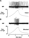
Spectrum of myelinated pulmonary afferents (III) cracking intermediate adapting receptors
- PMID: 33085910
- PMCID: PMC7792821
- DOI: 10.1152/ajpregu.00136.2020
Spectrum of myelinated pulmonary afferents (III) cracking intermediate adapting receptors
Abstract
Conventional one-sensor theory (one afferent fiber connects to a single sensor) categorizes the bronchopulmonary mechanosensors into the rapidly adapting receptors (RARs), slowly adapting receptors (SARs), or intermediate adapting receptors (IARs). RARs and SARs are known to sense the rate and magnitude of mechanical change, respectively; however, there is no agreement on what IARs sense. Some investigators believe that the three types of sensors are actually one group with similar but different properties and IARs operate within that group. Other investigators (majority) believe IARs overlap with the RARs and SARs and can be classified within them according to their characteristics. Clearly, there is no consensus on IARs function. Recently, a multiple-sensor theory has been advanced in which a sensory unit may contain many heterogeneous sensors, such as both RARs and SARs. There are no IARs. Intermediate adapting unit behavior results from coexistence of RARs and SARs. Therefore, the unit can sense both rate and magnitude of changes. The purpose of this review is to provide evidence that the multiple-sensor theory better explains sensory unit behavior.
Keywords: afferent; lung; receptor; sensory unit; vagus nerve.
Conflict of interest statement
No conflicts of interest, financial or otherwise, are declared by the author.
Figures


Similar articles
-
A historical perspective of pulmonary rapidly adapting receptors.Respir Physiol Neurobiol. 2021 May;287:103595. doi: 10.1016/j.resp.2020.103595. Epub 2020 Dec 9. Respir Physiol Neurobiol. 2021. PMID: 33309786 Review.
-
Spectrum of myelinated pulmonary afferents (II).Am J Physiol Regul Integr Comp Physiol. 2013 Nov 1;305(9):R1059-64. doi: 10.1152/ajpregu.00125.2013. Epub 2013 Sep 18. Am J Physiol Regul Integr Comp Physiol. 2013. PMID: 24049120 Free PMC article.
-
Spectrum of myelinated pulmonary afferents.Am J Physiol Regul Integr Comp Physiol. 2000 Dec;279(6):R2142-8. doi: 10.1152/ajpregu.2000.279.6.R2142. Am J Physiol Regul Integr Comp Physiol. 2000. PMID: 11080079
-
Deflation-activated receptors, not classical inflation-activated receptors, mediate the Hering-Breuer deflation reflex.J Appl Physiol (1985). 2016 Nov 1;121(5):1041-1046. doi: 10.1152/japplphysiol.00903.2015. Epub 2016 Sep 1. J Appl Physiol (1985). 2016. PMID: 27586839 Review.
-
Attenuation of pulmonary afferent input by vagal cooling in dogs.Respir Physiol. 1988 Apr;72(1):19-33. doi: 10.1016/0034-5687(88)90076-x. Respir Physiol. 1988. PMID: 3363233
Cited by
-
Mechanisms Involved in the Stimulatory and Inhibitory Effects of 5-Hydroxytryptamine on Vagal Mechanosensitive Afferents in Rat Lung.Front Physiol. 2022 Mar 21;13:813096. doi: 10.3389/fphys.2022.813096. eCollection 2022. Front Physiol. 2022. PMID: 35480033 Free PMC article.
-
A comparative study of bronchopulmonary slowly adapting receptors between rabbits and rats.Physiol Rep. 2022 Mar;10(6):e15069. doi: 10.14814/phy2.15069. Physiol Rep. 2022. PMID: 35343655 Free PMC article.
-
Paradoxical response of pulmonary slowly adapting units during constant pressure lung inflation.Am J Physiol Regul Integr Comp Physiol. 2021 Aug 1;321(2):R220-R227. doi: 10.1152/ajpregu.00116.2021. Epub 2021 Jun 30. Am J Physiol Regul Integr Comp Physiol. 2021. PMID: 34189947 Free PMC article.
-
The Coding Logic of Interoception.Annu Rev Physiol. 2024 Feb 12;86:301-327. doi: 10.1146/annurev-physiol-042222-023455. Epub 2023 Dec 7. Annu Rev Physiol. 2024. PMID: 38061018 Free PMC article. Review.
-
A multidimensional coding architecture of the vagal interoceptive system.Nature. 2022 Mar;603(7903):878-884. doi: 10.1038/s41586-022-04515-5. Epub 2022 Mar 16. Nature. 2022. PMID: 35296859 Free PMC article.
References
-
- Burgess PR, Perl ER. Cutaneous mechanoreceptors and nociceptors In: Handbook of Sensory Physiology. II. Somatosensory System, edited by Iggo A. New York: Springer-Verlag, 1973, p. 29–38.
Publication types
MeSH terms
Grants and funding
LinkOut - more resources
Full Text Sources
Miscellaneous

