. 2020 Oct 20;33(3):108275.
doi: 10.1016/j.celrep.2020.108275.

TNF Receptor 1 Promotes Early-Life Immunity and Protects against Colitis in Mice

Affiliations

TNF Receptor 1 Promotes Early-Life Immunity and Protects against Colitis in Mice

Cambrian Y Liu et al. Cell Rep. .

Abstract

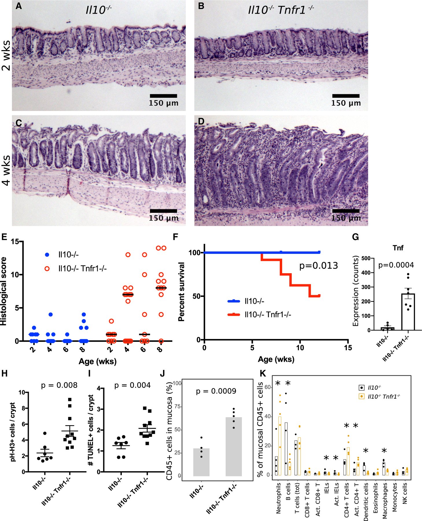
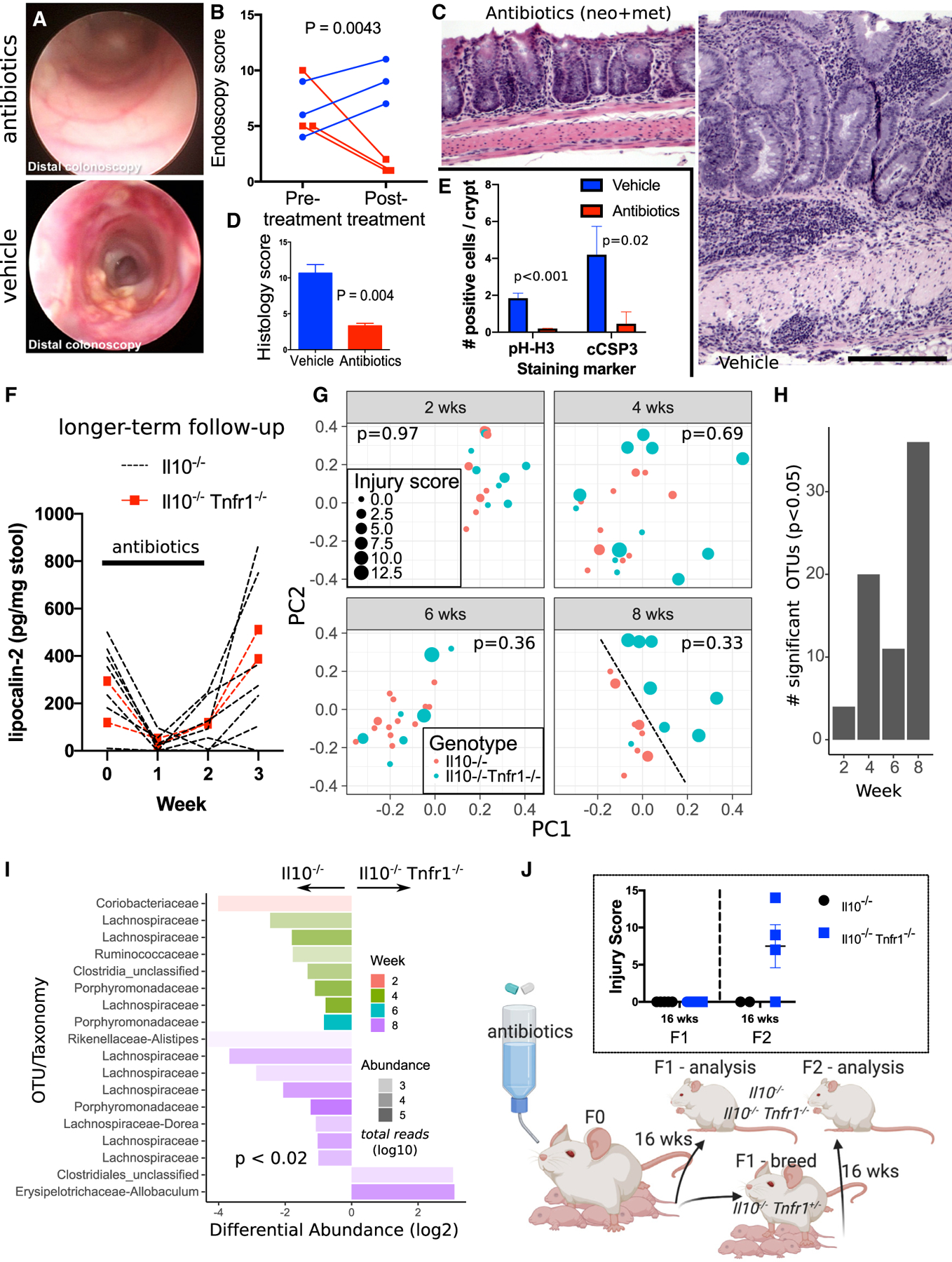
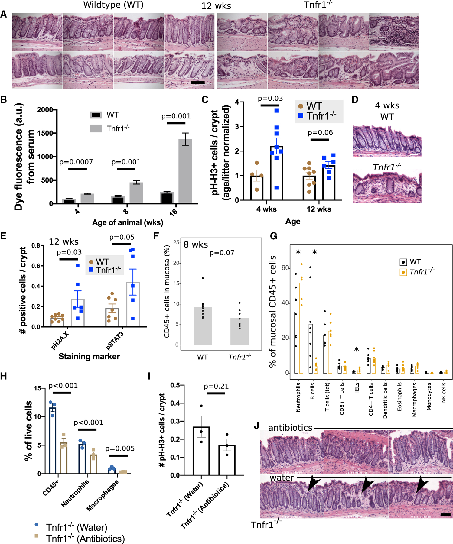
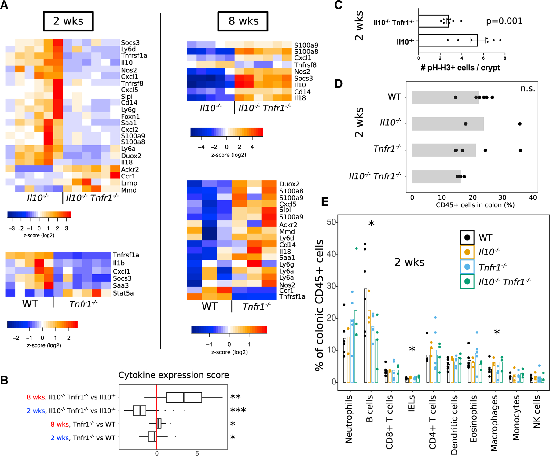
Neutralization of tumor necrosis factor (TNF) represents a widely used therapeutic strategy for autoimmune diseases including inflammatory bowel disease (IBD). However, the fact that many patients with IBD are non-responsive to anti-TNF therapies suggests the need for a better understanding of TNF signaling in IBD. Here, we show that co-deletion of TNF receptor 1 (TNFR1, Tnfrsf1a) in the Il10-/- spontaneous colitis model exacerbates disease, resulting in very-early-onset inflammation after weaning. The disease can be interrupted by treatment with antibiotics. The single deletion of TNFR1 induces subclinical colonic epithelial dysfunction and mucosal immune abnormalities, including accumulation of neutrophils and depletion of B cells. During the pre-disease period (before weaning), both Tnfr1-/- and Il10-/-Tnfr1-/- animals exhibit impaired expression of pro-inflammatory cytokines compared with wild-type and Il10-/- controls, respectively. Collectively, these results demonstrate the net anti-inflammatory functions of TNF/TNFR1 signaling through the regulation of colonic immune homeostasis in early life.

Keywords: IBD; antibiotics; barrier; microbiome; mucosa; tolerance; weaning reaction.


Conflict of interest statement

Declaration of Interests The authors declare no competing interests.

Figures

Figure 1.
Figure 1.. Il10−/− Tnfr1−/− Mice Develop Early-Onset, Severe Colitis at 4 Weeks Old
(A and B) Hematoxylin-and-eosin (H&E)-stained sections of pre-weaning 2-wk-old Il10/ (A) (n = 7) and Il10/ Tnfr1/ (B) (n = 8) mice show normal distal colon. (C and D) H&E sections of 4-week-old post-weaning mice reveal normal colon in the Il10/ genotype (C) (n = 6) but colitis in the Il10/ Tnfr1/ genotype (D) (n = 13). (E) Histological scoring of colitis severity (worst disease score is 15). Horizontal lines indicate median. (F) Kaplan-Meier survival curve indicates early mortality in Il10/ Tnfr1/ mice. (G) Nanostring analysis demonstrates upregulation of Tnf expression in Il10/ Tnfr1/ animals. (H and I) Crypts in 12-wk-old Il10/ Tnfr1/ mice exhibited increased numbers of proliferating (pH-H3+) (H) and apoptotic (TUNEL+) (I) cells. (J and K) Flow cytometric analysis of colonic mucosal scrapings demonstrates increased colonic mucosal infiltration of CD45+ hematopoietic cells (J) with increased immune cell subsets (K) in 8-wk-old Il10/ Tnfr1/ mice. Scale bars: (A)–(D) 100 μm. *p < 0.05. See also Figures S1, S2, and S3. Error bars: standard error.
Figure 2.
Figure 2.. Broad-Spectrum Antibiotic Treatment Improves Colitis in Il10−/− Tnfr1−/− Mice
(A–E) Improved endoscopic appearance (A), endoscopic score (B), histology (C and D), and crypt cell dynamics (E) were observed in 8-week-old antibiotic-treated Il10/ Tnfr1/ mice (n = 3) compared with controls (n = 3). (F) Fecal lipocalin levels diminished to nearly undetectable levels during antibiotic treatment of Il10/ (n = 7) and Il10/ Tnfr1/ (n = 2) mice but quickly rebounded. (G) 16S analysis of Il10/ Tnfr1/ and Il10/ cecal microbiomes collected from 2-, 4-, 6-, and 8-week-old Il10/ Tnfr1/ and Il10/ mice. Shown are weighted UniFrac distances plotted using multidimensional scaling. Each sample is coded by age (gray facets), genotype (color), and colitis score (size). At 8 weeks old, the genotypes could be qualitatively discriminated (dotted black line). The displayed p value for each age is computed by permuted analysis of variance using littermates, colitis severity, sex, and genotype conditioned on dam identity as model factors. (H) Bar graph displays the total number of significant operational taxonomic units (OTUs) that are differentially abundant in Il10/ versus Il10/ Tnfr1/ animals by age. (I) OTUs with taxonomic classifications plotted to indicate their relative abundance in Il10/ Tnfr1/ versus Il10/ samples. The transparency of the bars denotes the total abundance of the OTUs on a log scale (more transparent is less abundant). (J) Depiction of the multigenerational experiment analyzing the effects of maternal antibiotic treatment on colitis development, with associated colitis scoring shown in the inset. Scale bars: 200 μm. Error bars: standard error.
Figure 3.
Figure 3.. Adult Tnfr1−/− Mice Exhibit Epithelial Dysfunction and Alterations in Colonic Mucosal Immune Cell Representation
(A) Image montages show normal crypt structures in wild-type mice (n = 8) and focal abnormalities in 8-wk-old Tnfr1/ mice (n = 7). (B) Tnfr1/ mice at 4, 8, and 12 weeks old exhibited increased colonic permeability at 30 min after rectal instillation of 4 kDa FITC-dextran. The graph is representative of three different experiments. (C) Trending increase in pH-H3+ proliferative cells in knockout mice at 4 and 12 weeks old. (D) H&E images of the colonic mucosa at 4 weeks old demonstrate foci of altered crypt patterning in Tnfr1/ mice. (E) Adult 12-week-old Tnfr1/ colonic epithelium also had increased numbers of pH2A.X+ and pSTAT3+ cells. (F and G) Flow cytometry reveals a trend toward decreased hematopoietic cell numbers (F) in the colonic mucosa of 8-week-old Tnfr1/ mice. This was driven by increased neutrophils and a near-total loss of B cells (G). (H) Flow cytometric assessment of antibiotic-treated Tnfr1/ mice reveals reduced numbers of mucosal immune cells. (I) Changes to pH-H3 staining after antibiotic treatment were more modest and insignificant. (J) Qualitative reductions (arrowheads) in the density of inter-crypt cells were noted in H&E-stained sections of antibiotic-treated samples. Scale bars: (A) 100 μm; (D) and (J) 50 μm, *p < 0.05. Error bars: standard error.
Figure 4.
Figure 4.. TNFR1-Deficient Animals Show Early-Life Defects in Colonic Immune Response
(A) Heatmaps summarize cytokine-expression levels obtained from RNA-seq/NanoString analysis of colons from wild-type (WT), Tnfr1/, Il10/, and Il10/ Tnfr1/ animals at 2 weeks (pre-disease) and 8 weeks (post-disease onset) old. Overall cytokine expression was decreased in TNFR1-deficient animals at 2 weeks of age but increased in those animals at 8 weeks old. Note that reads for Il10 were still detected in Il10/ animals because of continued production of mRNA encoding of an early-termination codon. (B) The boxplot shows a summarized score of cytokine expression changes across comparisons of genotypes and ages. The lower and upper hinges represent the 25th and 75th percentiles. Whiskers denote the range of the data, excepting outlying points beyond 150% of the interquartile range. All values were obtained from RNA-seq except for the comparison of Il10/ Tnfr1/ versus Il10/ mice at 8 weeks old (NanoString). Significance of scores was tested using the t test against 0 (no change). (C) Il10/ Tnfr1/ animals at 2 weeks old show reduced pH-H3+ cell counts per crypt. (D and E) Flow cytometric analysis of whole colons collected from 2-week-old mice shows overall similarity of colonic hematopoietic (CD45+) cell counts (D) but loss of B cells in TNFR1-deficient animals (E). Significance was evaluated using ANOVA. *p < 0.05. See also Figure S4. Error bars: standard error.

References

    1. Al Nabhani Z, Dulauroy S, Marques R, Cousu C, Al Bounny S, Dejardin F, Sparwasser T, Berard M, Cerf-Bensussan N, and Eberl G (2019). A weaning reaction to microbiota is required for resistance to immunopathologies in the adult. Immunity 50, 1276–1288.e1275. - PubMed
    1. Atreya R, Zimmer M, Bartsch B, Waldner MJ, Atreya I, Neumann H, Hildner K, Hoffman A, Kiesslich R, Rink AD, et al. (2011). Antibodies against tumor necrosis factor (TNF) induce T-cell apoptosis in patients with inflammatory bowel diseases via TNF receptor 2 and intestinal CD14+ macrophages. Gastroenterology 141, 2026–2038. - PubMed
    1. Bank S, Skytt Andersen P, Burisch J, Pedersen N, Roug S, Galsgaard J, Ydegaard Turino S, Brodersen JB, Rashid S, Kaiser Rasmussen B, et al. (2014). Polymorphisms in the inflammatory pathway genes TLR2, TLR4, TLR9, LY96, NFKBIA, NFKB1, TNFA, TNFRSF1A, IL6R, IL10, IL23R, PTPN22, and PPARG are associated with susceptibility of inflammatory bowel disease in a Danish cohort. PLoS ONE 9, e98815. - PMC - PubMed
    1. Begue B, Verdier J, Rieux-Laucat F, Goulet O, Morali A, Canioni D, Hugot JP, Daussy C, Verkarre V, Pigneur B, et al. (2011). Defective IL10 signaling defining a subgroup of patients with inflammatory bowel disease. Am. J. Gastroenterol 106, 1544–1555. - PubMed
    1. Ben-Horin S, Kopylov U, and Chowers Y (2014). Optimizing anti-TNF treatments in inflammatory bowel disease. Autoimmun. Rev 13, 24–30. - PubMed

Publication types

MeSH terms