

Development of a Smartphone-Based Skin Simulation Model for Medical Education
- PMID: 33086367
- PMCID: PMC8580374
- DOI: 10.1097/SIH.0000000000000509
Development of a Smartphone-Based Skin Simulation Model for Medical Education
Abstract
Introduction: Teaching dermatology to medical students entails a series of lectures, pictures, and hands-on skin examinations to convey a sense of skin features and textures, often by use of simulated skin models. However, such methods can often lack accurate visual and tactile texture representation of skin lesions. To facilitate learning, we have developed a smartphone-based skin simulation model, which provides a configurable visual and tactile sense of a lesion by using the ubiquitous availability of smartphone-based mobile platforms.
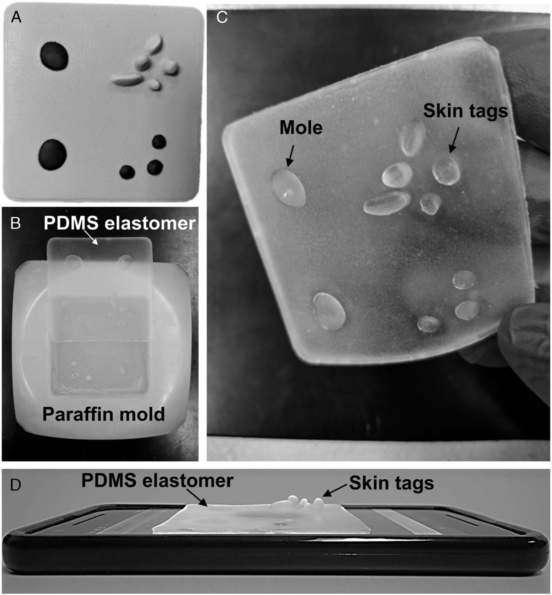
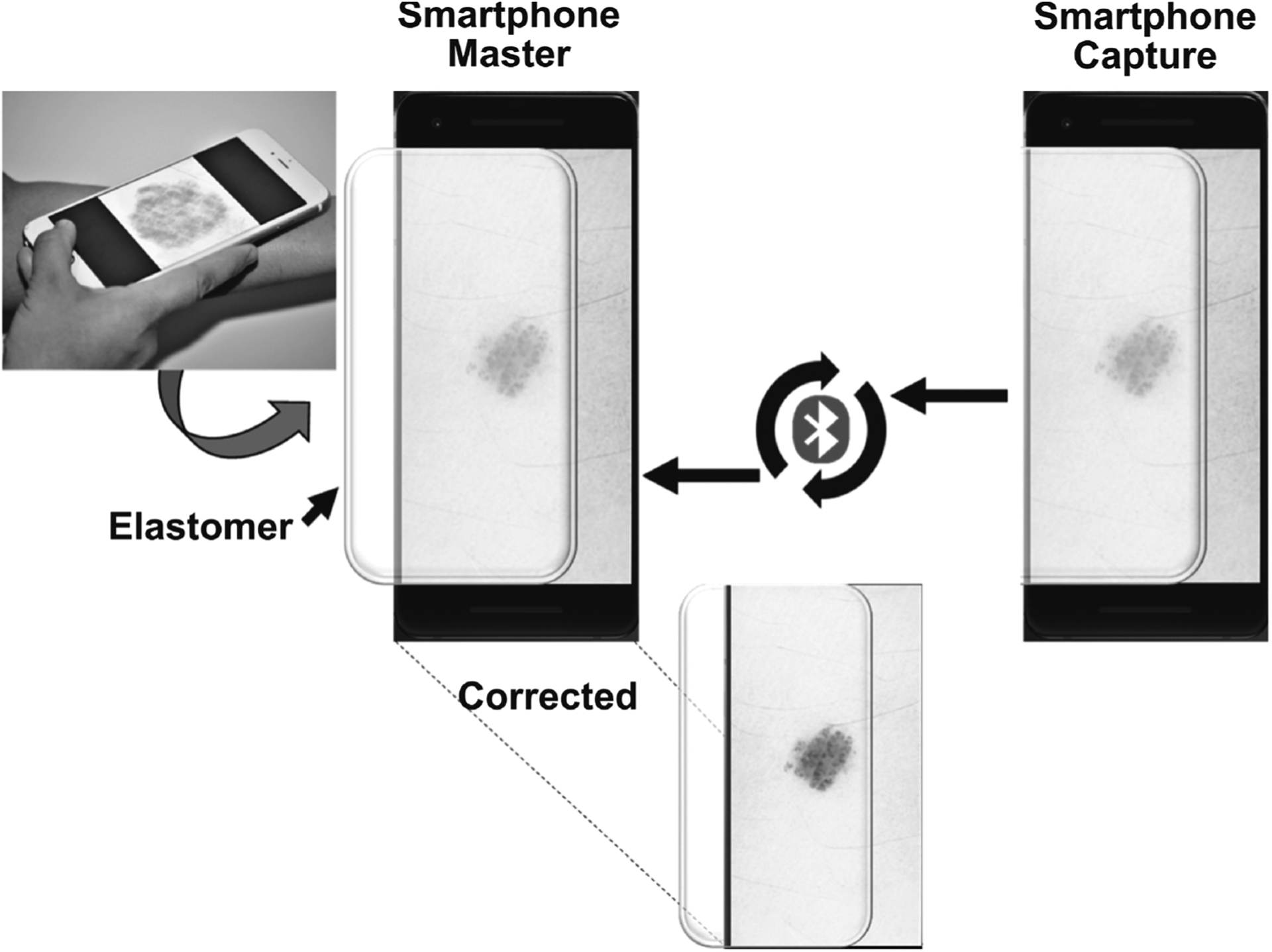
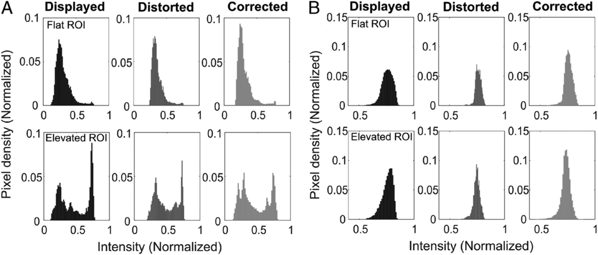
Methods: A polydimethylsiloxane (PDMS) overlay was used as a configurable translucent elastomer material to model the stiffness and texture of skin. A novel custom smartphone-based app was developed to capture images of various skin lesions, which were subsequently displayed on a tablet or second smartphone, over which the PDMS model skin elastomer was placed. Using the local Bluetooth connection between mobile devices, an iterative feedback algorithm corrected the visual distortion caused by the optical scattering of the translucent elastomer, enabling better virtual visualization of the lesion.
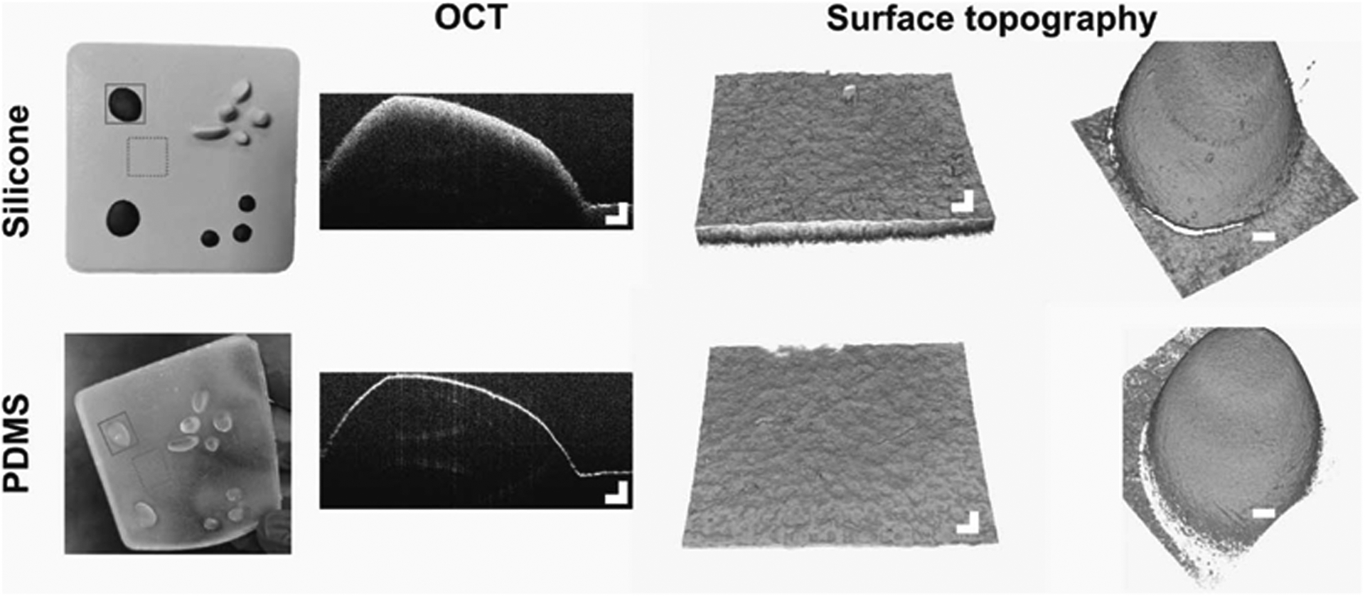
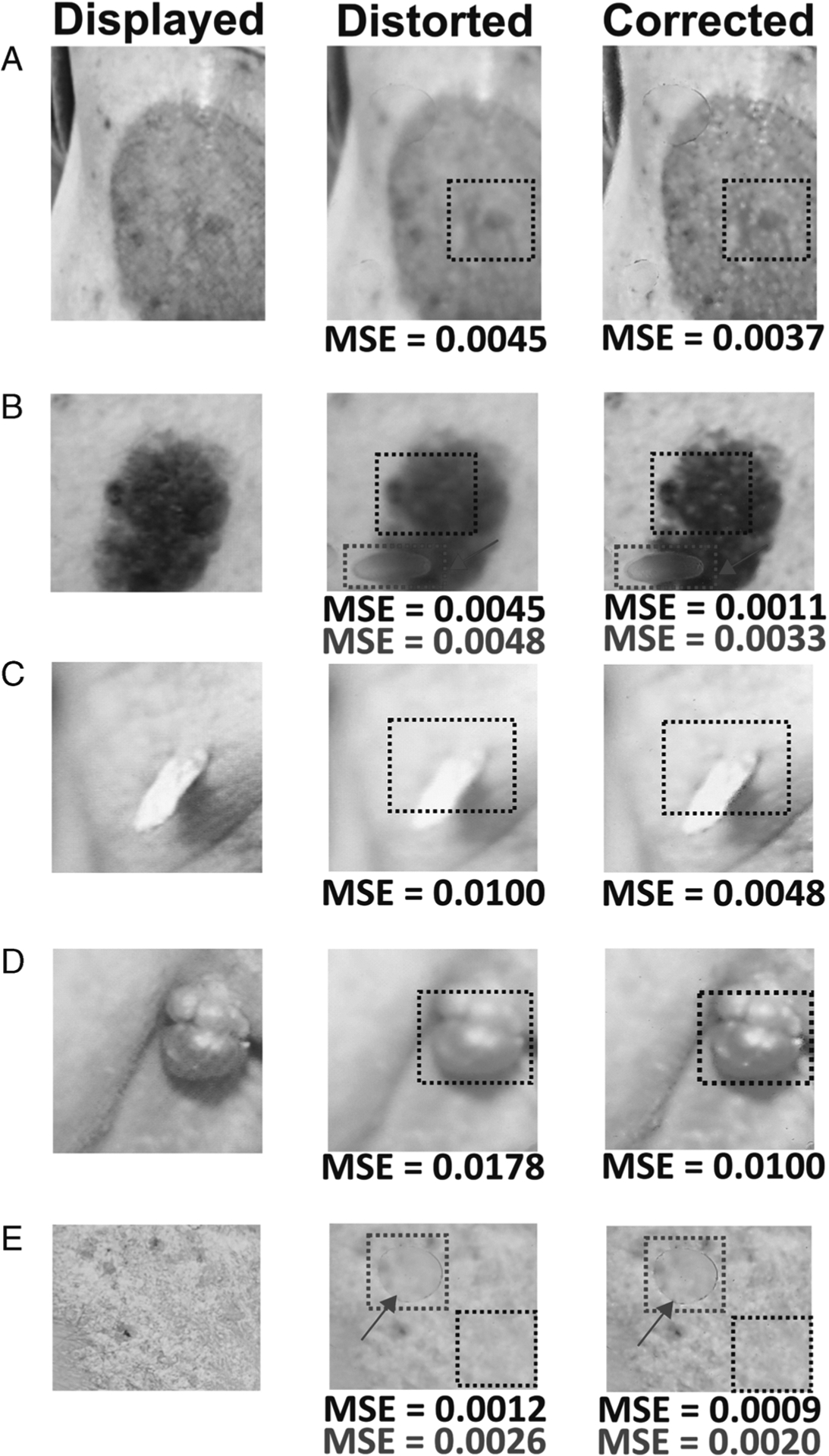
Results: The developed smartphone-based app corrected the distortion of images projected through the simulated skin elastomer. Surface topography of the developed PDMS elastomer provided a more accurate representation of skin texture.
Conclusions: In this investigation, we developed a smartphone-based skin lesion visualization app with a simulated skin elastomer for training/education in not only dermatology but also all general medical specialties that examine the skin. This technique has the potential to advance the educational experience by giving students the ability to see, touch, and feel pragmatic skin textures and lesions.
Copyright © 2020 Society for Simulation in Healthcare.
Conflict of interest statement
The authors declare no conflict of interest.
Figures
References
-
- Dąbrowska AK, Rotaru GM, Derler S, et al. Materials used to simulate physical properties of human skin. Skin Res Technol 2016;22:3–14. - PubMed
-
- Garcia C, Poletti E. Surgical pearl: a model to practice the Mohs surgical technique. J Am Acad Dermatol 2006;55:313–314. - PubMed
-
- Chen TM, Mellette JR. Surgical pearl: tomato—an alternative model for shave biopsy training. J Am Acad Dermatol 2006;54:517–518. - PubMed
-
- Wanitphakdeedecha R, Nguyen TH, Chen TM. The banana: a surgery training model to refine blade control for Mohs layer removal and skin incisions. Dermatol Surg 2008;34:1088–1090. - PubMed
-
- Bastos EM, Silva RDP. Proposal of a synthetic ethylene-vinyl acetate bench model for surgical foundations learning: suture training. Acta Cir Bras 2011;26:149–152. - PubMed
MeSH terms
Grants and funding
LinkOut - more resources
Full Text Sources
