. 2020 Oct 19;10(10):840.
doi: 10.3390/diagnostics10100840.

Characterization of Uterine Motion in Early Gestation Using MRI-Based Motion Tracking

Affiliations

Characterization of Uterine Motion in Early Gestation Using MRI-Based Motion Tracking

Thomas Martin et al. Diagnostics (Basel). .

Abstract

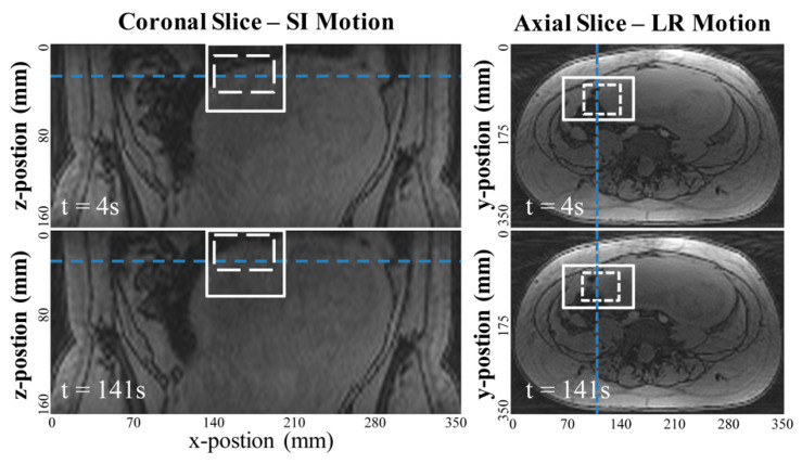
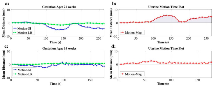
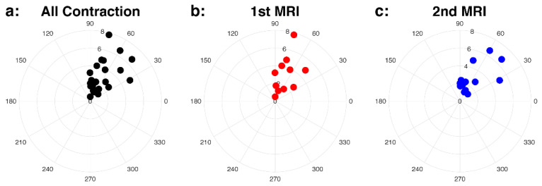
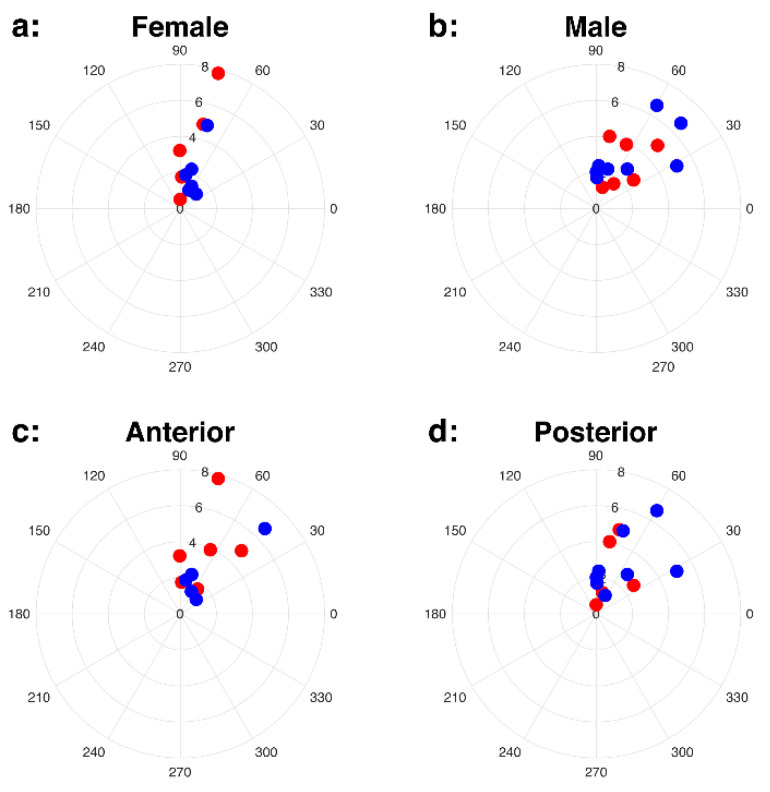
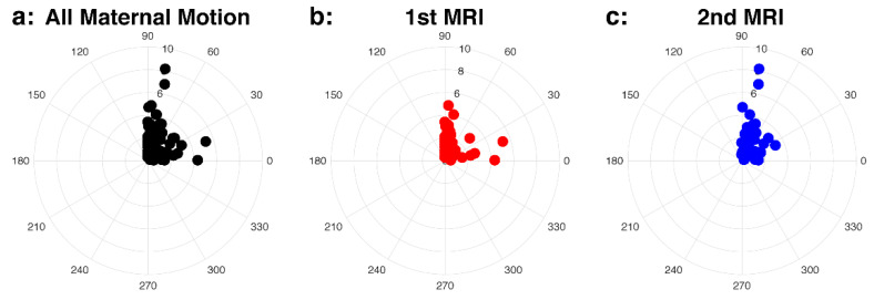
Magnetic resonance imaging (MRI) is a promising non-invasive imaging technique that can be safely used to study placental development and function. However, studies of the human placenta performed by MRI are limited by uterine motion and motion in the uterus during MRI remains one of the major limiting factors. Here, we aimed to investigate the characterization of uterine activity during MRI in the second trimester of pregnancy using MRI-based motion tracking. In total, 46 pregnant women were scanned twice (first scan between 14 and 18 weeks and second scan between 19 and 24 weeks), and 20 pregnant subjects underwent a single MRI between 14 and 18 weeks GA, resulting in 112 MRI scans. An MRI-based algorithm was used to track uterine motion in the superior-inferior and left-right directions. Uterine contraction and maternal motion cases were separated by the experts, and unpaired Wilcoxon tests were performed within the groups of gestational age (GA), fetal sex, and placental location in terms of the overall intensity measures of the uterine activity. In total, 22.3% of cases had uterine contraction during MRI, which increased from 18.6% at 14-18 weeks to 26.4% at 19-24 weeks GA. The dominant direction of the uterine contraction and maternal motion was the superior to the inferior direction during early gestation.

Keywords: MRI motion tracking; human pregnancy; maternal motion; placenta MRI; uterine contraction.


Conflict of interest statement

The authors declare no conflict of interest.

Figures

Figure 1
Figure 1
Example images measuring the uterine motion in mm with the image-based template matching algorithm. The coronal reformatted images were used to measure superior-inferior (SI) motion and axial images were used to measure left-right (LR) motion. The template matching algorithm searches for the target ROI (dotted white square) within the search region (solid squares) for each dynamic time frame, using a least-squares comparison metric based on normalized image intensity.
Figure 2
Figure 2
Examples of uterine motion time plots obtained by combining the two motion plots for superior-inferior (blue) and left-right (green) directions. Examples of uterine contraction (a,b) and of maternal motion (c,d) are shown. The area under the curve of the uterine contraction is shown as the shaded area.
Figure 3
Figure 3
Intensity (AUCnorm) and direction (θ) of the uterine contraction in the polar coordinates for all cases (a), first (1st) MRI, colored as red, (b), and second (2nd) MRI, colored as blue, (c) cases. The polar plots show the dominant direction is SI.
Figure 4
Figure 4
Uterine contraction for female (a) and male (b) fetal sex, and anterior (c) and posterior (d) placentas. First (1st) and second (2nd) MRI scans are colored as red (1st MRI) and blue (2nd MRI). The scatter plots in the polar coordinates show that the dominant direction of motion is SI.
Figure 5
Figure 5
Intensity (Umax) and direction (θ) of the maternal motion in the polar coordinates for all cases (a), first (1st) MRI (b), and second (2nd) MRI (c) cases.

Similar articles

Cited by

References

    1. Derwig I.E., Akolekar R., Zelaya F., Gowland P., Barker G.J., Nicolaides K.H. Association of placental volume measured by MRI and birth weight percentile. J. Magn. Reson. Imaging. 2011;34:1125–1130. doi: 10.1002/jmri.22794. - DOI - PubMed
    1. Thomson A.M., Billewicz W.Z., Hytten F.E. The weight of the placenta in relation to birthweight. BJOG Int. J. Obstet. Gynaecol. 1969;76:865–872. doi: 10.1111/j.1471-0528.1969.tb15722.x. - DOI - PubMed
    1. Damodaram M., Story L., Eixarch E., Patel A., McGuinness A., Allsop J., Wyatt-Ashmead J., Kumar S., Rutherford M. Placental MRI in Intrauterine Fetal Growth Restriction. Placenta. 2010;31:491–498. doi: 10.1016/j.placenta.2010.03.001. - DOI - PubMed
    1. Li S., Hoskins P., Anderson T., McDicken W. Measurement of mean velocity during pulsatile flow using time-averaged maximum frequency of doppler ultrasound waveforms. Ultrasound Med. Boil. 1993;19:105–113. doi: 10.1016/0301-5629(93)90002-6. - DOI - PubMed
    1. Angtuaco T.L., Shah H.R., Mattison D.R., Quirk J.G. MR imaging in high-risk obstetric patients: A valuable complement to US. Radiographics. 1992;12:91–109. doi: 10.1148/radiographics.12.1.1734485. - DOI - PubMed