. 2020 Oct 19;21(20):7724.
doi: 10.3390/ijms21207724.

Effect of Silymarin Supplementation on Physical Performance, Muscle and Myocardium Histological Changes, Bodyweight, and Food Consumption in Rats Subjected to Regular Exercise Training

Affiliations

Effect of Silymarin Supplementation on Physical Performance, Muscle and Myocardium Histological Changes, Bodyweight, and Food Consumption in Rats Subjected to Regular Exercise Training

Nancy Vargas-Mendoza et al. Int J Mol Sci. .

Abstract

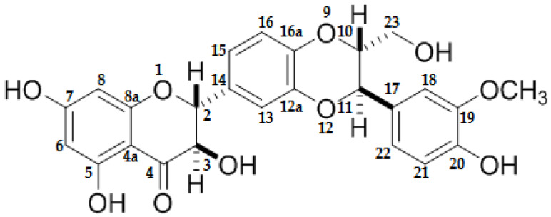
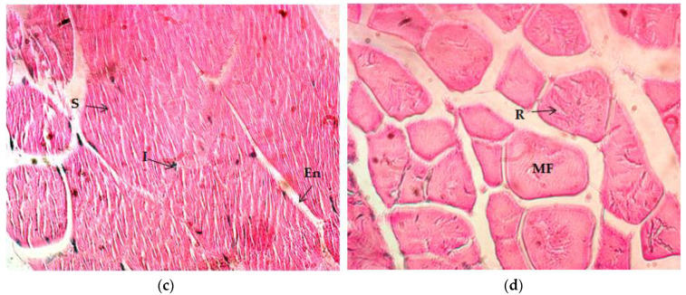
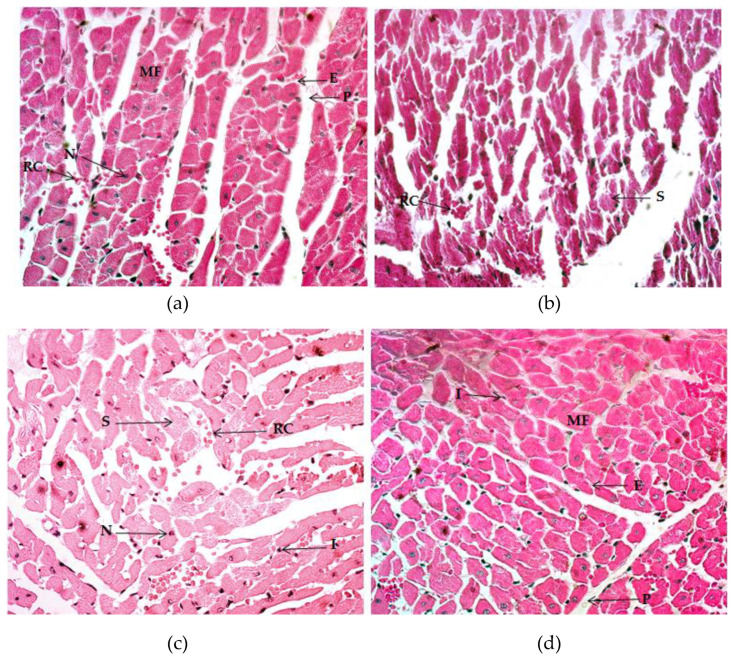
(1) Background: Regular exercise induces physiological and morphological changes in the organisms, but excessive training loads may induce damage and impair recovery or muscle growth. The purpose of the study was to evaluate the impact of Silymarin (SM) consumption on endurance capacity, muscle/cardiac histological changes, bodyweight, and food intake in rats subjected to 60 min of regular exercise training (RET) five days per week. (2) Methods: Male Wistar rats were subjected to an eight-week RET treadmill program and were previously administered SM and vitamin C. Bodyweight and food consumption were measured and registered. The maximal endurance capacity (MEC) test was performed at weeks one and eight. After the last training session, the animals were sacrificed, and samples of quadriceps/gastrocnemius and cardiac tissue were obtained and process for histological analyzes. (3) Results: SM consumption improved muscle recovery, inflammation, and damaged tissue, and promoted hypertrophy, vascularization, and muscle fiber shape/appearance. MEC increased after eight weeks of RET in all trained groups; moreover, the SM-treated group was enhanced more than the group with vitamin C. There were no significant changes in bodyweight and in food and nutrient consumption along the study. (5) Conclusion: SM supplementation may enhance physical performance, recovery, and muscle hypertrophy during the eight-week RET program.

Keywords: exercise training; muscle; myocardium; myofibers; silymarin.


Conflict of interest statement

The authors declare no conflict of interest.

Figures

Figure 1
Figure 1
Silybin structure the major compound of silymarin.
Figure 2
Figure 2
Quadriceps muscle histology (Hematoxylin and Eosin (H&E) stain (40×). (a) Image of the control group. (b) Image corresponding to the exercise-training groups without supplementation. (c) Image of the exercise-training groups plus vitamin C. (d) Image of the exercise-training group plus silymarin. MF: muscle fiber; En: endomysium; Ep: epimysium; Pe: perimysium; S: splitting; I: inflammation.
Figure 2
Figure 2
Quadriceps muscle histology (Hematoxylin and Eosin (H&E) stain (40×). (a) Image of the control group. (b) Image corresponding to the exercise-training groups without supplementation. (c) Image of the exercise-training groups plus vitamin C. (d) Image of the exercise-training group plus silymarin. MF: muscle fiber; En: endomysium; Ep: epimysium; Pe: perimysium; S: splitting; I: inflammation.
Figure 3
Figure 3
Histology of the gastrocnemius muscle, Hematoxylin and Eosin (H&E) stain (40×). (a) Image of the control group. (b) Image of the exercise-training group without supplementation. (c) Image of the exercise-training groups plus vitamin C. (d) Image of the exercise-training group plus silymarin. MF: muscle fiber; En: endomysium; Ep: epimysium; S: splitting; I: inflammation.
Figure 3
Figure 3
Histology of the gastrocnemius muscle, Hematoxylin and Eosin (H&E) stain (40×). (a) Image of the control group. (b) Image of the exercise-training group without supplementation. (c) Image of the exercise-training groups plus vitamin C. (d) Image of the exercise-training group plus silymarin. MF: muscle fiber; En: endomysium; Ep: epimysium; S: splitting; I: inflammation.
Figure 4
Figure 4
Histology of the myocardium, Hematoxylin and Eosin (H&E) stain (40×). (a) Image of the control group. (b) Image of the exercise-training group without supplementation. (c) Image of the exercise-training groups plus vitamin C. (d) Image of the exercise-training group plus silymarin. MF: muscle fiber; E: endocardium; P: pericardium; RC: red cells; N: nuclei; S: splitting; I: inflammation.

Similar articles

Cited by

References

    1. Frontera W.R., Ochala J. Skeletal muscle: A brief review of structure and function. Calcif. Tissue Int. 2015;96:183–195. doi: 10.1007/s00223-014-9915-y. - DOI - PubMed
    1. Rennie M.J., Wackerhage H., Spangenburg E.E., Booth F.W. Control of the size of the human muscle mass. Annu. Rev. Physiol. 2004;66:799–828. doi: 10.1146/annurev.physiol.66.052102.134444. - DOI - PubMed
    1. Marcotte G.R., West D.W., Baar K. The molecular basis for load-induced skeletal muscle hypertrophy. Calcif. Tissue Int. 2015;96:196–210. doi: 10.1007/s00223-014-9925-9. - DOI - PMC - PubMed
    1. Barany M. ATPase activity of myosin correlated with speed of muscle shortening. J. Gen. Physiol. 1967;50:197–218. doi: 10.1085/jgp.50.6.197. - DOI - PMC - PubMed
    1. Schiaffino S. Fibre types in skeletal muscle: A personal account. Acta Physiol. 2010;199:451–463. doi: 10.1111/j.1748-1716.2010.02130.x. - DOI - PubMed