

Proteomics of Extracellular Vesicles: Update on Their Composition, Biological Roles and Potential Use as Diagnostic Tools in Atherosclerotic Cardiovascular Diseases
- PMID: 33086718
- PMCID: PMC7588996
- DOI: 10.3390/diagnostics10100843
Proteomics of Extracellular Vesicles: Update on Their Composition, Biological Roles and Potential Use as Diagnostic Tools in Atherosclerotic Cardiovascular Diseases
Abstract
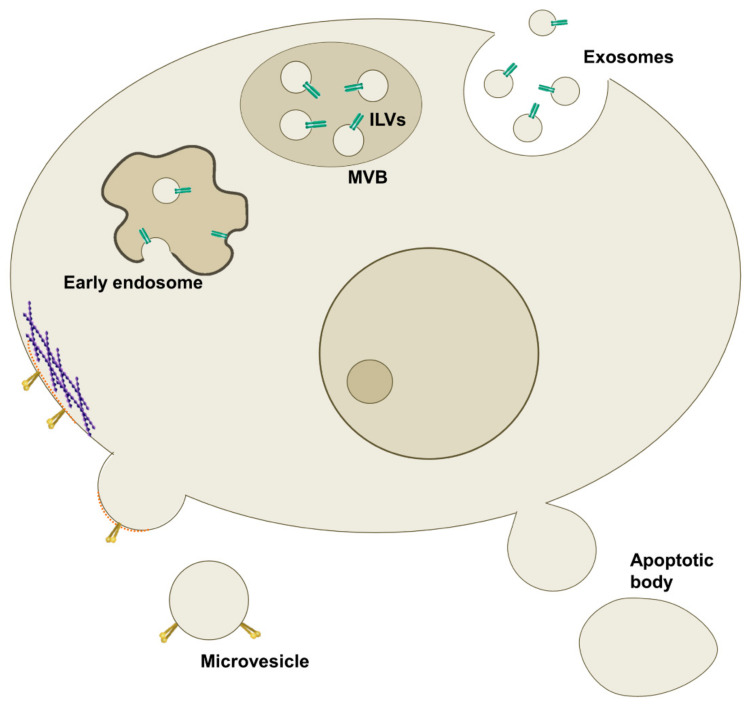
Extracellular vesicles (EVs) are lipid-bound vesicles released from cells under physiological and pathological conditions. Basing on biogenesis, dimension, content and route of secretion, they can be classified into exosomes, microvesicles (MVs) and apoptotic bodies. EVs have a key role as bioactive mediators in intercellular communication, but they are also involved in other physiological processes like immune response, blood coagulation, and tissue repair. The interest in studying EVs has increased over the years due to their involvement in several diseases, such as cardiovascular diseases (CVDs), and their potential role as biomarkers in diagnosis, therapy, and in drug delivery system development. Nowadays, the improvement of mass spectrometry (MS)-based techniques allows the characterization of the EV protein composition to deeply understand their role in several diseases. In this review, a critical overview is provided on the EV's origin and physical properties, as well as their emerging functional role in both physiological and disease conditions, focusing attention on the role of exosomes in CVDs. The most important cardiac exosome proteomic studies will be discussed giving a qualitative and quantitative characterization of the exosomal proteins that could be used in future as new potential diagnostic markers or targets for specific therapies.
Keywords: biomarkers; cardiovascular diseases; exosomes; extracellular vesicles; mass spectrometry; proteins.
Conflict of interest statement
The authors declare no conflict of interest.
Figures


Similar articles
-
Extracellular Vesicles in Cardiovascular Theranostics.Theranostics. 2017 Sep 26;7(17):4168-4182. doi: 10.7150/thno.21274. eCollection 2017. Theranostics. 2017. PMID: 29158817 Free PMC article. Review.
-
A Protocol for Isolation, Purification, Characterization, and Functional Dissection of Exosomes.Methods Mol Biol. 2021;2261:105-149. doi: 10.1007/978-1-0716-1186-9_9. Methods Mol Biol. 2021. PMID: 33420988
-
Extracellular vesicles characteristics and emerging roles in atherosclerotic cardiovascular disease.Metabolism. 2018 Aug;85:213-222. doi: 10.1016/j.metabol.2018.04.008. Epub 2018 May 1. Metabolism. 2018. PMID: 29727628 Review.
-
Biology and Role of Extracellular Vesicles (EVs) in the Pathogenesis of Thrombosis.Int J Mol Sci. 2019 Jun 11;20(11):2840. doi: 10.3390/ijms20112840. Int J Mol Sci. 2019. PMID: 31212641 Free PMC article. Review.
-
Exosomes: Nomenclature, Isolation, and Biological Roles in Liver Diseases.Biomol Ther (Seoul). 2023 May 1;31(3):253-263. doi: 10.4062/biomolther.2022.161. Biomol Ther (Seoul). 2023. PMID: 37095734 Free PMC article. Review.
Cited by
-
Role of exosomal microRNAs in cancer therapy and drug resistance mechanisms: focus on hepatocellular carcinoma.Front Oncol. 2022 Jul 15;12:940056. doi: 10.3389/fonc.2022.940056. eCollection 2022. Front Oncol. 2022. PMID: 35912267 Free PMC article. Review.
-
Isolation and Identification of Plasma Extracellular Vesicles Protein Biomarkers.Methods Mol Biol. 2023;2660:207-217. doi: 10.1007/978-1-0716-3163-8_14. Methods Mol Biol. 2023. PMID: 37191799
-
Circulating Extracellular Vesicles As Biomarkers and Drug Delivery Vehicles in Cardiovascular Diseases.Biomolecules. 2021 Mar 5;11(3):388. doi: 10.3390/biom11030388. Biomolecules. 2021. PMID: 33808038 Free PMC article. Review.
-
Development of stem cell therapy for atherosclerosis.Mol Cell Biochem. 2024 Apr;479(4):779-791. doi: 10.1007/s11010-023-04762-8. Epub 2023 May 13. Mol Cell Biochem. 2024. PMID: 37178375 Review.
-
Exosomal Composition, Biogenesis and Profiling Using Point-of-Care Diagnostics-Implications for Cardiovascular Disease.Front Cell Dev Biol. 2022 Jun 1;10:853451. doi: 10.3389/fcell.2022.853451. eCollection 2022. Front Cell Dev Biol. 2022. PMID: 35721503 Free PMC article. Review.
References
Publication types
Grants and funding
LinkOut - more resources
Full Text Sources
Other Literature Sources
