Case Reports
. 2020 Oct 19;10(10):847.
doi: 10.3390/diagnostics10100847.

Persistent Left Superior Vena Cava with Absent Right Superior Vena Cava and Discrete Subaortic Stenosis Diagnosed in a Patient with Sick Sinus Syndrome: A Case Report and Brief Review of the Literature

Affiliations
Case Reports

Persistent Left Superior Vena Cava with Absent Right Superior Vena Cava and Discrete Subaortic Stenosis Diagnosed in a Patient with Sick Sinus Syndrome: A Case Report and Brief Review of the Literature

Irina Demșa et al. Diagnostics (Basel). .

Abstract

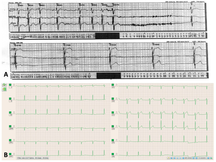
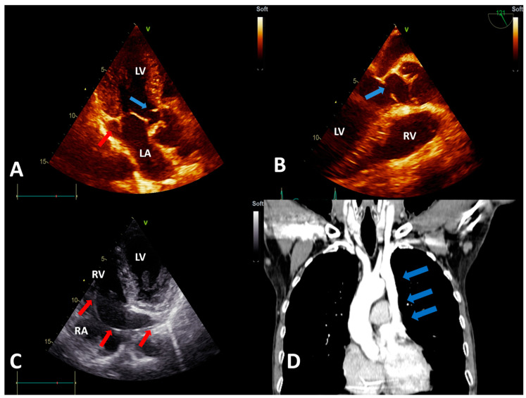
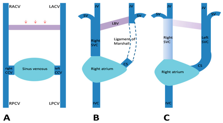
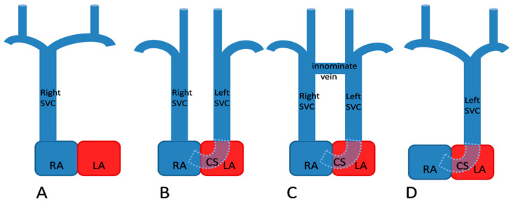
A persistent left superior vena cava (PLSVC) is the most frequent anomaly of the venous drainage system. While both a right and left superior vena cava (SVC) are usually present, a unique, left-sided SVC, also known as an isolated PLSVC, accounts for only 10-20% of cases. It is frequently associated with arrhythmias and other congenital cardiac anomalies. Though it is usually an asymptomatic condition, it may pose significant problems whenever central venous access is needed. We report a case of an isolated PLSVC that was diagnosed incidentally during pacemaker implantation for sinus node dysfunction. The venous anomaly was associated with subvalvular aortic stenosis determined by a subaortic membrane; this particular association of congenital cardiovascular anomalies is a rare finding, with only a few cases reported in the literature. We aim to highlight the clinical and practical implications of this condition, as well as to discuss the embryonic development and diagnostic methods of this congenital defect.

Keywords: absent right superior vena cava; dilated coronary sinus; discrete subaortic stenosis; pacemaker implantation; persistent left superior vena cava.


Conflict of interest statement

The authors declare no conflict of interest.

Figures

Figure 1
Figure 1
(A) Electrocardiographic Holter monitoring showing the tachycardia–bradycardia syndrome. (B) Electrocardiogram showing the sinus rhythm, benign early repolarization pattern, the increased voltage of the QRS complex, and subtle secondary repolarization changes in lead V6 and the inferior leads.
Figure 2
Figure 2
(A) Transthoracic echocardiography (TTE), apical 3-chamber view showing the subaortic membrane (blue arrow) and the dilated coronary sinus (red arrow). (B) Transesophageal echocardiography view showing the subaortic membrane (blue arrow). (C) TTE, modified apical 4-chamber view showing the pacemaker lead (red arrows) entering the right atrium via the dilated coronary sinus (visualized in longitudinal section). (D) Computed tomography angiography showing the persistent left superior vena cava (blue arrows), with an absent right superior vena cava. Abbreviations: LA, left atrium; LV, left ventricle; RA, right atrium; and RV, right ventricle.
Figure 3
Figure 3
Embryological development of a persistent left superior vena cava (SVC). (A) The venous drainage system of the embryo, with transverse anastomosis (red arrows) forming between the anterior cardinal veins. (B) Normal regression of the proximal segment of the left anterior cardinal vein (LACV) and the formation of the ligament of Marshall. (C) When this obliteration fails to occur, a left SVC develops. Abbreviations: CCV, common cardinal vein; CS, coronary sinus; IVC, inferior vena cava; JV, internal jugular vein; LBV, left brachiocephalic vein; LPCV, left posterior cardinal vein; RACV, right anterior cardinal vein; RPCV, right posterior cardinal vein; and SV, subclavian vein (Figure modified from Figure 5 in [17]).
Figure 4
Figure 4
Anatomic types of persistent left superior vena cava (SVC). (A) Normal, right-sided SVC (B) Double SVC without any anastomosis (C) Double SVC with a transverse anastomosis between the two venae cavae (D) Left SVC with absent right SVC. Abbreviations: CS, coronary sinus; LA, left atrium; and RA, right atrium (Figure modified from Figure 36.8 in [18]).

References

    1. Campbell M., Deuchar D.C. The left-sided superior vena cava. Br. Heart J. 1954;16:423–439. doi: 10.1136/hrt.16.4.423. - DOI - PMC - PubMed
    1. Kochav J. Persistent Left Superior Vena Cava. In: DeFaria Yeh D., Bhatt A., editors. Adult Congenital Heart Disease in Clinical Practice. Springer; Cham, Switzerland: 2018. pp. 143–150. - DOI
    1. Bartram U., van Praagh S., Levine J.C., Hines M., Bensky A.S., van Praagh R. Absent right superior vena cava in visceroatrial situs solitus. Am. J. Cardiol. 1997;80:175–183. doi: 10.1016/S0002-9149(97)00314-7. - DOI - PubMed
    1. Biffi M., Boriani G., Frabetti L., Bronzetti G., Branzi A. Left superior vena cava persistence in patients undergoing pacemaker or cardioverter-defibrillator implantation: A 10-year experience. Chest. 2001;120:139–144. doi: 10.1378/chest.120.1.139. - DOI - PubMed
    1. Couvreur T., Ghaye B. Left superior vena cava. In: Rémy-Jardin M., Remy J., editors. Integrated Cardiothoracic Imaging with MDCT. Springer; Berlin/Heidelberg, Germany: 2009. pp. 289–305. - DOI

Publication types

LinkOut - more resources