

Dose-Dependent Effects of Zoledronic Acid on Human Periodontal Ligament Stem Cells: An In Vitro Pilot Study
- PMID: 33086890
- PMCID: PMC7784504
- DOI: 10.1177/0963689720948497
Dose-Dependent Effects of Zoledronic Acid on Human Periodontal Ligament Stem Cells: An In Vitro Pilot Study
Abstract
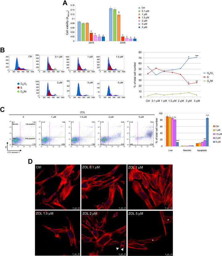
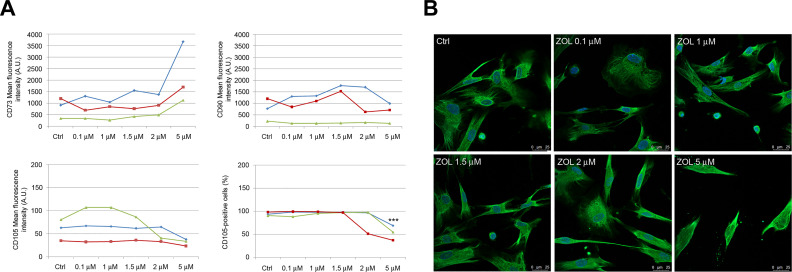
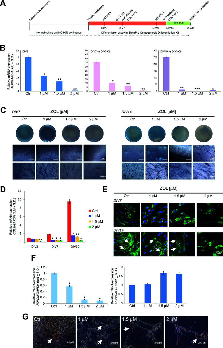
Bisphosphonates (BPs) are widely used to treat several metabolic and oncological diseases affecting the skeletal system. Despite BPs' well-known therapeutic potential, they also displayed important side effects, among which is BPs-related osteonecrosis of the jaw, by targeting osteoclast activities, osteoblast, and osteocyte behavior. The aim of this study is to evaluate the biological effects of zoledronic acid (ZOL) in an in vitro model of periodontal ligament stem cells (PDLSCs) by using an experimental setting that resembles the in vivo conditions. PDLSCs were treated with different concentrations of ZOL ranging from 0.1 to 5 μM. The effects of ZOL exposure were evaluated on cell viability via 3-[4,5-Dimethylthiaoly]-2,5-diphenyltetrazolium bromide (MTT), cell cycle analysis, apoptosis detection, and immunofluorescence. Quantitative real-time polymerase chain reaction (PCR), colorimetric detection of alkaline phosphatase activity, and Alizarin Red S staining were performed to investigate the osteogenic potential of PDLSCs exposed to ZOL. MTT analysis showed that the viability of PDLSCs exposed to ZOL concentration ≥1.5 μM for 3 and 6 days was significantly lower (P < 0.001) than that of untreated cells. The percentage of apoptotic cells was significantly higher in PDLSCs exposed for 4 days to ZOL at 2 μM (P < 0.01) and 5 μM (P < 0.001) when compared to the control. Moreover, ZOL treatment (3 days) accounted for alterations in cell cycle distribution, with an increase in the proportion of cells in G0/G1 phase and a reduction in the proportion of cells in S phase. Chronic exposure (longer than 7 days) of PDLSCs to ZOL accounted for the downregulation of ALP, RUNX2, and COL1 genes at all tested concentrations, which fit well with the reduced alkaline phosphatase activity reported after 7 and 14 days of treatment. Reduced Col1 deposition in the extracellular matrix was reported after 14 days of treatment. Increased calcium deposits were observed in treated cells when compared to the control cultures. In conclusion, chronic exposure to 1 μM ZOL induced significant reduction of osteogenic differentiation, while ZOL concentrations ≥1.5 μM are required to impair PDLSCs viability and induce apoptosis.
Keywords: cell cultures; human periodontal ligament stem cells; osteogenic differentiation; zoledronic acid.
Conflict of interest statement
Figures
Similar articles
-
Periostin promotes migration and osteogenic differentiation of human periodontal ligament mesenchymal stem cells via the Jun amino-terminal kinases (JNK) pathway under inflammatory conditions.Cell Prolif. 2017 Dec;50(6):e12369. doi: 10.1111/cpr.12369. Epub 2017 Aug 23. Cell Prolif. 2017. PMID: 28833827 Free PMC article.
-
Enamel matrix derivative enhances the proliferation and osteogenic differentiation of human periodontal ligament stem cells on the titanium implant surface.Organogenesis. 2017 Jul 3;13(3):103-113. doi: 10.1080/15476278.2017.1331196. Epub 2017 Jun 9. Organogenesis. 2017. PMID: 28598248 Free PMC article.
-
Simvastatin induces the osteogenic differentiation of human periodontal ligament stem cells.Fundam Clin Pharmacol. 2014 Oct;28(5):583-92. doi: 10.1111/fcp.12050. Epub 2013 Nov 8. Fundam Clin Pharmacol. 2014. PMID: 24112098
-
Lipopolysaccharide differentially affects the osteogenic differentiation of periodontal ligament stem cells and bone marrow mesenchymal stem cells through Toll-like receptor 4 mediated nuclear factor κB pathway.Stem Cell Res Ther. 2014 May 27;5(3):67. doi: 10.1186/scrt456. Stem Cell Res Ther. 2014. PMID: 24887697 Free PMC article.
-
Downregulation of lncRNA DANCR promotes osteogenic differentiation of periodontal ligament stem cells.BMC Dev Biol. 2020 Jan 14;20(1):2. doi: 10.1186/s12861-019-0206-8. BMC Dev Biol. 2020. PMID: 31931700 Free PMC article.
Cited by
-
A Review of Novel Strategies for Human Periodontal Ligament Stem Cell Ex Vivo Expansion: Are They an Evidence-Based Promise for Regenerative Periodontal Therapy?Int J Mol Sci. 2023 Apr 25;24(9):7798. doi: 10.3390/ijms24097798. Int J Mol Sci. 2023. PMID: 37175504 Free PMC article. Review.
-
Choosing the Right Partner for Medication Related Osteonecrosis of the Jaw: What Central European Dentists Know.Int J Environ Res Public Health. 2021 Apr 22;18(9):4466. doi: 10.3390/ijerph18094466. Int J Environ Res Public Health. 2021. PMID: 33922326 Free PMC article.
-
Osteonecrosis of the Jaws in Patients with Hereditary Thrombophilia/Hypofibrinolysis-From Pathophysiology to Therapeutic Implications.Int J Mol Sci. 2022 Jan 7;23(2):640. doi: 10.3390/ijms23020640. Int J Mol Sci. 2022. PMID: 35054824 Free PMC article. Review.
-
Medication-Related Osteonecrosis of the Jaw: A Critical Narrative Review.J Clin Med. 2021 Sep 24;10(19):4367. doi: 10.3390/jcm10194367. J Clin Med. 2021. PMID: 34640383 Free PMC article. Review.
-
Evaluation of in vitro osteoblast and osteoclast differentiation from stem cell: a systematic review of morphological assays and staining techniques.PeerJ. 2024 Jul 25;12:e17790. doi: 10.7717/peerj.17790. eCollection 2024. PeerJ. 2024. PMID: 39071131 Free PMC article.
References
-
- Van Acker HH, Anguille S, Willemen Y, Smits EL, Van Tendeloo VF. Bisphosphonates for cancer treatment: mechanisms of action and lessons from clinical trials. Pharmacol Ther. 2016;158:24–40. - PubMed
-
- Widler L, Jahnke W, Green JR. The chemistry of bisphosphonates: from antiscaling agents to clinical therapeutics. Anticancer Agents Med Chem. 2012;12(2):95–101. - PubMed
-
- Coxon FP, Thompson K, Rogers MJ. Recent advances in understanding the mechanism of action of bisphosphonates. Curr Opin Pharmacol. 2006;6(3):307–312. - PubMed
-
- Russell RGG, Watts NB, Ebetino FH, Rogers MJ. Mechanisms of action of bisphosphonates: similarities and differences and their potential influence on clinical efficacy. Osteoporos Int. 2008;19(6):733–759. - PubMed
-
- Dunford JE. Molecular targets of the nitrogen containing bisphosphonates: the molecular pharmacology of prenyl synthase inhibition. Curr Pharm Des. 2010;16(27):2961–2969. - PubMed
Publication types
MeSH terms
Substances
LinkOut - more resources
Full Text Sources
