. 2020 Nov 3;9(21):e017748.
doi: 10.1161/JAHA.120.017748. Epub 2020 Oct 22.

Potential Consequences of the Red Blood Cell Storage Lesion on Cardiac Electrophysiology

Affiliations

Potential Consequences of the Red Blood Cell Storage Lesion on Cardiac Electrophysiology

Marissa Reilly et al. J Am Heart Assoc. .

Abstract

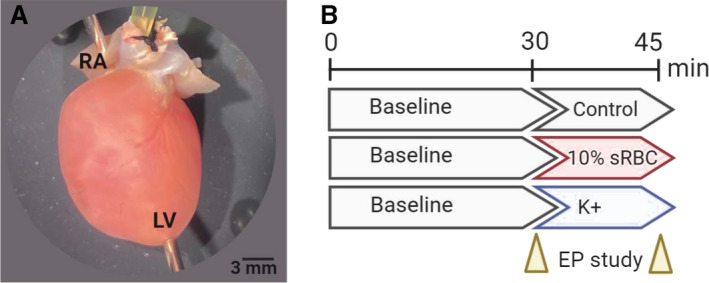
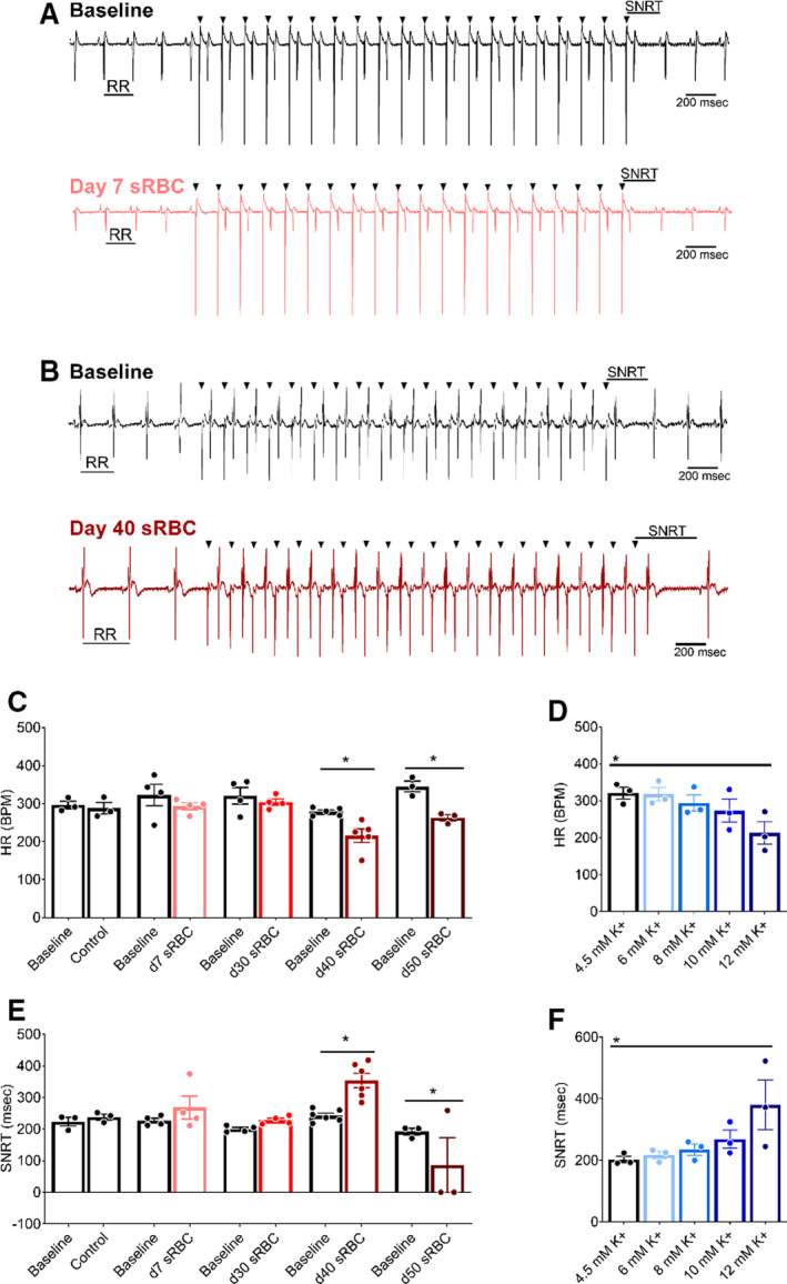
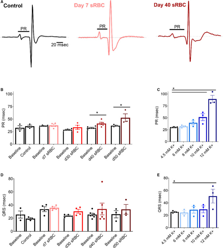
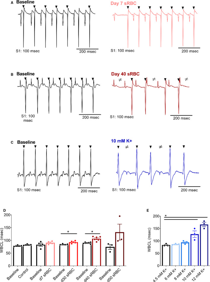
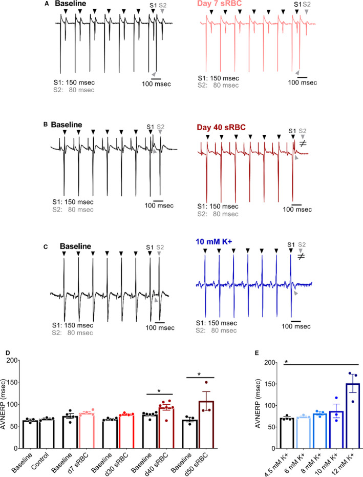
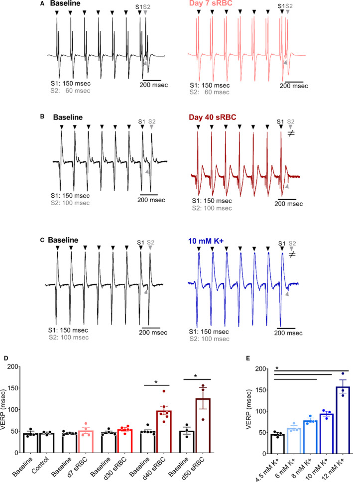
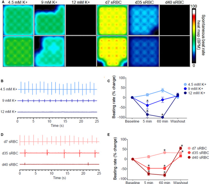
Background The red blood cell (RBC) storage lesion is a series of morphological, functional, and metabolic changes that RBCs undergo following collection, processing, and refrigerated storage for clinical use. Since the biochemical attributes of the RBC unit shifts with time, transfusion of older blood products may contribute to cardiac complications, including hyperkalemia and cardiac arrest. We measured the direct effect of storage age on cardiac electrophysiology and compared it with hyperkalemia, a prominent biomarker of storage lesion severity. Methods and Results Donor RBCs were processed using standard blood-banking techniques. The supernatant was collected from RBC units, 7 to 50 days after donor collection, for evaluation using Langendorff-heart preparations (rat) or human induced pluripotent stem cell-derived cardiomyocytes. Cardiac parameters remained stable following exposure to "fresh" supernatant from red blood cell units (day 7: 5.8±0.2 mM K+), but older blood products (day 40: 9.3±0.3 mM K+) caused bradycardia (baseline: 279±5 versus day 40: 216±18 beats per minute), delayed sinus node recovery (baseline: 243±8 versus day 40: 354±23 ms), and increased the effective refractory period of the atrioventricular node (baseline: 77±2 versus day 40: 93±7 ms) and ventricle (baseline: 50±3 versus day 40: 98±10 ms) in perfused hearts. Beating rate was also slowed in human induced pluripotent stem cell-derived cardiomyocytes after exposure to older supernatant from red blood cell units (-75±9%, day 40 versus control). Similar effects on automaticity and electrical conduction were observed with hyperkalemia (10-12 mM K+). Conclusions This is the first study to demonstrate that "older" blood products directly impact cardiac electrophysiology, using experimental models. These effects are likely caused by biochemical alterations in the supernatant from red blood cell units that occur over time, including, but not limited to hyperkalemia. Patients receiving large volume and/or rapid transfusions may be sensitive to these effects.

Keywords: cardiac electrophysiology; hyperkalemia; red blood cell storage lesion.


Conflict of interest statement

None.

Figures

Figure 1
Figure 1. Heart preparation and experimental timeline
A, Isolated, intact rodent heart with retrograde Langendorff‐perfusion via an aortic cannula. Pacing electrodes were attached to the RA and apex of the LV to perform an EP. B, Experimental timeline included 30‐minutes perfusion with KH media, containing 4.5 mM K+ (control), which commenced with an EP protocol. Thereafter, the media remained unchanged (control), supplemented with 10% sRBC, or supplemented with increasing potassium concentrations. The EP study was repeated again after 15–20 minutes, and results were compared with baseline. Timeline created using biorender.com. EP indicates electrophysiology study; KH, Krebs‐Henseleit media; LV, left ventricle; RA, right atria; and sRBC, supernatant from red blood cell units.
Figure 2
Figure 2. RBC storage age is associated with heart rate slowing and sinus node dysfunction
A, Biosignals recorded from isolated hearts perfused with media supplemented with 10% sRBC collected from a day 7 unit, or (B) day 40 unit. ECGs were recorded during sinus rhythm (RR interval highlighted), followed by train of atrial paces (black arrowheads denote pacing spikes). Each atrial pace results in a ventricular response. SNRT was measured from the last pacing spike to resumption of sinus rhythm. C, Stable heart rate following exposure to RBC units aged 7–30 d, but bradycardia observed with sRBC collected from units aged ≥ 40 d. D, HR slowing observed at highest potassium concentration tested (12 mM K+). E, Exposure to day 40 sRBC resulted in slowed sinus node recovery, or complete cessation of sinus function with day 50 sRBC. F, Increased SNRT also observed at highest potassium concentration tested (12 mM K+). Mean±SEM, *P<0.05, n=3–6. BPM indicates beats per minute; HR, heart rate; RBC, red blood cell; SNRT, sinus node recovery time; and sRBC, supernatant from red blood cell units.
Figure 3
Figure 3. RBC storage age is associated with slowed atrioventricular conduction
A, Pseudo‐ECGs recorded during sinus rhythm from isolated hearts perfused with control media (left), media supplemented with 10% sRBC collected from a day 7 unit (middle) or day 40 unit (right). PR interval time is denoted. (B,C) Atrioventricular conduction slows in the presence of day 40 and day 50 sRBC, or 10–12 mM K+. (D,E) Exposure to sRBC units had no measurable effect on ventricular depolarization time (QRS) during sinus rhythm. Mean±SEM, *P<0.05, n=3–6. RBC indicates red blood cell; and sRBC, supernatant from red blood cell units.
Figure 4
Figure 4. RBC storage age is associated with increased refractoriness of the AV node
A, Biosignals recorded with atrial pacing (S1‐S1) to measure WBCL in isolated hearts in the presence of day 7 sRBC, (B) day 40 sRBC, or (C) 10 mM K+. D, Slowed AV node conduction following exposure to sRBC from older units, but not “fresh” day 7 units. E, Slowed AV conduction following exposure to 10–12 mM K+. Arrowheads denote ventricular response to atrial pacing at S1 (black) pacing cycle length. ≠ denotes failed conduction. Mean±SEM, *P<0.05, n≥3. AV indicates atrioventricular; sRBC, supernatant from red blood cell units; and WBCL, Wenckebach cycle length.
Figure 5
Figure 5. RBC storage age is associated with an increased AV node effective refractory period
A, Biosignals recorded with atrial pacing (S1–S2) to pinpoint AVNERP in the presence of day 7 sRBC, (B) day 40 sRBC, or (C) 10 mM K+. D, AVNERP did not change after exposure to day 7–30 sRBC, but increased with day 40 and day 50 sRBC exposure. E, AVNERP increased with severe hyperkalemia. Arrowheads denote ventricular response to atrial pacing at S1 (black) or S2 (gray) pacing cycle length. ≠ denotes failed conduction. Mean±SEM, *P<0.05, n=3–6. AVNERP indicates atrioventricular node effective refractory period; RBC, red blood cell; sRBC, supernatant from red blood cell units.
Figure 6
Figure 6. RBC storage age is associated with increased ventricular refractoriness
A, Biosignals recorded with ventricular pacing (S1–S2) to pinpoint the VERP in isolated hearts perfused with media supplemented with 10% sRBC collected from a day 7 unit, (B) day 40 unit, or (C) 10 mM K+. D, Ventricular refractoriness was unchanged after exposure to day 7–30, but increased with day 40–50 sRBC and (E) media supplemented with 8–12 mM K+. Arrowheads denote ventricular response to pacing at S1 (black) or S2 (gray) pacing cycle length. ≠ denotes failed conduction. Mean±SD, *P<0.05, n=3–6. RBC indicates red blood cell; sRBC, supernatant from red blood cell units; and VERP, ventricular effective refractory period.
Figure 7
Figure 7. Reduced automaticity in hiPSC‐CM
A, Microelectrode array heat map shows 16‐electrode recordings from hiPSC‐CM treated with control media (4.5 mM K+), media with increasing potassium concentrations (9–12 mM K+), or 10% sRBC collected from RBC units aged 7–40 days. The heat map corresponds to the spontaneous beating rate. B, Biosignals recorded from hiPSC‐CM show a decline in beating rate with elevated potassium concentrations. C, Percent change in beating rate following treatment with elevated potassium concentrations, compared with baseline. D, Biosignals show a decline in the beating rate with “older” sRBC samples (day 35–40) but not “fresh” sRBC samples (day 7). E, Percent change in beating rate following sRBC treatment, compared with baseline. Mean±SEM, n=12–24, *Significantly different from baseline, P<0.05. BPM indicates beats per minute; hiPSC‐CM, human induced pluripotent stem cell–derived cardiomyocytes; RBC, red blood cell; and sRBC, supernatant from red blood cell units.

Similar articles

Cited by

References

    1. Jonas RA. Comprehensive Surgical Management of Congenital Heart Disease. London: Hodder Education Group; 2004.
    1. Punjabi PP, Taylor KM. The science and practice of cardiopulmonary bypass: From cross circulation to ECMO and SIRS. Glob Cardiol Sci Pract. 2013;2013:249–260. - PMC - PubMed
    1. Speiss BD. Transfusion and outcome in heart surgery. Ann Thorac Surg. 2002;74:986–987. - PubMed
    1. Chung K-W, Basavaraju SV, Mu Y, van Santen KL, Haass KA, Henry R, Berger J, Kuehnert MJ. Declining blood collection and utilization in the United States. Transfusion. 2016;56:2184. - PMC - PubMed
    1. Jones JM, Sapiano MRP, Savinkina AA, Haass KA, Baker ML, Henry RA, Berger JJ, Basavaraju SV. Slowing decline in blood collection and transfusion in the United States - 2017. Transfusion. 2020;60(Suppl 2):S1–S9. - PMC - PubMed

Publication types