. 2020 Nov;26(11):577-589.
doi: 10.1089/ten.TEC.2020.0217. Epub 2020 Nov 13.

Role of Curcuminoids and Tricalcium Phosphate Ceramic in Rat Spinal Fusion

Affiliations

Role of Curcuminoids and Tricalcium Phosphate Ceramic in Rat Spinal Fusion

Daniel A Ryan et al. Tissue Eng Part C Methods. 2020 Nov.

Abstract

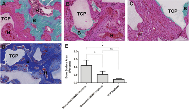
Despite considerable research effort, there is a significant need for safe agents that stimulate bone formation. Treatment of large or complex bone defects remains a challenge. Implantation of small molecule-induced human bone marrow-derived mesenchymal stromal cells (hBMSCs) on an appropriate tricalcium phosphate (TCP) scaffold offers a robust system for noninvasive therapy for spinal fusion. To show the efficacy of this approach, we identified a small molecule curcuminoid that when combined with TCP ceramic in the presence of hBMSCs selectively induced growth of bone cells: after 8- or 25-day incubations, alkaline phosphatase was elevated. Treatment of hBMSCs with curcuminoid 1 and TCP ceramic increased osteogenic target gene expression (i.e., Runx2, BMP2, Osteopontin, and Osteocalcin) over time. In the presence of curcuminoid 1 and TCP ceramic, osteogenesis of hBMSCs, including proliferation, differentiation, and mineralization, was observed. No evidence of chondrogenic or adipogenic potential using this protocol was observed. Transplantation of curcuminoid 1-treated hBMSC/TCP mixtures into the spine of immunodeficient rats showed that it achieved spinal fusion and provided greater stability of the spinal column than untreated hBMSC-TCP implants or TCP alone implants. On the basis of histological analysis, greater bone formation was associated with curcuminoid 1-treated hBMSC implants manifested as contiguous growth plates with extensive hematopoietic territories. Stimulation of hBMSCs by administration of small molecule curcuminoid 1 in the presence of TCP ceramic afforded an effective noninvasive strategy that increased spinal fusion repair and provided greater stability of the spinal column after 8 weeks in immunodeficient rats. Impact statement Bone defects only slowly regenerate themselves in humans. Current procedures to restore spinal defects are not always effective. Some have side effects. In this article, a new method to produce bone growth within 8 weeks in rats is presented. In the presence of tricalcium phosphate ceramic, curcuminoid-1 small molecule-stimulated human bone marrow-derived mesenchymal stromal cells showed robust bone cell growth in vitro. Transplantation of this mixture into the spine showed efficient spinal fusion in rats. The approach presented herein provides an efficient biocompatible scaffold for delivery of a potentially clinically useful system that could be applicable in patients.

Keywords: bone growth; bone marrow-derived mesenchymal stem cells; curcumins; tricalcium phosphate ceramic.


Conflict of interest statement

No competing financial interests exist.

Figures

FIG. 1.
FIG. 1.
Naturally-occuring and synthetic curcumins tested for ALP activity in hBMSC osteogenic differentiation. (A) Curcumins synthesized and tested for osteogenic differentiation. (B) Effect of curcumins 1–8 (500 nM) and curcumin extract (500 nM, weighted average concentration of curcumins) on ALP activity after an 8-day incubation with hBMSCs. Data are mean fold-change ALP activity ± SD (n = 3) normalized to turmeric extract. *p < 0.05, ** p < 0.01. (C) Shows compounds 1 and 2 selected for advanced studies. ALP, alkaline phosphatase; hBMSC, human bone marrow-derived mesenchymal stromal cell.
FIG. 2.
FIG. 2.
Effect of 1 or 2 on alkaline phosphatase activity in hBMSCs incubated in the presence or absence of TCP granules. hBMSCs were incubated with compounds 1 or 2 (500 nM), TCP granules alone (5 mg/mL), or compounds 1 or 2 (500 nM) in the presence of TCP granules. ALP activity was measured after 8 and 25 days, and results were normalized to vehicle (i.e., DMSO)-treated cells on day 8. (A) ALP activity from hBMSCs treated with BisVal-BDC 1 in the presence or absence of TCP granules. (B) ALP activity from hBMSCs treated with BisVal-CyBDC 2 in the presence or absence of TCP granules. Data are mean ± SD (n = 3). *p < 0.05, **p < 0.01, ***p < 0.001. TCP, tricalcium phosphate.
FIG. 3.
FIG. 3.
Effect of 1 or 2 on osteogenic gene expression in hBMSCs incubated in the presence or absence of TCP granules. hBMSCs were incubated with compounds 1 or 2 (500 nM) and TCP granules (5 mg/mL). Runx2, BMP2, OPN, and ON gene expression was determined by qPCR after 8 days. Relative gene expression levels were normalized to vehicle (i.e., DMSO)-treated cells and expressed as fold-change in expression levels. Data were mean with error bars for SEM (n = 4). *p < 0.05, **p < 0.01, ***p < 0.001.
FIG. 4.
FIG. 4.
Effect of 1 or 2 on Ca2+ Mineral Deposition in hBMSCs in the presence or absence of TCP granules. hBMSCs stained with Alizarin Red S after incubation with 1 or 2 (500 nM) in the presence or absence of TCP granules (5 mg/mL) for 25 days. Staining intensity in the images was quantified by densitometry and normalized to vehicle (i.e., DMSO)-treated cells. (A) Vehicle (i.e., DMSO)-treated cells. (B) BisVal-BDC 1. (C) BisVal-CyBDC 2. (D) TCP granules. (E) Compound 1 in the presence of TCP granules. (F) Compound 2 in the presence of TCP granules. (G) Densitometry analysis of (A–C) staining. (H) Densitometry analysis of (D–F) staining. Data are mean ± SD (n = 6). ***p < 0.001. Color images are available online.
FIG. 5.
FIG. 5.
Effect of compound 1 on induction of hBMSC Wnt gene expression in the presence or absence of TCP granules: Mechanistic studies. hBMSCs were incubated with compound 1 (500 nM) in the presence or absence of TCP granules (5 mg/mL), and expression of Wnt inducible target genes was determined after 8 days in culture by quantifying mRNA with qPCR. Gene expression was normalized to vehicle (i.e., DMSO)-treated cells. (A) Runx2, (B) BMP2, (C) Axin 2, (D) Wnt3a, (E) Wnt5a, (F) Sox9. Data are mean ± SD (n = 3).
FIG. 6.
FIG. 6.
TCP-collagen scaffold used for posterolateral spinal fusion. (A) Shaped TCP-collagen scaffolds were prepared with dimensions (millimeters) to fit the void space between L4 and L5 transverse processes using a μCT image of an adult rat lumbar spine as reference. (B) Representative 6-week radiograph of a rat that received bilateral BisVal-BDC (1)-stimulated hBMSC implants on the shaped TCP-collagen scaffold. (C) Confocal fluorescent microscopy image of compound 1-stimulated hBMSCs that were stained with Live-Dead Stain 6 h after adsorption of cells to the scaffold. Image taken at 10 × magnification. Color images are available online.
FIG. 7.
FIG. 7.
Effect of implants on L4-L5 spinal fusion in spinal segments. (A) Removed rat spines containing implants were subjected to constant bending forces and visually ranked by blinded trained observers. Implants were ranked as fused (bridging, secure fusion mass), partially fused (bridging fusion mass that showed limited motion), or nonfused (not bridging or unsecure implants). Categorical rankings were expressed as a percent of the total number of animals in each group. (B) Representative μCT image of L4-L5 vertebral disc height measurements used for determining stability of the lumbar spinal segments under a controlled lateral displacement. The distance of the left length and right length of the L4-L5 disc height was determined by μCT at the neutral position and then under a constant left (+10 mm) and right (−10 mm) lateral displacement. Deviation in disc height was calculated from the averaged differences in disc height from the neutral position that represented the absolute sum of contraction and elongation deviations. (C) Quantification of absolute deviation in disc heights (μm) with constant lateral displacement shown in a Box and Whisker Plot with 75th percentile (box), minimum and maximum values (whiskers), and median (line) with n = 7/group. * p < 0.05, ns = not significant (i.e., p > 0.1). Compound 1-treated hBMSC-TCP scaffold implants (stimulated hBMSC implants), nontreated hBMSC TCP scaffold implants (untreated hBMSC implants), and TCP scaffold implants were described above.
FIG. 8.
FIG. 8.
Histology of L4-L5 fusion mass from implants (A–D). Representative histological sections labeled: B, bone; TCP, tricalcium phosphate; H, hematopoietic territories. Goldner's Trichrome stained sections of (A). Compound 1-treated hBMSC-TCP scaffold implants (20 × ), (B). Untreated hBMSC-TCP scaffold implants (20 × ) and (C). TCP scaffold alone implants (i.e., no cells) (20 × ). (D). Masson's Trichrome stained decalcified section of compound 1-stimulated hBMSC-TCP scaffold implants (10 × ). Scale bar = 100 μm in images. (E). Quantitative densitometry analysis of de novo bone surface area in nondecalcified stained sections expressed as bone surface area as a percent of the total histological section. Data are the averaged values showing standard error of the mean (n = 7/group). ^, almost significant; ns, not significant. p < 0.10, *p < 0.05. Color images are available online.

Similar articles

References

    1. Betz R.R. Limitations of autograft and allograft: new synthetic solutions. Orthopedics 25, s561, 2002 - PubMed
    1. Pape H.C., Evans A., and Kobbe P.. Autologous bone graft: properties and techniques. J Orthop Trauma 24, S35, 2010 - PubMed
    1. Holtzclaw D., Toscano N., Eisenlohr L., and Callan D.. The safety of bone allografts used in dentistry: a review. J Am Dent Assoc 139, 1192, 2008 - PubMed
    1. Yuan H., Fernandes H., Habibovic P., et al. . Osteoinductive ceramics as a synthetic alternative to autologous bone grafting. Proc Natl Acad Sci U S A 107, 13614, 2010 - PMC - PubMed
    1. Bianco P., Cao X., Frenette P.S., et al. . The meaning, the sense and the significance: translating the science of mesenchymal stem cells into medicine. Nat Med 19, 35, 2013 - PMC - PubMed

Publication types

MeSH terms