. 2020 Oct 21;39(1):223.
doi: 10.1186/s13046-020-01735-3.

Activation of the KDM5A/miRNA-495/YTHDF2/m6A-MOB3B axis facilitates prostate cancer progression

Affiliations

Activation of the KDM5A/miRNA-495/YTHDF2/m6A-MOB3B axis facilitates prostate cancer progression

Chen Du et al. J Exp Clin Cancer Res. .

Abstract

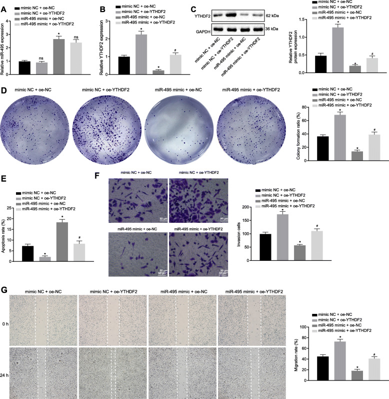
Background: Accumulating evidence supports that lysine-specific demethylase 5 (KDM5) family members act as oncogenic drivers. This study was performed to elucidate the potential effects of KDM5A on prostate cancer (PCa) progression via the miR-495/YTHDF2/m6A-MOB3B axis.

Methods: The expression of KDM5A, miR-495, YTHDF2 and MOB3B was validated in human PCa tissues and cell lines. Ectopic expression and knockdown experiments were developed in PCa cells to evaluate their effects on PCa cell proliferation, migration, invasion and apoptosis. Mechanistic insights into the interaction among KDM5A, miR-495, YTHDF2 and MOB3B were obtained after dual luciferase reporter, ChIP, and PAR-CLIP assays. Me-RIP assay was used to determine m6A modification level of MOB3B mRNA in PCa cells. Mouse xenograft models of PCa cells were also established to monitor the tumor growth.

Results: KDM5A was highly expressed in human PCa tissues and cell lines. Upregulated KDM5A stimulated PCa cell proliferation, migration and invasion, but reduced cell apoptosis. Mechanistically, KDM5A, as a H3K4me3 demethylase, bound to the miR-495 promoter, which led to inhibition of its transcription and expression. As a target of miR-495, YTHDF2 could inhibit MOB3B expression by recognizing m6A modification of MOB3B mRNA and inducing mRNA degradation. Furthermore, KDM5A was found to downregulate MOB3B expression, consequently augmenting PCa cell proliferation, migration and invasion in vitro and promoting tumor growth in vivo via the miR-495/YTHDF2 axis.

Conclusion: In summary, our study highlights the potential of histone demethylase KDM5A activity in enhancing PCa progression, and suggests KDM5A as a promising target for PCa treatment.

Keywords: Invasion; KDM5A; MOB3B; Migration; Prostate cancer; YTHDF2; m6A modification; microRNA-145.


Conflict of interest statement

The author declares no competing interest exists.

Figures

Fig. 1
Fig. 1
KDM5A is highly expressed in the PCa tissues and cells, and it predicts a poor prognosis. a, KDM5A mRNA expression in PCa and adjacent normal tissues of patients detected by RT-qPCR. N = 78 for PCa patients. b, Western blot analysis and immunohistochemistry of KDM5A protein in PCa and adjacent normal tissues of patients. N = 78 for PCa patients. c, Corelation of KDM5A expression with DFS and OS analyzed using the Kaplan-Meier method. d, KDM5A mRNA expression in RWPE-1, LNCaP, C42, DU145 and PC3 cells detected by RT-qPCR. e and f, Western blot analysis and quantification of KDM5A protein in RWPE-1, LNCaP, C42, DU145 and PC3 cells. * p < 0.05 vs. RWPE-1 cells. Data in panel A and B were analyzed by paired t-test and those in panel C-E were analyzed by one-way ANOVA with Tukey’s post hoc test
Fig. 2
Fig. 2
KDM5A stimulates PCa cell proliferation, migration and invasion while reducing cell apoptosis. a and b, The silencing efficiency of KDM5A in PC3 cells detected by RT-qPCR and Western blot analysis. c, KDM5A mRNA expression in oe-KDM5A-transfected cells detected by RT-qPCR. d, Western blots of KDM5A protein in oe-KDM5A-transfected cells. e, PC3 cell proliferation following oe- or sh-KDM5A transfection measured by colony formation assay. f, PC3 cell apoptosis following oe- or sh-KDM5A transfection measured by flow cytometry. g, PC3 cell invasion following oe- or sh-KDM5A transfection measured by Transwell assay. Scale bar = 50 μm. h, PC3 cell migration following oe- or sh-KDM5A transfection measured by Scratch test. * p < 0.05 vs. cells transfected with sh-NC or oe-NC. Data in panel A, B and E-H were analyzed by one-way ANOVA with Tukey’s post hoc test, and data in panel C and D were analyzed by unpaired t-test
Fig. 3
Fig. 3
KDM5A potentiates PCa cell proliferation, invasion and migration while suppressing cell apoptosis by inhibiting miR-495. a, miR-495 expression in cancer and adjacent normal tissues of 78 patients detected by RT-qPCR. b, Correlation between miR-495 and KDM5A in cancer and adjacent normal tissues of 78 patients. c, miR-495 expression in RWPE-1, LNCaP, C42, DU145 and PC3 cells detected by RT-qPCR. d, The binding of KDM5A to the promoter of miR-495 confirmed by dual luciferase reporter assay. e, miR-495 expression in oe-KDM5A-transfected PC3 cells. f, The binding of KDM5A to the promoter of miR-495 verified by ChIP assay. g, Western blots of H3K4me3 in the antibody-transcription factor complex pulled down by KDM5A antibody. h, KDM5A mRNA expression in PC3 cells following varied transfection detected by RT-qPCR. i, Western blot analysis of KDM5A protein in PC3 cells following varied transfection. j, miR-495 expression in PC3 cells following varied transfection detected by RT-qPCR. k, PC3 cell proliferation following varied transfection measured by Colony formation assay. l, PC3 cell apoptosis following varied transfection measured by flow cytometry. m, PC3 cell invasion following varied transfection measured by Transwell assay. Scale bar = 50 μm. n, PC3 cell migration following varied transfection measured by Scratch test. * p < 0.05 vs. adjacent normal tissues, RWPE-1 cells, or cells transfected with oe-NC or oe-NC + mimic NC. # p < 0.05 vs. cells transfected with oe-KDM5A + mimic NC. ns p > 0.05 vs. cells transfected with oe-NC + miR-495 mimic or oe-KDM5A + mimic NC. Data in panel A were analyzed by paired t-test, in panel D-G by unpaired t-test and those in panel H-J were analyzed by one-way ANOVA with Tukey’s post hoc test
Fig. 4
Fig. 4
miR-495 targets YTHDF2 and suppresses its expression. a, Predicted binding sites between miR-495 and YTHDF2 via bioinformatics analysis. b, The mRNA expression of YTHDF2 in PCa and adjacent normal tissues detected by RT-qPCR. c, Correlation between miR-495 and YTHDF2 in PCa tissues and adjacent normal tissues of the patients. d, Western blot analysis of YTHDF2 protein in cancer and adjacent normal tissues of the patients. e, The binding of miR-495 to the mRNA of YTHDF2 confirmed by dual luciferase reporter assay. f and i, miR-495 expression in miR-495 mimic- or miR-495 inhibitor-transfected cells detected by RT-qPCR. G and j, YTHDF2 mRNA expression in miR-495 mimic- or miR-495 inhibitor-transfected cells detected by RT-qPCR. h and k, Western blot analysis and quantification of YTHDF2 protein in miR-495 mimic- or miR-495 inhibitor-transfected cells. * p < 0.05 vs. adjacent normal tissues, or cells transfected with mimic NC or inhibitor NC. Data in panel B and D were analyzed using paired t-test, and those in panel E-K were analyzed using unpaired t-test
Fig. 5
Fig. 5
Upregulated YTHDF2 promotes the inhibited proliferation, invasion and migration while diminishing the augmented cell apoptosis evoked by miR-495 in PCa cells. a, miR-495 expression in cells following varied transfection detected by RT-qPCR. b, The mRNA expression of YTHDF2 in cells following varied transfection detected by RT-qPCR. c, Western blots of YTHDF2 protein in cells following varied transfection. d, PC3 cell proliferation following varied transfection measured by Colony formation assay. e, PC3 cell apoptosis following varied transfection measured by flow cytometry. f, PC3 cell invasion following varied transfection measured by Transwell assay. Scale bar = 50 μm. g, PC3 cell migration following varied transfection measured by Scratch test. * p < 0.05 vs. cells transfected with mimic NC + oe-NC. # p < 0.05 vs. cells transfected with miR-495 mimic + oe-NC. ns p > 0.05 vs. cells transfected with mimic NC + oe-NC or miR-495 mimic + oe-NC. Data in panel A-G were analyzed by one-way ANOVA with Tukey’s post hoc test
Fig. 6
Fig. 6
YTHDF2 recognizes the m6A modification of MOB3B mRNA, evokes mRNA degradation and suppresses MOB3B expression. a, MOB3B mRNA expression in cancer and adjacent normal tissues of 75 patients detected by RT-qPCR. b, Correlation between MOB3B and YTHDF2 in cancer and adjacent normal tissues of 75 patients. c, Western blot analysis of MOB3B protein in cancer and adjacent normal tissues of 75 patients. d, m6A modification level of MOB3B in cancer and adjacent normal tissues assessed using Me-RIP. e, The binding of YTHDF2 to the MOB3B mRNA in cancer and adjacent normal tissues confirmed by PAR-CLIP assay. f, MOB3B mRNA expression in oe-YTHDF2-transfected PC3 cells detected by RT-qPCR. g, Western blot analysis of MOB3B protein in oe-YTHDF2-transfected PC3 cells. h, m6A modification in MOB3B in oe-YTHDF2-transfected PC3 cells assessed using Me-RIP. i, The binding of YTHDF2 to the MOB3B mRNA in oe-YTHDF2-transfected PC3 cells confirmed by PAR-CLIP assay. * p < 0.05 vs. adjacent normal tissues or cells transfected with oe-NC. Data in panel A and C-E were analyzed by paired t-test and those in panel F-I were analyzed by unpaired t-test
Fig. 7
Fig. 7
KDM5A represses MOB3B and then elicits proliferation, migration and invasion of PCa cells while reducing cell apoptosis via the miR-495/YTHDF2 axis. a, Correlation analysis of KDM5A and MOB3B in PCa tissues. b, The mRNA expression of KDM5A, YTHDF2 and MOB3B in PC3 cells with varied transfection detected by RT-qPCR. c, Western blots of KDM5A, YTHDF2 and MOB3B proteins in cells with varied transfection. d, miR-495 expression in cells following varied transfection detected by RT-qPCR. e, The m6A modification level of MOB3B in cells following varied transfection assessed using Me-RIP. f, PC3 cell proliferation following varied transfection measured by Colony formation assay. g, PC3 cell apoptosis following varied transfection measured by flow cytometry. h, PC3 cell invasion following varied transfection measured by Transwell assay. Scale bar = 50 μm. i, PC3 cell migration following varied transfection measured by Scratch test (Scale bar = 200 μm). * p < 0.05 vs. cells transfected with oe-NC. # p < 0.05 or ns p > 0.05 vs. cells transfected with oe-KDM5A + oe-NC. Data in panel a-h were analyzed by one-way ANOVA with Tukey’s post hoc test
Fig. 8
Fig. 8
KDM5A promotes tumorigenicity by downregulating MOB3B expression in nude mice via the miR-495/YTHDF2 axis. a-c, Xenograft tumors and quantitative analysis of tumor size and volume after oe-KDM5A or oe-KDM5A + oe-MOB3B treatment. Scale bar = 10 mm in panel A. d, mRNA expression of KDM5A, YTHDF2 and MOB3B in mice following oe-KDM5A or oe-KDM5A + oe-MOB3B treatment detected by RT-qPCR. e, Western blots of KDM5A, YTHDF2 and MOB3B proteins in mice following oe-KDM5A or oe-KDM5A + oe-MOB3B treatment. f, miR-495 expression in mice following oe-KDM5A or oe-KDM5A + oe-MOB3B treatment detected by RT-qPCR. g, m6A modification level of MOB3B in mice following oe-KDM5A or oe-KDM5A + oe-MOB3B treatment assessed using Me-RIP. * p < 0.05 vs. cells transfected with oe-NC. # p < 0.05 or ns p > 0.05 vs. cells transfected with oe-KDM5A + oe-NC. Data in panel A and C-G were analyzed by one-way ANOVA with Tukey’s post hoc test, and those in panel B were analyzed by repeated measures ANOVA with Bonferroni’s post hoc test

References

    1. Pernar CH, Ebot EM, Wilson KM, Mucci LA. The epidemiology of prostate cancer. Cold Spring Harb Perspect Med. 2018;8. - PMC - PubMed
    1. Bashir MN. Epidemiology of prostate cancer. Asian Pac J Cancer Prev. 2015;16:5137–5141. doi: 10.7314/APJCP.2015.16.13.5137. - DOI - PubMed
    1. Litwin MS, Tan HJ. The diagnosis and treatment of prostate cancer: a review. JAMA. 2017;317:2532–2542. doi: 10.1001/jama.2017.7248. - DOI - PubMed
    1. Hou J, Wu J, Dombkowski A, Zhang K, Holowatyj A, Boerner JL, et al. Genomic amplification and a role in drug-resistance for the kdm5a histone demethylase in breast cancer. Am J Transl Res. 2012;4:247–256. - PMC - PubMed
    1. Gale M, Sayegh J, Cao J, Norcia M, Gareiss P, Hoyer D, et al. Screen-identified selective inhibitor of lysine demethylase 5a blocks cancer cell growth and drug resistance. Oncotarget. 2016;7:39931–39944. doi: 10.18632/oncotarget.9539. - DOI - PMC - PubMed

MeSH terms