Review
. 2020 Sep:93:102908.
doi: 10.1016/j.dnarep.2020.102908.

Human DNA ligases in replication and repair

Affiliations
Review

Human DNA ligases in replication and repair

Annahita Sallmyr et al. DNA Repair (Amst). 2020 Sep.

Abstract

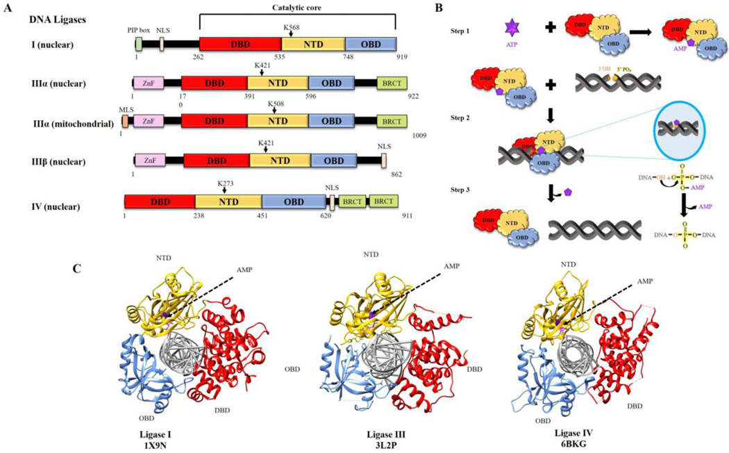
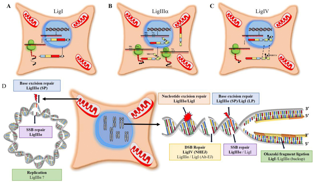
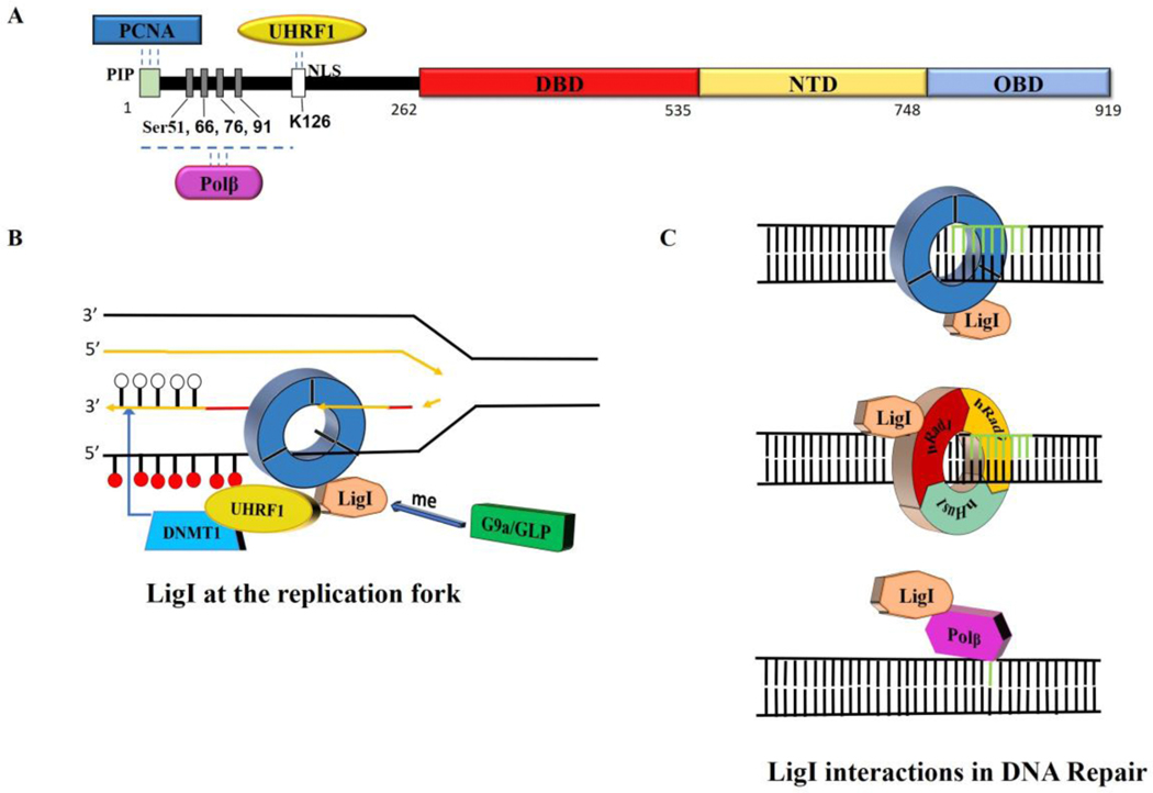
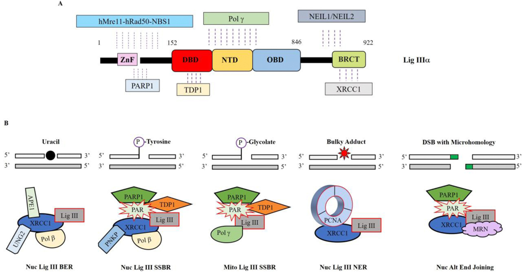
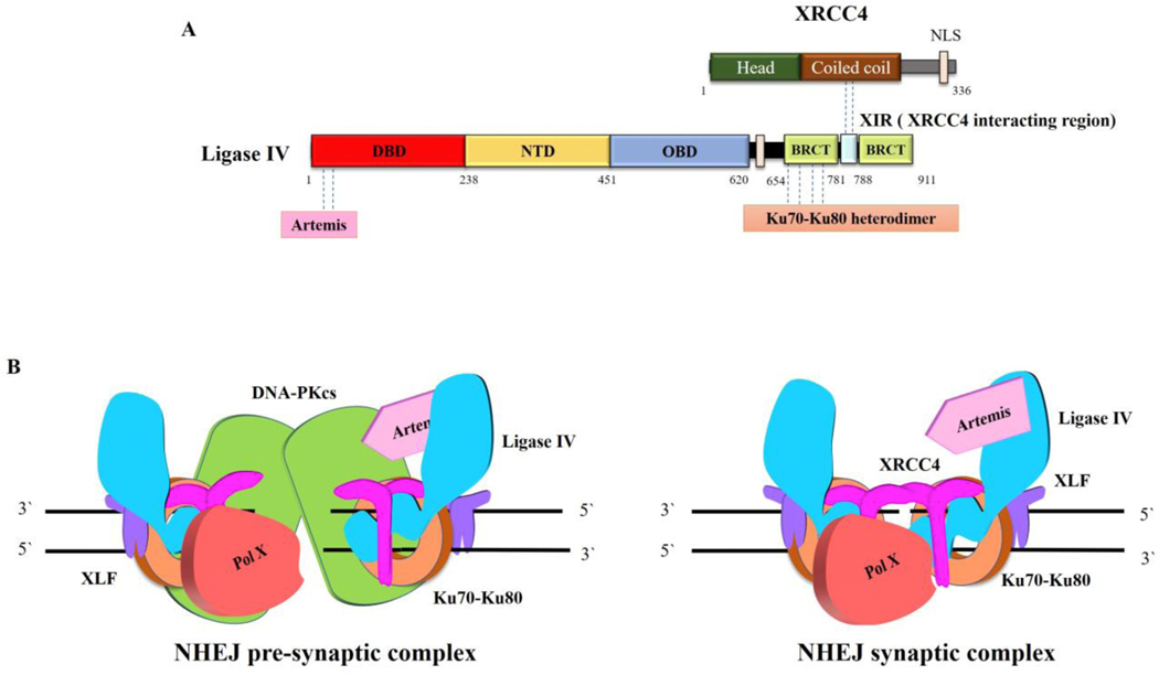
To ensure genome integrity, the joining of breaks in the phosphodiester backbone of duplex DNA is required during DNA replication and to complete the repair of almost all types of DNA damage. In human cells, this task is accomplished by DNA ligases encoded by three genes, LIG1, LIG3 and LIG4. Mutations in LIG1 and LIG4 have been identified as the causative factor in two inherited immunodeficiency syndromes. Moreover, there is emerging evidence that DNA ligases may be good targets for the development of novel anti-cancer agents. In this graphical review, we provide an overview of the roles of the DNA ligases encoded by the three human LIG genes in DNA replication and repair.

Keywords: DNA joining; Okazaki fragments; alternative end joining; base excision repair; non-homologous end joining; nucleotide excision repair; single strand break reapir.


Figures

Fig. 1.
Fig. 1.. DNA ligases encoded by the three human LIG genes; ligation reaction mechanism and structure of catalytic regions complexed with nicked DNA.
A. Human DNA ligases (I, IIII and IV) share a conserved catalytic region comprised of DNA binding domain (DBD, red), nucleotidyl transferase domain containing the lysine residues that form the phosphoramidite bond with AMP (NTD, yellow) and oligonucleotide binding or OB-fold containing domain (OBD, blue). PCNA interacting peptide box (PIP box, light green), Zinc finger DNA binding domain (Znf, pink), BRCA1 C terminus domain (BRCT, lime green), mitochondrial localization signal (MLS, brown) and nuclear localization signal (NLS, white) are indicated. B. Schematic diagram of ligation reaction; step 1: Formation of ligase-adenylate using ATP. Step 2: Ligase-adenylate engages nicked DNA and transfers AMP to 5’phospahe termini, forming DNA-adenylate. Step 3: Non-adenylated Ligase interacts with DNA-adenylate to form phosphodiester bond. C. Crystal structures of DNA ligase catalytic regions with nicked DNA. Images were generated using the software Chimera (http://www.cgl.ucsf.edu/chimera) (23) and structures from RCSB PDB.
Fig. 2.
Fig. 2.. Subcellular localization and functions of human DNA ligases.
A. After translation of LigI mRNA in the cytoplasm, LigI polypeptide is directed to the nucleus via an N-terminal NLS (white). B. Two different AUG codons in LigIIIα mRNA are used to initiate translation. Translation initiation at the first AUG generates a LigIIIα polypeptide with an N-terminal MLS (brown) that enables LigIIIIα but not XRCC1 to enter mitochondria. An internal AUG, which is preferentially utilized to initiate translation by the ribosome, generates a LigIIIα polypeptide lacking the MLS. This polypeptide, which also lacks a NLS, forms a complex with XRCC1 and is targeted to the nucleus via the XRCC1 NLS (white). C. Both LigIV and its partner protein XRCC4 have NLSs (white). There are contradictory reports as to whether LigIV is responsible for the nuclear localization of XRCC4 or vice versa. D. The participation of human DNA ligases in nuclear (right panel) and mitochondrial (left panel) DNA replication and repair is indicated with the major enzyme are bolded. While the original proposed mechanism of mitochondrial DNA replication does not require multiple ligation events, other mechanisms have been proposed.
Fig. 3.
Fig. 3.. LigI protein partners.
A. Linear schematic of LigI polypeptide showing the N-terminal binding sites for PCNA, Polβ and UHRF1 as well phosphorylated serine residues and an acetylated lysine residue. B. Interaction of LigI with PCNA is critical for the recruitment of LIgI to replication foci and the efficient joining of Okazaki fragments. Methylation of LigI by G9a is required for an interaction with UHRF1/DNMT1 that enables DNMT1 to methylate the newly synthesized DNA at the replication fork (white circles unmethylated cytosine; red circles, methylated cytosine. C. Upper panel, interaction of LigI with PCNA is critical for the repair of non-bulky lesions by the long patch BER pathway and the S-phase specific subpathway of NER. Middle panel, interaction of LigI with the hRad9-hRad1-hHus1 clamp stimulates DNA joining during long patch BER and may also occur in NER. Lower panel, LigI interacts with Polβ within a BER complex, suggesting a role for LigI in short patch BER.
Fig. 4.
Fig. 4.. LigIIIα protein partners.
A. Linear schematic of LigIIIα polypeptide showing the binding sites for hMre11-Rad50-Nbs1, PARP1, TDP1, Pol γ, NEIL1/NEIL2 and XRCC1. B. LigIIIα/XRCC1 complexes involved in nuclear short patch base excision repair (BER), short patch single strand break repair (SSBR), nucleotide excision repair subpathway operating throughout the cell cycle (NER) and alternative (ALT) end-joining. In these complexes, XRCC1 acts as a flexible scaffold that links end-processing enzymes with the LigIII catalytic region. While similar complexes lacking XRCC1, such as the predicted mitochondrial SSBR complex, are likely to be involved in mitochondrial DNA repair, DNA metabolism in this organelle is less well characterized than in the nucleus.
Fig. 5.
Fig. 5.. LigIV protein partners.
A. Linear schematic of LigIV polypeptide showing the binding sites for Artemis, Ku70/Ku80 and XRCC4. B. The Ku heterodimer ring (orange) threads onto DSB ends and then recruits other core NHEJ components, DNA PKcs (green), Ligase IV (blue)/XRCC4 (magenta) and XLF (purple) to form a pre-synaptic complex in which the DNA ends are brought together but no aligned (left panel). Other factors, such as Artemis (cyan) and DNA polymerase family X members μ or λ (red), that may be needed to modify ends prior to ligation are also recruited. In the pre-synaptic complex, the DNA ends are not accessible for processing and ligation. There is predicted to be a transition involving autophosphorylation of DNA PKcs that results in a conformational change in DNA PKcs and/or the its departure from the complex, resulting in the formation of the synaptic complex (right panel), in which the DNA ends are aligned and accessible to interactions with Ligase IV and end processing enzymes.

Similar articles

Cited by

References

    1. Ellenberger T. and Tomkinson AE (2008) Eukaryotic DNA ligases: structural and functional insights. Annu Rev Biochem, 77, 313–338. - PMC - PubMed
    1. Tomkinson AE and Sallmyr A. (2013) Structure and function of the DNA ligases encoded by the mammalian LIG3 gene. Gene, 531, 150–157. - PMC - PubMed
    1. Ho CK, Wang LK, Lima CD and Shuman S. (2004) Structure and mechanism of RNA ligase. Structure (Camb), 12, 327–339. - PubMed
    1. Shuman S. and Schwer B. (1995) RNA capping enzyme and DNA ligase: a superfamily of covalent nucleotidyl transferases. Mol Microbiol, 17, 405–410. - PubMed
    1. Tomkinson AE, Naila T. and Khattri Bhandari S. (2020) Altered DNA ligase activity in human disease. Mutagenesis, 35, 51–60. - PMC - PubMed

Publication types