Comparative Study
. 2021 Jan 15;27(2):554-565.
doi: 10.1158/1078-0432.CCR-20-1422. Epub 2020 Oct 21.

Altered Gemcitabine and Nab-paclitaxel Scheduling Improves Therapeutic Efficacy Compared with Standard Concurrent Treatment in Preclinical Models of Pancreatic Cancer

Affiliations
Comparative Study

Altered Gemcitabine and Nab-paclitaxel Scheduling Improves Therapeutic Efficacy Compared with Standard Concurrent Treatment in Preclinical Models of Pancreatic Cancer

Adam R Wolfe et al. Clin Cancer Res. .

Abstract

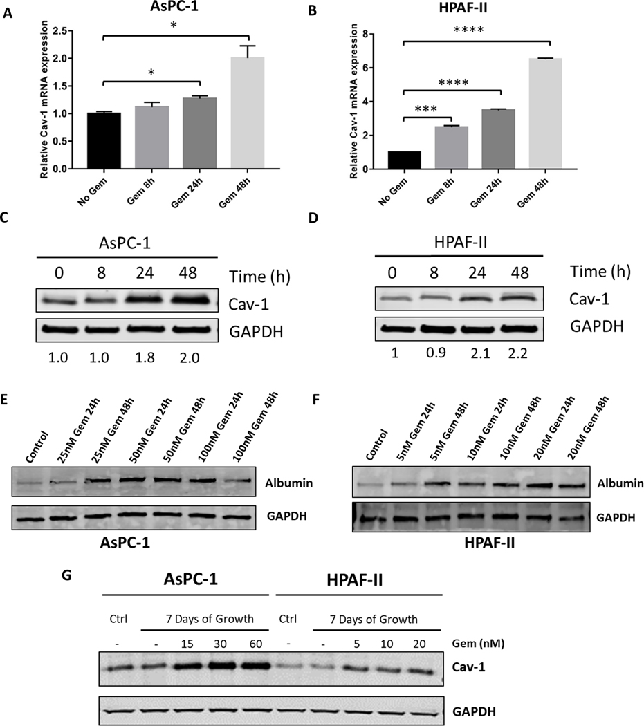
Purpose: Concurrent gemcitabine and nab-paclitaxel treatment is one of the preferred chemotherapy regimens for metastatic and locally advanced pancreatic ductal adenocarcinoma (PDAC). Previous studies demonstrate that caveolin-1 (Cav-1) expression is critical for nab-paclitaxel uptake into tumors and correlates with response. Gemcitabine increases nab-paclitaxel uptake by increasing Cav-1 expression. Thus, we hypothesized that pretreatment with gemcitabine would further enhance the sensitivity of PDAC to nab-paclitaxel by increasing Cav-1 expression and nab-paclitaxel uptake.

Experimental design: We investigated the sensitivity of different gemcitabine and nab-paclitaxel treatment regimens in a panel of PDAC cell lines and orthotopic xenograft models. The sensitivity of different treatment regimens was compared with the standard concurrent treatment.

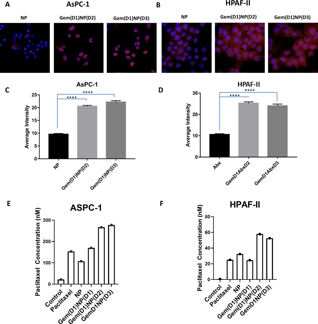
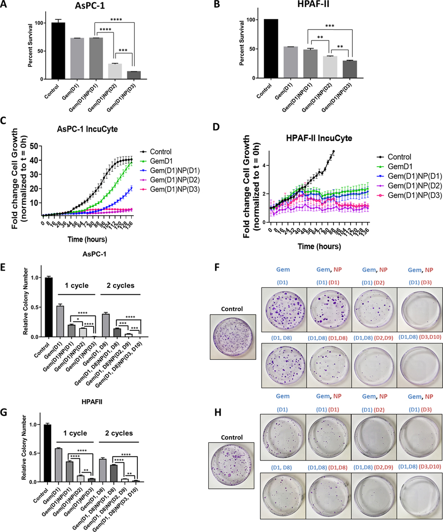
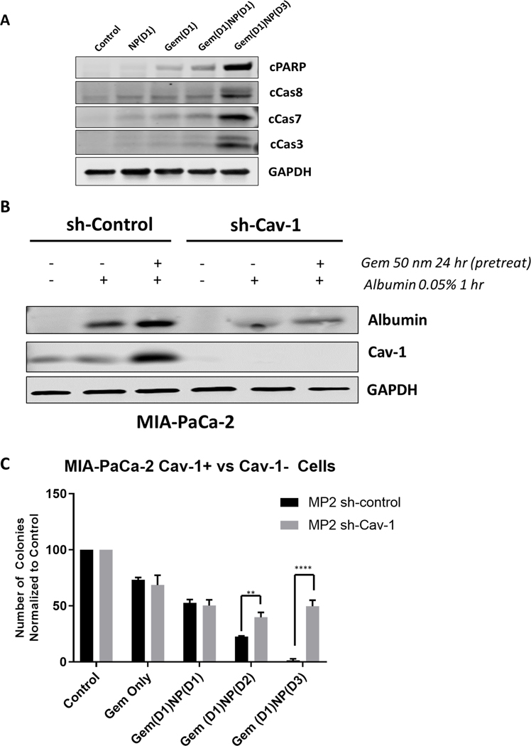
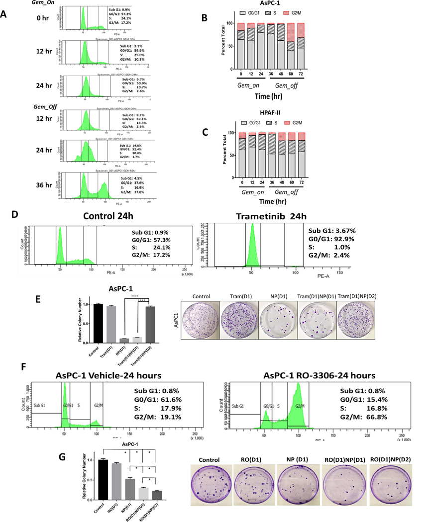
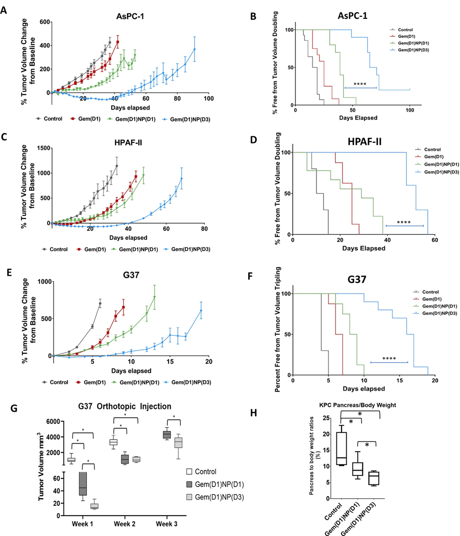
Results: Pretreatment with gemcitabine before nab-paclitaxel increased Cav-1 and albumin uptake and significantly decreased proliferation and clonogenicity compared with concurrent treatment, which correlated with increased levels of apoptosis. Cav-1 silencing reduced the uptake of albumin, and therapeutic advantage was observed when cells were pretreated with gemcitabine prior to nab-paclitaxel. In addition, we observed that pretreatment with gemcitabine resulted in partial synchronization of cells in the G2-M-phase at the time of nab-paclitaxel treatment, providing another mechanism for the benefit of altered scheduling. In heterotopic and orthotopic xenograft models, the altered schedule of gemcitabine prior to nab-paclitaxel significantly delayed tumor growth compared with concurrent delivery without added toxicity.

Conclusions: Pretreatment with gemcitabine significantly increased nab-paclitaxel uptake and correlated with an increased treatment efficacy and survival benefit in preclinical models, compared with standard concurrent treatment. These results justify preclinical and clinical testing of this altered scheduling combination.


Conflict of interest statement

Conflict of Interest: None

Figures

Figure. 1
Figure. 1
Gemcitabine treatment of PDAC cells increases Cav-1 expression and albumin uptake in vitro. A-D. AsPC-1 and HPAF-II cells were treated with gemcitabine (50 nM) for 8, 24, 48 hours and Cav-1 expression was measured by qRT-PCR (A,B) and western blot (C,D). Band quantitation with Image J software shown underneath (C,D). E-F. AsPC-1 and HPAF-II cells were treated with increasing doses of gemcitabine (25–100 nM for AsPC-1 and 5–20 nM for HPAF-II) for 24 hours or 48 hours followed by measurement of intracellular human albumin expression by western blot. G. Western blot of AsPC-1 and HPAF-II cells treated with gemcitabine for 7 days at increasing gemcitabine concentrations compared to control (Ctrl) lysates made prior to treatment with 7 days of gemcitabine. *p < 0.05, **p < 0.01, ***p < 0.001, ****p<0.0001.
Figure. 2
Figure. 2
Gemcitabine pre-treatment of PDAC cells increases nab-paclitaxel uptake in vitro A. AsPC-1 and B. HPAF-II cells +/− pre-treatment with gemcitabine (50 nM) for 24 hours were pulsed with NP on day 2 (Gem(D1)NP(D2)) or day 3 (Gem(D1)NP(D3)) for one hour followed by immunofluorescence analysis of human albumin. Representative set of images of NP alone or cells pre-treated with gemcitabine for 24 hours are shown. C-D. Fluorescence intensity quantification was performed on Image J software based on raw integrated density of individual cells. E-F. Intracellular concentration of paclitaxel measured by mass spectrometry was determined in ASPC-1 and HPAF-II cells after treatment with vehicle (control), paclitaxel (100 nM), or NP (100 nM) for one hour. In the last 3 conditions, gemcitabine (50 nM) was delivered concurrent [Gem(D1)NP(D1)], 24 hours [Gem(D1)NP(D2)] or 48 [Gem(D1)NP(D3)] hours prior to NP as indicated. ****p<0.0001.
Figure 3.
Figure 3.
Gemcitabine delivered prior to NP increases the therapeutic efficacy of the drug combination in vitro. A-B. AsPC-1 and HPAF-II cells were treated with vehicle control or gemcitabine (Gem, 50 nM) for a total of 24 hours with either NP (1 nM) delivered on same day [Gem(D1)NP(D1)], NP added on day 2 [Gem(D1)NP(D2)], or NP treated on day 3 with a 24 hour gap of no treatments [Gem(D1)NP(D3)]. NP and gemcitabine were removed after 24 hours of treatment for each condition. Following 72 hours from the start of treatments, proliferation was measured by the alamarBlue assay. Cell growth displayed as fold-change from control condition (vehicle alone). C-D. AsPC-1 and HPAF-II cells tracked for 136 hours measuring confluence using the IncuCyte live-cell imaging system. Treatment schedules were kept the same as described in the alamarBlue assay (A-B). E-H. Extended time course colony forming assays (3 weeks) with corresponding plate images normalized to the control treatment (vehicle) are shown for AsPC-1 (E-F) and HPAF-II cells (G-H) for the different treatment schedules using 1 or 2 cycles of treatment. *p < 0.05, **p < 0.01, ***p < 0.001, ****p<0.0001.
Figure 4.
Figure 4.
Loss of Cav-1 leads to reduced albumin uptake and response to gemcitabine and NP. A. Western blot of apoptotic markers in AsPC-1 cells. Note that levels of cleaved PARP, cleaved caspase 8, 7, and 3 are highest in the Gem(D1)NP(D3) cells. Lysates were collected at 24 hours following start of last treatment. B. MIA-PaCa-2 control (scrambled shRNA) and shCav-1 (shRNA to Cav-1) stably transduced cells were treated with DMSO or gemcitabine (50 nM) for 24 hours followed by a pulse treatment of 0.05% weight/volume of human serum albumin (HSA) in cell culture medium for 1 hour prior to cell lysis. Loss of Cav-1 resulted in reduction in albumin uptake as measured by western blot with a HSA-specific primary antibody. C. Colony forming assays of MIA-PaCa-2 stable control and shCav-1 cells treated with either gemcitabine alone (50 nM), gemcitabine and NP (1 nM) treatment both on day 1 for 24 hours [Gem(D1)NP(D1)], gemcitabine on day 1 and NP on day 2 [Gem(D1)NP(D2)], or gemcitabine on day 1, no treatment on day 2, and NP on day 3 [Gem(D1)NP(D3)]. **p < 0.01, ****p<0.0001.
Figure 5.
Figure 5.
Gemcitabine treatment results in cell cycle synchronization into G2/M phase after gemcitabine removal. A. Cell cycle analysis of AsPC-1 cells by propidium iodide staining following gemcitabine treatment (50 nM) for 24 hours and following removal of gemcitabine at 60 hours (36 hours after gemcitabine removal). B. Representative bar graphs of cell cycle percentages at 12 hour time points during gemcitabine treatment for AsPC-1 and C) HPAF-II cells shows increase of G2/M proportion after 24–36 hours after gemcitabine removal (at time 48–60 hr). D. Trametinib treatment for 24 hours arrests AsPC-1 cells in the G0/G1 phase of the cell cycle. E. Pre-treatment of AsPC-1 cells with trametinib for 24 hours followed by 24 hour NP (1 nM) treatment [Tram(D1)NP(D2)] results in abrogation of colony forming inhibition observed with the treatment using NP alone or trametinib and NP treatment on the same day [Tram(D1)NP(D1)]. F. RO-3306 (10 μM) treatment for 24 hours arrests cells in the G2/GM phase of the cell cycle after staining with propidium iodide. G. Pre-treatment of AsPC-1 cells with RO-3306 for 24 hours, followed by NP treatment [RO(D1)NP(D2)] for 24 hours results in increased cell colony forming inhibition compared with RO-3306 alone [RO(D1)], NP alone [NP(D1)], or RO-3306 and NP treatment on the same day [RO(D1)NP(D1)]. *p < 0.05, **p < 0.01, ***p < 0.001, ****p<0.0001.
Figure 6.
Figure 6.
Altered scheduling enhances the therapeutic efficacy of gemcitabine plus nab-paclitaxel in vivo. Tumor xenografts were formed by subcutaneously injecting 2.0 × 10⁶ AsPC-1, HPAF-II, and 1.0 × 10⁶ G37 cells into the flanks of athymic nude mice. Once tumors reached the initial starting volume (100–200 mm3), mice were randomized to the 4 groups shown, with gemcitabine I.P. (50 mg/kg) always dosed on days 1,5,9 and NP I.V. (20 mg/kg) dosed on days 1,5,9 or 3,7,11. Tumor growth was measured up to 100 days. Tumor growth was significantly reduced in the Gem(D1)NP(D3) schedule compared to the concurrent Gem(D1)NP(D1) schedule for A. AsPC-1, C. HPAF-II, and E. G37 cell lines. Tumor doubling time was significantly prolonged for the Gem(D1)NP(D3) schedule compared to the concurrent Gem(D1)NP(D1) schedule for B. AsPC-1, D. HPAF-II, and F. G37 cell lines. For each cell line, at least n=8–10 mice per study group were used. G. 1 × 10⁴ G37 cells were implanted into the pancreata of athymic nude mice. One week post injection mice were randomized to three groups: control (saline), gemcitabine and NP delivered concurrently for 3 cycles on days 1,5,9 [Gem(D1)NP(D1)], or gemcitabine delivered on days 1,5,9 and NP on days 3,7,11 [Gem(D1)NP(D3]. Tumor volumes were measured starting one week following treatment conclusion by trans-abdominal ultrasound. H. KPC-Luc cells were injected into the pancreata of athymic nude mice and randomized one week post injection to the same treatment groups as described in the G37 orthotopic experiment. Pancreas to body ratios were recorded at the time of necropsy (n=4–6 mice per group). *p < 0.05, **p < 0.01, ***p < 0.001, ****p<0.0001.

References

    1. Siegel RL, Miller KD, and Jemal A. Cancer statistics, 2020. CA: A Cancer Journal for Clinicians. 2020;70(1):7–30. - PubMed
    1. Rahib L, Smith BD, Aizenberg R, Rosenzweig AB, Fleshman JM, and Matrisian LM. Projecting Cancer Incidence and Deaths to 2030: The Unexpected Burden of Thyroid, Liver, and Pancreas Cancers in the United States. Cancer Research. 2014;74(11):2913–21. - PubMed
    1. Katz MHG, Wang H, Fleming JB, Sun CC, Hwang RF, Wolff RA, et al. Long-Term Survival After Multidisciplinary Management of Resected Pancreatic Adenocarcinoma. Annals of Surgical Oncology. 2009;16(4):836. - PMC - PubMed
    1. Burris HA 3rd, Moore MJ, Andersen J, Green MR, Rothenberg ML, Modiano MR, et al. Improvements in survival and clinical benefit with gemcitabine as first-line therapy for patients with advanced pancreas cancer: a randomized trial. J Clin Oncol. 1997;15(6):2403–13. - PubMed
    1. Oettle H, Neuhaus P, Hochhaus A, Hartmann JT, Gellert K, Ridwelski K, et al. Adjuvant chemotherapy with gemcitabine and long-term outcomes among patients with resected pancreatic cancer: the CONKO-001 randomized trial. Jama. 2013;310(14):1473–81. - PubMed

Publication types

MeSH terms