. 2020 Dec 1;26(23):6266-6276.
doi: 10.1158/1078-0432.CCR-20-2066. Epub 2020 Oct 21.

Electronic DNA Analysis of CSF Cell-free Tumor DNA to Quantify Multi-gene Molecular Response in Pediatric High-grade Glioma

Affiliations

Electronic DNA Analysis of CSF Cell-free Tumor DNA to Quantify Multi-gene Molecular Response in Pediatric High-grade Glioma

Amy K Bruzek et al. Clin Cancer Res. .

Abstract

Purpose: Pediatric high-grade glioma (pHGG) diagnosis portends poor prognosis and therapeutic monitoring remains difficult. Tumors release cell-free tumor DNA (cf-tDNA) into cerebrospinal fluid (CSF), allowing for potential detection of tumor-associated mutations by CSF sampling. We hypothesized that direct, electronic analysis of cf-tDNA with a handheld platform (Oxford Nanopore MinION) could quantify patient-specific CSF cf-tDNA variant allele fraction (VAF) with improved speed and limit of detection compared with established methods.

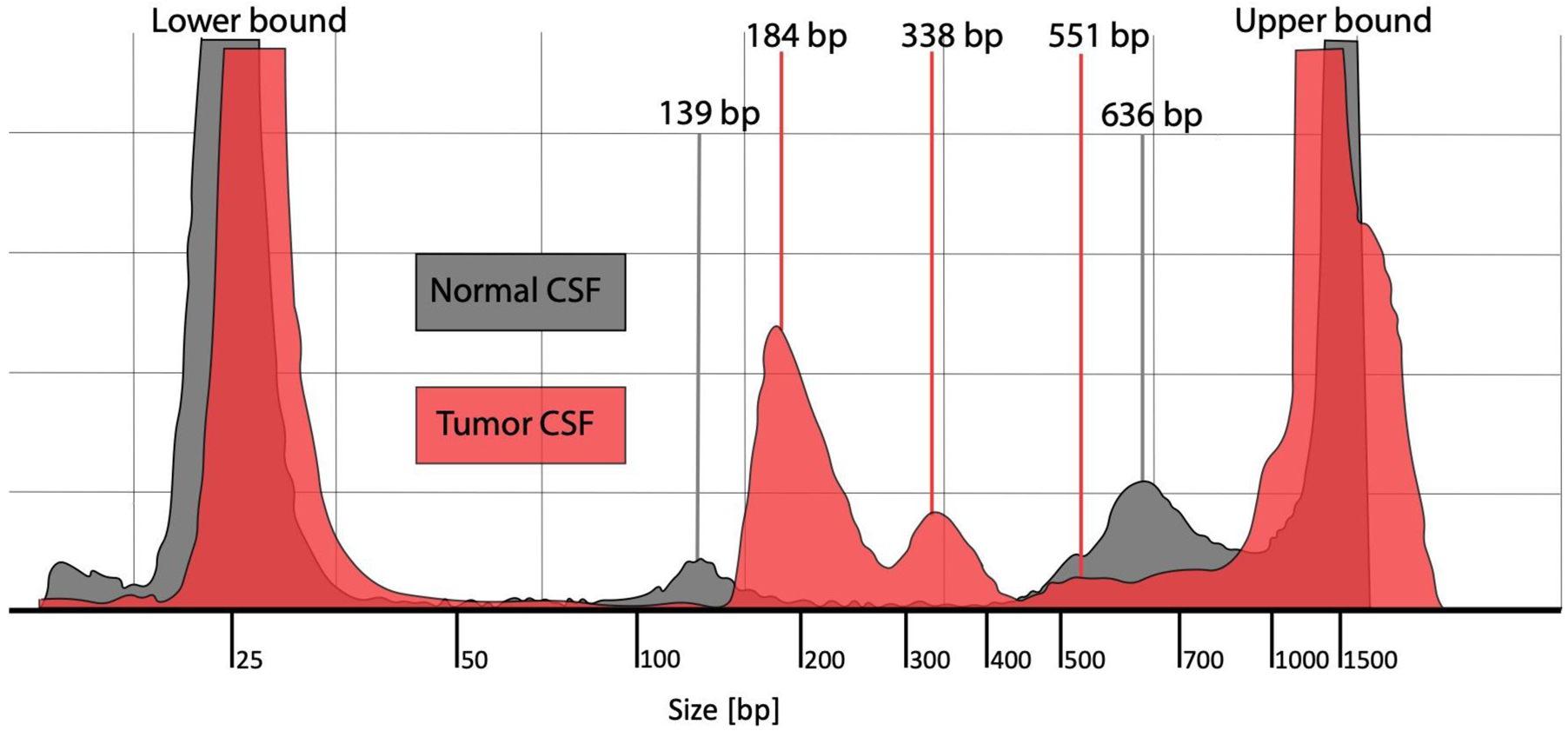
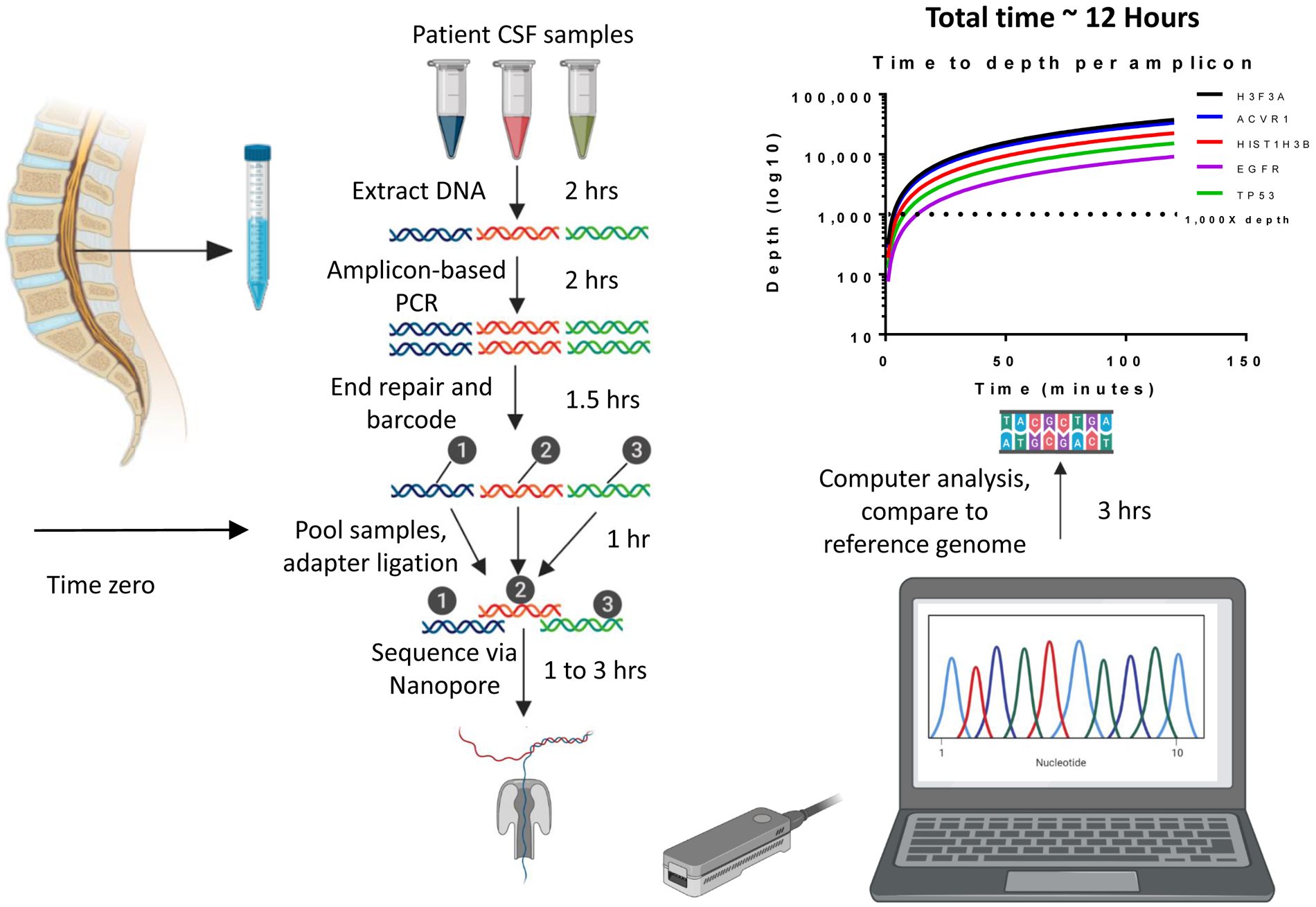
Experimental design: We performed ultra-short fragment (100-200 bp) PCR amplification of cf-tDNA for clinically actionable alterations in CSF and tumor samples from patients with pHGG (n = 12) alongside nontumor CSF (n = 6). PCR products underwent rapid amplicon-based sequencing by Oxford Nanopore Technology (Nanopore) with quantification of VAF. Additional comparison to next-generation sequencing (NGS) and droplet digital PCR (ddPCR) was performed.

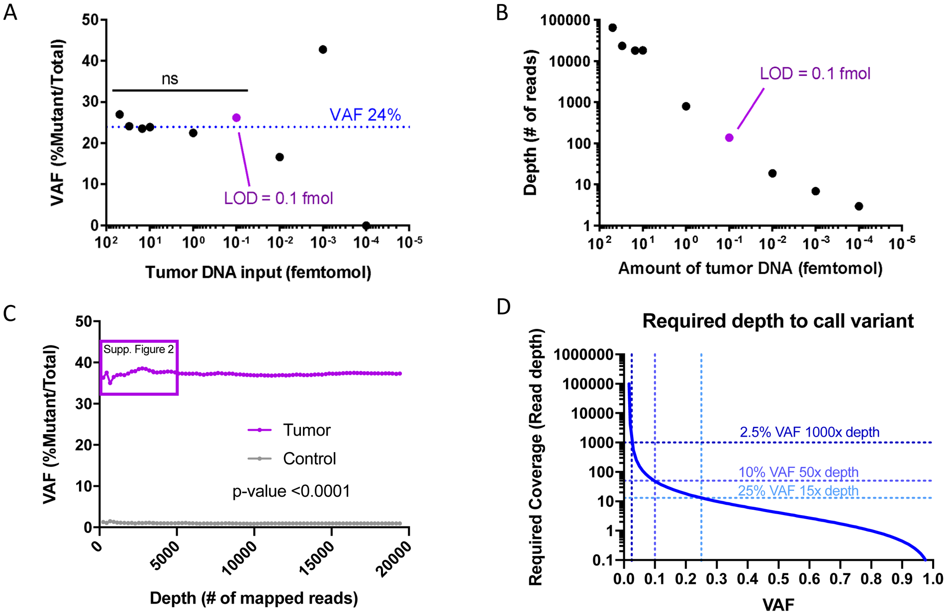
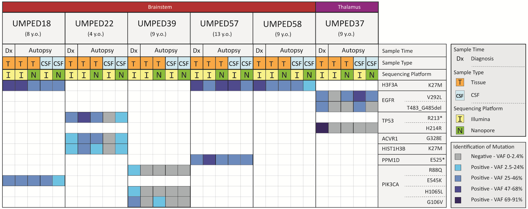
Results: Nanopore demonstrated 85% sensitivity and 100% specificity in CSF samples (n = 127 replicates) with 0.1 femtomole DNA limit of detection and 12-hour results, all of which compared favorably with NGS. Multiplexed analysis provided concurrent analysis of H3.3A (H3F3A) and H3C2 (HIST1H3B) mutations in a nonbiopsied patient and results were confirmed by ddPCR. Serial CSF cf-tDNA sequencing by Nanopore demonstrated correlation of radiological response on a clinical trial, with one patient showing dramatic multi-gene molecular response that predicted long-term clinical response.

Conclusions: Nanopore sequencing of ultra-short pHGG CSF cf-tDNA fragments is feasible, efficient, and sensitive with low-input samples thus overcoming many of the barriers restricting wider use of CSF cf-tDNA diagnosis and monitoring in this patient population.


Conflict of interest statement

Disclosure of Potential Conflicts of Interest:

None of the authors have potential conflicts of interest.

Figures

Figure 1.
Figure 1.. CSF cf-tDNA fragment analysis
Fragment analysis of cell-free DNA from normal CSF (gray color) and from tumor CSF from a patient with DIPG (red color). The figure shows DNA fragment sizing and quantification using TapeStation High Sensitivity DNA ScreenTape Bioanalyzer for each of the two sample types. Abbreviations: CSF = cerebrospinal fluid, DIPG = diffuse intrinsic pontine glioma, bp = base pairs
Figure 2.
Figure 2.. Limit of Detection and Error Rate of Nanopore CSF cf-tDNA Sequencing
A) Limit of detection by fmol of tumor DNA input onto the Nanopore flow cell needed to detect stable reference VAF of 24% H3F3A K27M obtained by Illumina sequencing at diagnosis. The purple dot shows the limit of detection. B) The graph shows read depth versus input DNA, demonstrating the depth required to call VAF of 24% at each amount of input DNA. The purple dot is the limit of detection. C) The graph shows the error rate of Nanopore in the negative control sample (gray line) as well as the VAF for H3F3A K27M for the tumor sample (purple line). D) Cochran’s equation was used to estimate the number of samples (reads) required to call various VAF’s where the lower bound of the 95% confidence interval is equal to the maximum observed false positive rate. Abbreviations: VAF = Variant Allele Fraction, fmol = femtomol, Supp. = supplemental
Figure 3.
Figure 3.. Nanopore sequencing efficiency analysis
CSF was drawn via lumbar puncture at time zero of processing. After DNA extraction, the sample(s) underwent amplification with pediatric HGG-specific genes, followed by sequencing on the MinION device. Raw sequencing output was basecalled using Guppy, then reads were aligned using Minimap2 to the human reference genome hg19. Integrated Genome Viewer was used to calculate variant allele fraction of aligned amplicons. Time required to sequence to a depth of 1000X ranged from 4 minutes to 14 minutes, with several genes shown as examples. The total time to results (variant allele fraction of SNPs) was less than 12 hours. Samples were run in triplicate.
Figure 4.
Figure 4.. Comparison of Nanopore and NGS sequencing of pHGG tumor and CSF cf-tDNA
Each UMPED number is a pediatric patient who had Illumina-based tissue sequencing at time of diagnosis and who donated tissue and CSF at time of death (age at time of death shown beneath the UMPED number). Anatomical location of the patient’s HGG is shown across the top while the tumor-driving mutations found at time of diagnosis are shown on the right. Sample type (tissue or CSF) and whether it was from diagnosis or autopsy is indicated beneath the age at death. Each sample is marked as being sequenced by Illumina or Nanopore and its corresponding column indicates if it was positive or negative for the mutation given for that row. For samples where mutation was found, the VAF was determined. Darker shades of blue indicate higher VAF quartiles. Samples were run in triplicate for Nanopore sequencing. Abbreviations: del = deletion, * = stop codon, VAF = variant allele fraction
Figure 5.
Figure 5.. Clinical trial schema for serial CSF cf-tDNA molecular response assessment
A) The ONC201 clinical trial timeline. B) Axial brain T1 with contrast MRI images at each time point on the ONC201 trial for patient UMPED77 (dotted line denotes estimate tumor border). Total tumor area is noted (in mm2, shown in purple). C) Nanopore sequencing results for H3F3A K27M VAF (orange) and tumor area (blue) for each CSF collection time point for the patient in 5B. D) Axial brain T1 with contrast MRI images at each time point on the ONC201 trial for patient UMPED60. Total tumor area is noted (in mm2, shown in purple). E) Nanopore sequencing results (shown as VAF) for four key tumor-driving mutations (identified from surgical biopsy) for the patient in 5D, as well as tumor area for each of the clinical trial time points. All samples were run in triplicate for Nanopore sequencing.

References

    1. Sturm D, Witt H, Hovestadt V, Khuong-Quang DA, Jones DT, Konermann C, et al. Hotspot mutations in H3F3A and IDH1 define distinct epigenetic and biological subgroups of glioblastoma. Cancer Cell 2012;22(4):425–37 doi 10.1016/j.ccr.2012.08.024. - DOI - PubMed
    1. Mackay A, Burford A, Carvalho D, Izquierdo E, Fazal-Salom J, Taylor KR, et al. Integrated Molecular Meta-Analysis of 1,000 Pediatric High-Grade and Diffuse Intrinsic Pontine Glioma. Cancer Cell 2017;32(4):520–37 e5 doi 10.1016/j.ccell.2017.08.017. - DOI - PMC - PubMed
    1. Hoffman LM, Veldhuijzen van Zanten SEM, Colditz N, Baugh J, Chaney B, Hoffmann M, et al. Clinical, Radiologic, Pathologic, and Molecular Characteristics of Long-Term Survivors of Diffuse Intrinsic Pontine Glioma (DIPG): A Collaborative Report From the International and European Society for Pediatric Oncology DIPG Registries. J Clin Oncol 2018;36(19):1963–72 doi 10.1200/JCO.2017.75.9308. - DOI - PMC - PubMed
    1. Lulla RR, Saratsis AM, Hashizume R. Mutations in chromatin machinery and pediatric high-grade glioma. Sci Adv 2016;2(3):e1501354 doi 10.1126/sciadv.1501354. - DOI - PMC - PubMed
    1. Lu VM, Alvi MA, McDonald KL, Daniels DJ. Impact of the H3K27M mutation on survival in pediatric high-grade glioma: a systematic review and meta-analysis. J Neurosurg Pediatr 2018;23(3):308–16 doi 10.3171/2018.9.PEDS18419. - DOI - PubMed

Publication types

MeSH terms