. 2020 Oct 21;6(43):eaba9337.
doi: 10.1126/sciadv.aba9337. Print 2020 Oct.

Natural polymorphism of Ym1 regulates pneumonitis through alternative activation of macrophages

Affiliations

Natural polymorphism of Ym1 regulates pneumonitis through alternative activation of macrophages

Wenhua Zhu et al. Sci Adv. .

Abstract

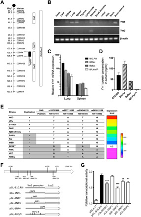
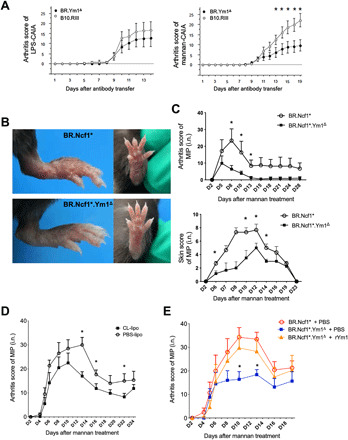
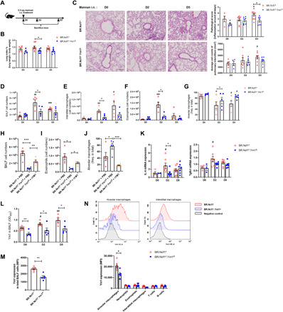
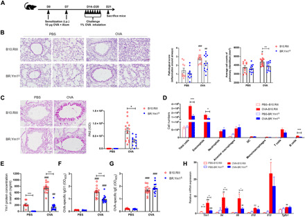
We have positionally cloned the Ym1 gene, with a duplication and a promoter polymorphism, as a major regulator of inflammation. Mice with the RIIIS/J haplotype, with the absence of Ym1 expression, showed reduced susceptibility to mannan-enhanced collagen antibody-induced arthritis and to chronic arthritis induced by intranasal exposure of mannan. Depletion of lung macrophages alleviated arthritis, whereas intranasal supplement of Ym1 protein to Ym1-deficient mice reversed the disease, suggesting a key role of Ym1 for inflammatory activity by lung macrophages. Ym1-deficient mice with pneumonitis had less eosinophil infiltration, reduced production of type II cytokines and IgG1, and skewing of macrophages toward alternative activation due to enhanced STAT6 activation. Proteomics analysis connected Ym1 polymorphism with changed lipid metabolism. Induced PPAR-γ and lipid metabolism in Ym1-deficient macrophages contributed to cellular polarization. In conclusion, the natural polymorphism of Ym1 regulates alternative activation of macrophages associated with pulmonary inflammation.


Figures

Fig. 1
Fig. 1. Polymorphisms of Ym1 lead to the variance of gene expression.
(A) Genetic map of mouse chromosome 3 and congenic fragment. Cia5, Cia21, and Cia22 loci were identified by linkage analysis and partial advanced intercross. The smallest congenic fragment covering the Ym1 gene was minimized as shown. Ym1 expression in different mouse strains was assessed (n = 3 to 5 for each strain). (B) Gene expression of Ym1 and Ym2 in wild-type B10.RIII mice was assessed by PCR with β-actin as a housekeeping gene. (C) Ym1 expression in lung and spleen of indicated strains was detected by RT-qPCR. (D) Circulating Ym1 levels in different strains were detected by ELISA. (E) Natural polymorphisms for indicated inbred and wild-derived mouse strains. Data of Ym1 expression in different strains were from the ImmGen database. N.D., no data. (F) Promoters from B10.RIII and RIIIS/J mice were cloned into pGL4.17 plasmids (named as pGL-B10.RIII and pGL-RIIIS/J). Site mutation was done using pGL-B10.RIII to convert SNP1 to SNP4 sites to RIIIS/J genotypes (named as pGL-SNP1, pGL-SNP2, pGL-SNP3, and pGL-SNP4). (G) Constructed reporter gene plasmids were transfected into HEK293T cells, and relative luciferase activities were detected. All values are expressed as means ± SEM. **P < 0.01 and ***P < 0.001.
Fig. 2
Fig. 2. Ym1 deficiency in congenic mice protects arthritis development induced by mannan through macrophages.
(A) B10.RIII and BR.Ym1Δ congenic mice (n = 11 to 18 per group) were injected intravenously with four monoclonal antibodies at day 0 and boosted with LPS (25 μg) or mannan (2 mg) at day 5 to induce CAIA. The arthritis severity was scored. BR.Ncf1* and BR.Ncf1*.Ym1Δ mice (n = 6 per group) were administrated with 20 mg of mannan intranasally (i.n.) to induce psoriasis and chronic arthritis. (B) Arthritic joint phenotype and psoriasis-like skin lesions in the hind paws. Photo credit: Wenhua Zhu, Xi’an Jiaotong University Health Science Center. (C) The arthritis and psoriasis severity was scored. The area under the arthritis scoring curve until D28 between the two strains was analyzed statistically (P < 0.05). (D) Clodronate-liposome (CL-lipo) or PBS-liposome (PBS-lipo) was treated intranasally to BR.Ncf1* mice (n = 6 per group) to deplete macrophages. Two days later, mice were administrated with mannan intranasally to induce arthritis. (E) BR.Ncf1* and BR.Ncf1*.Ym1Δ mice (n = 7 per group) were administrated with mannan intranasally with or without Ym1 protein supplement to induce arthritis. Disease severity was scored using a macroscopic scoring system. All values are expressed as means ± SEM. *P < 0.05.
Fig. 3
Fig. 3. Ym1 deficiency reduces pulmonary inflammation in mannan intranasally induced arthritis mice.
(A) BR.Ncf1* and BR.Ncf1*.Ym1Δ mice (n = 5 to 8 per group) were administrated with 0.5 mg of mannan intranasally (i.n.) to induce pulmonary inflammation. Lung tissues and BALF were collected at days 0, 2, and 5. (B) Lung weight normalized by body weight was calculated. (C) Lung sections were stained by hematoxylin and eosin (H&E), and peribronchial infiltration and interstitial infiltration of inflammatory cells were evaluated. Then, total cells, eosinophils, interstitial macrophages, and alveolar macrophages from mannan intranasally treated mice (D to G) or from mannan intranasally treated mice with Ym1 protein (1 μg per mouse) supplement (H to J) were analyzed by flow cytometry. (K) mRNA expression of Il4 and Tgfb1 in lung tissues was detected by RT-qPCR. In addition, Ym1 concentration (L) in BALF at days 0, 2, and 5 was determined by ELISA. Besides, Ym1 expression in total BALF cells (M) and different types of immune cells (N) in mannan-treated mice at day 2 was analyzed by flow cytometry. The representative histograms of Ym1 in alveolar macrophages and interstitial macrophages (N) were shown. All values are expressed as means ± SEM. # indicates the comparison among different time points in each mouse strain, and * indicates the comparison between the two mouse strains. */#P < 0.05, **/##P < 0.01, and ***/###P < 0.001.
Fig. 4
Fig. 4. Low expression of Ym1 alleviates the severity of AIPI.
(A) B10.RIII and BR.Ym1Δ mice were sensitized and challenged by OVA to induce AIPI (n = 10 to 13 per group). (B) Lung tissues were stained with H&E to observe pathological changes, and peribronchial infiltration and interstitial infiltration of inflammatory cells were evaluated. (C) Lung sections were stained with periodic acid–Schiff (PAS) to detect mucus production, and the integral optical density (IOD) of staining was calculated. (D) Immune cells in BALF were analyzed by flow cytometry. Serum concentrations of Ym1 (E), OVA-specific IgG1 (F), and OVA-specific IgE (G) were determined by ELISA. (H) mRNA expression of Ym1 and type II cytokines including Il4, Il5, Il10, Il13, and Tgfb1 in lung tissues was detected by RT-qPCR. All values are expressed as means ± SEM. # indicates the comparison between the OVA group and the PBS group in each mouse strain, and * indicates the comparison between the two mouse strains. */#P < 0.05, **/##P < 0.01, and ***/###P < 0.001.
Fig. 5
Fig. 5. Ym1 negatively regulates AAMs.
The polarization markers of alveolar macrophages (AM) in mannan intranasally (i.n.) treated mice (A) or mice with rYm1 protein supplement (B) and in mice of the AIPI model (C) were detected by flow cytometry. Bone marrow–derived macrophages (BMMs; M0) from B10.RIII, BR.Ym1Δ, BR.Ncf1*, and BR.Ncf1*.Ym1Δ mice (n = 3 per group) were treated with LPS and IFN-γ or IL-4 and IL-13 to polarize cells to CAMs or AAMs. (D) Ym1 mRNA expression was detected by RT-qPCR. (E) Arg1 and MRC1 expression in AAMs was detected by Western blotting, and representative blot was shown. In addition, the mRNA expression (F) of AAM markers including Ym1, Arg1, MRC1, and Fizz1 at different time points (0, 3, 6, 12, 24, and 48 hours) after IL-4 and IL-13 stimulation was detected by RT-qPCR. Protein expression (G) of Ym1, Arg1, and MRC1 and the activation of STAT6 (H) were detected by Western blotting, and the statistics are shown in fig. S6. All values are expressed as means ± SEM. In (A) to (C), # indicates the comparison in the same mouse strain, and * indicates the comparison between the two mouse strains. */#P < 0.05, **P < 0.01, and ***/###P < 0.001.
Fig. 6
Fig. 6. Ym1 regulates lipid metabolism and PPAR-γ to orchestrate macrophage polarization.
Protein profiles of thioglycollate-elicited macrophages from B10.RIII and BR.Ym1Δ mice were determined by label-free mass spectrum analysis (n = 4). (A) Volcano plot comparison showed the differentially expressed proteins in macrophages. (B) Expression of differentially expressed proteins relevant to lipid metabolism. Then, BMMs (M0) derived from BR.Ncf1* and BR.Ncf1*.Ym1Δ mice were polarized to AAMs, and the expression (C) of PPAR-α, PPAR-γ, and PPAR-δ was detected by Western blotting. Antagonists of PPAR-γ (T0070907) and CPT1 (Etomoxir) were used during AAM polarization, and expression (D) of MRC1 and Arg1 was detected by Western blotting. (E) In addition, BMMs were transfected with si-Stat6 (pool of si-Stat6-1, si-Stat6-2, and si-Stat6-3) and si-Pparg-3 siRNA to knock down STAT6 and PPAR-γ or negative control siRNA (si-NC). After AAM polarization, the expression of STAT6, PPAR-γ, MRC1, and Arg1 was detected by Western blotting, and the statistics are shown in fig. S8. (F) Illustration of the role of Ym1 on regulating alveolar macrophages in pulmonary inflammation. All values are expressed as means ± SEM. *P < 0.05, **P < 0.01, and ***P < 0.001.

Similar articles

Cited by

References

    1. Martinez F. D., Genes, environments, development and asthma: A reappraisal. Eur. Respir. J. 29, 179–184 (2007). - PubMed
    1. Yau A. C., Holmdahl R., Rheumatoid arthritis: Identifying and characterising polymorphisms using rat models. Dis. Model. Mech. 9, 1111–1123 (2016). - PMC - PubMed
    1. Olofsson P., Holmberg J., Tordsson J., Lu S., Akerstrom B., Holmdahl R., Positional identification of Ncf1 as a gene that regulates arthritis severity in rats. Nat. Genet. 33, 25–32 (2003). - PubMed
    1. Olsson L. M., Johansson A. C., Gullstrand B., Jonsen A., Saevarsdottir S., Ronnblom L., Leonard D., Wettero J., Sjowall C., Svenungsson E., Gunnarsson I., Bengtsson A. A., Holmdahl R., A single nucleotide polymorphism in the NCF1 gene leading to reduced oxidative burst is associated with systemic lupus erythematosus. Ann. Rheum. Dis. 76, 1607–1613 (2017). - PubMed
    1. Zhao J., Ma J., Deng Y., Kelly J. A., Kim K., Bang S. Y., Lee H. S., Li Q. Z., Wakeland E. K., Qiu R., Liu M., Guo J., Li Z., Tan W., Rasmussen A., Lessard C. J., Sivils K. L., Hahn B. H., Grossman J. M., Kamen D. L., Gilkeson G. S., Bae S. C., Gaffney P. M., Shen N., Tsao B. P., A missense variant in NCF1 is associated with susceptibility to multiple autoimmune diseases. Nat. Genet. 49, 433–437 (2017). - PMC - PubMed

Publication types