. 2020 Dec 22;95(2):e01391-20.
doi: 10.1128/JVI.01391-20. Print 2020 Dec 22.

Eukaryotic Translation Elongation Factor 1 Delta Inhibits the Nuclear Import of the Nucleoprotein and PA-PB1 Heterodimer of Influenza A Virus

Affiliations

Eukaryotic Translation Elongation Factor 1 Delta Inhibits the Nuclear Import of the Nucleoprotein and PA-PB1 Heterodimer of Influenza A Virus

Qingxia Gao et al. J Virol. .

Erratum in

Abstract

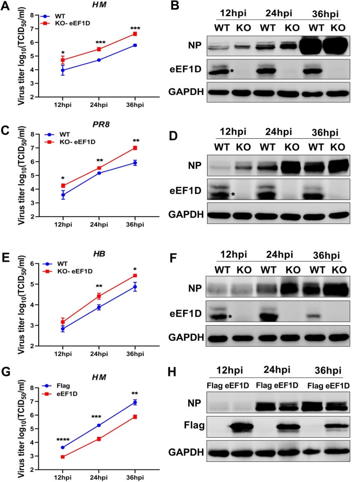
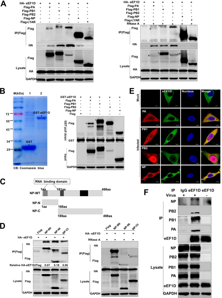
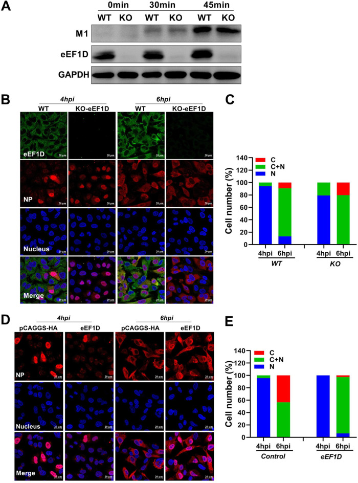
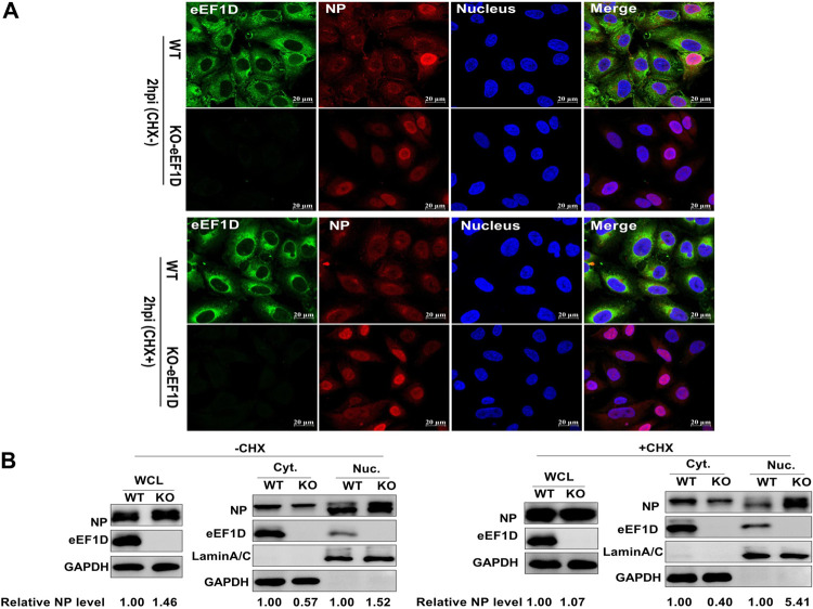
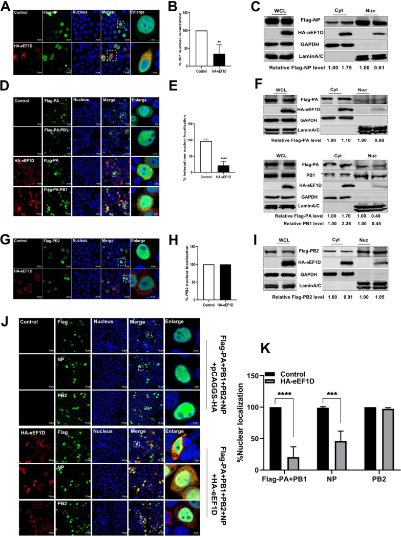
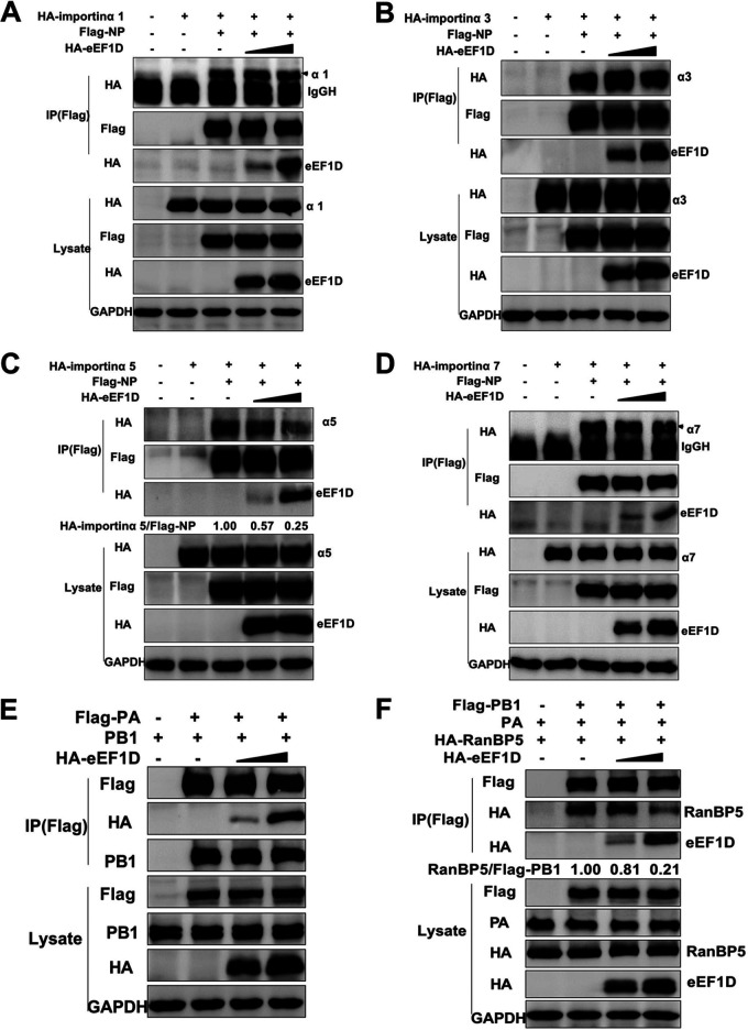
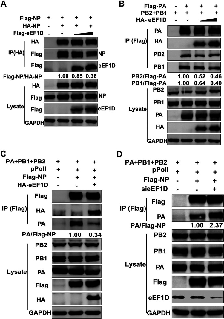
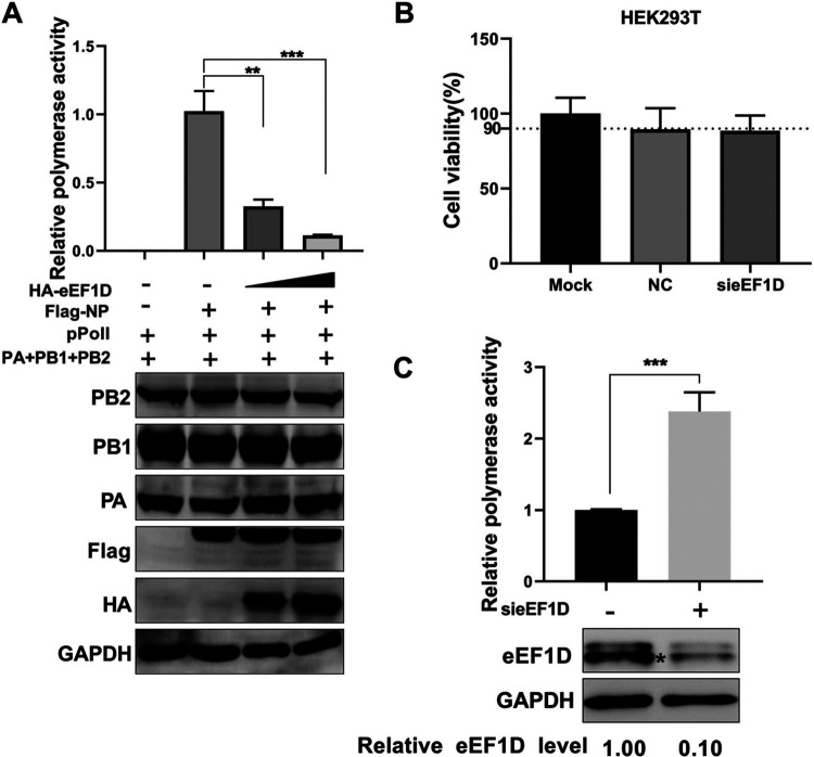
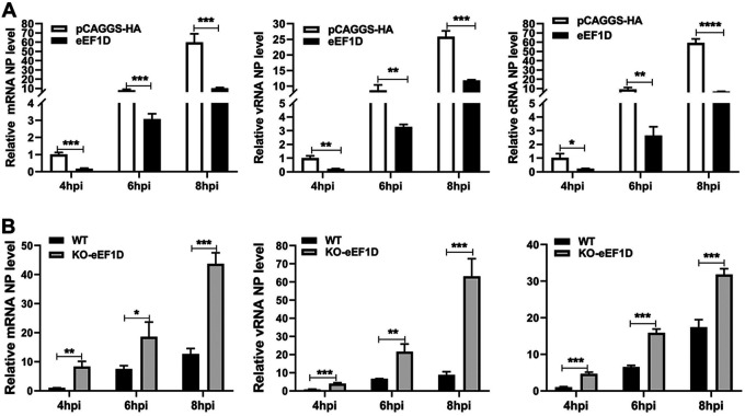
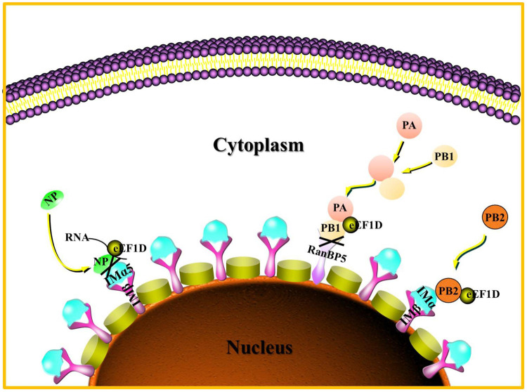
The viral ribonucleoprotein (vRNP) of the influenza A virus (IAV) is responsible for the viral RNA transcription and replication in the nucleus, and its functions rely on host factors. Previous studies have indicated that eukaryotic translation elongation factor 1 delta (eEF1D) may associate with RNP subunits, but its roles in IAV replication are unclear. Herein, we showed that eEF1D was an inhibitor of IAV replication because knockout of eEF1D resulted in a significant increase in virus yield. eEF1D interacted with RNP subunits polymerase acidic protein (PA), polymerase basic 1 (PB1), polymerase basic 2 (PB2), and also with nucleoprotein (NP) in an RNA-dependent manner. Further studies revealed that eEF1D impeded the nuclear import of NP and PA-PB1 heterodimer of IAV, thereby suppressing the vRNP assembly, viral polymerase activity, and viral RNA synthesis. Together, our studies demonstrate eEF1D negatively regulating the IAV replication by inhibition of the nuclear import of RNP subunits, which not only uncovers a novel role of eEF1D in IAV replication but also provides new insights into the mechanisms of nuclear import of vRNP proteins.IMPORTANCE Influenza A virus is the major cause of influenza, a respiratory disease in humans and animals. Different from most other RNA viruses, the transcription and replication of IAV occur in the cell nucleus. Therefore, the vRNPs must be imported into the nucleus for viral transcription and replication, which requires participation of host proteins. However, the mechanisms of the IAV-host interactions involved in nuclear import remain poorly understood. Here, we identified eEF1D as a novel inhibitor for the influenza virus life cycle. Importantly, eEF1D impaired the interaction between NP and importin α5 and the interaction between PB1 and RanBP5, which impeded the nuclear import of vRNP. Our studies not only reveal the molecular mechanisms of the nuclear import of IAV vRNP but also provide potential anti-influenza targets for antiviral development.

Keywords: NP; PA-PB1 heterodimer; eEF1D; influenza A virus; vRNP.


Figures

FIG 1
FIG 1
eEF1D suppresses influenza A virus replication. (A to F) Effect of eEF1D knocked out on IAV replication. KO-eEF1D A549 cells were obtained by using the CRISPR/Cas9 system. WT-A549 cells and KO-eEF1D A549 cells were infected with PR8 H1N1 virus (MOI, 0.01), HM H5N1 virus (MOI, 0.01), or HB H1N1 virus (MOI, 0.1). Cell supernatants and lysates were harvested at 12 hpi, 24 hpi, and 36 hpi. Virus titers were determined by TCID50 assay on MDCK cells, as shown in panels A, C, and E. eEF1D protein expression and viral proteins expression were detected by Western blotting, as shown in panels B, D, and F (mean ± SD of three independent experiments. *, P < 0.05; **, P < 0.01; ***, P < 0.001; two-tailed Student's t test). (G and H) Effect of eEF1D overexpressed on IAV replication. A549 cells were transfected with Flag-eEF1D or vector as negative control for 24 h followed by infection with HM H5N1 virus (MOI, 0.01). Cell supernatants and lysates were harvested at 12 hpi, 24 hpi, and 36 hpi. Virus titers were determined by TCID50 assay on MDCK cells (mean ± SD of three independent experiments. *, P < 0.05; **, P < 0.01; ***, P < 0.001; two-tailed Student's t test).
FIG 2
FIG 2
Interactions between eEF1D and RNP subunits. (A) HEK293T cells were cotransfected with HA-eEF1D and Flag-PA, Flag-PB1, Flag-PB2,Flag-NP, or Flag-LYAR followed by lysing at 24 h posttransfection. Cell lysates were treated with RNase A (100 U) at 37°C for 1 h or no treatment before being mixed together. The co-IP assay was carried out using an anti-Flag antibody followed by Western blotting. (B) GST pulldown assay of purified GST-eEF1D and lysates of HEK293T cells expressing Flag-PA, Flag-PB1, Flag-PB2, or Flag-NP. Purified GST (lane 1) and GST-eEF1D (lane 2) proteins were detected by using Coomassie blue (CB) staining. (C) Schematic diagram of the NP truncated segments. (D) HEK293T cells in 6-well plates were cotransfected with empty vector, NP-Wt, NP-Nt, or NP-Ct, along with HA-eEF1D using Lipofectamine 2000. At 24 h posttransfection, the cells were lysed, and the collected lysates were untreated or treated with the RNase A (100 U) at 37°C for 1 h. The co-IP assay using an anti-Flag antibody followed by Western blot analyses. (E) Colocalization of endogenous eEF1D and RNP components in infected cells. A549 cells were mock infected or infected with the PR8 H1N1 virus (MOI, 2) for 12 hpi, and confocal microscopy was performed using an anti-eEF1D mouse antibody (green) and anti-PA, anti-PB1, anti-PB2, and anti-NP rabbit antibody (red). DAPI was used to stain for the nucleus (blue). Samples were examined with a confocal microscope (LSM 880; Zeiss). Images are representative of three independent experiments. Scale bar, 10 μM. (F) Interactions between endogenous eEF1D and RNP components during infection. HEK293T cells in 10-cm dishes were infected and mock infected with the PR8 H1N1 virus (MOI, 2) for 12 hpi. The HEK293T cells were lysed by 500 μl IP lysis buffer and immunoprecipitated using an anti-eEF1D mouse antibody or mouse control IgG.
FIG 3
FIG 3
eEF1D affects the nucleocytoplasmic distribution of vRNP. (A) Effect of eEF1D on IAV internalization. WT-A549 cells or KO-eEF1D A549 cells were incubated with PR8 H1N1 virus at an MOI of 10 for 0, 30, and 45 min (at 37°C) without trypsin in the medium. Then, cells were washed with PBS-HCl (pH 1.3) to remove attached virions from the surface and lysed for Western blotting. (B) Localization of the NP proteins in PR8-infected WT-A549 cells and KO-eEF1D A549 cells. WT-A549 cells and KO-eEF1D A549 cells were infected with PR8 H1N1 at an MOI of 5. At 4 and 6 hpi, cells were fixed, permeabilized, and stained with rabbit anti-NP (red), mouse anti-eEF1D (green), and DAPI (blue). (D) Localization of the NP proteins in PR8-infected control cells and eEF1D-overexpressed cells. Coverslips coated with A549 cells were cotransfected vector (pCAGGS-HA) or eEF1D. At 24 h posttransfection, cells were infected with PR8 virus at an MOI of 5. At 4 and 6 hpi, cells were fixed, permeabilized, and stained with rabbit anti-NP (red) and DAPI (blue). (C and E) Quantitative analysis of NP localization in infected cells. At least 100 cells in each group were scored. N, predominantly nuclear; N+C, nuclear and cytoplasmic; C, predominantly cytoplasmic.
FIG 4
FIG 4
Knockout of endogenous eEF1D facilitates nuclear import of vRNPs. (A) Increased translocation of NP to nuclear in eEF1D knockout cells during infection using confocal microscopy. WT-A549 cells and KO-eEF1D A549 cells were infected with PR8 H1N1 virus (MOI, 10) at the same time. Cells were fixed at 2 h postinfection with CHX or without CHX treatment followed by immunostaining the NP (red), eEF1D (green), and nucleus (blue). The images were acquired under a confocal microscope with ×63 oil lens. Scale bar, 20 μM. For these experiments, fluorescence was examined with a confocal microscope (LSM 880; Zeiss). Images are representative of three independent experiments. (B) Western blot analysis of the distribution of NP in the cytoplasmic and nuclear fractions in virus-infected WT- and KO-eEF1D cells. WT-A549 and KO-eEF1D A549 cells were infected with PR8 H1N1 virus (MOI = 10) at the same time. Cells were harvested at 2 hpi and subjected to cellular fraction and Western blotting. Western densitometry analysis was performed using ImageJ. Lamin A/C and GAPDH were used as a loading control for nuclear and cytoplasmic fractions, respectively. The relative NP levels (NP/GAPDH or lamin A/C) are shown at the bottom of panel B. The relative expression levels of NP (NP/GAPDH) were also detected in the whole-cell lysates (WCLs).
FIG 5
FIG 5
eEF1D affects the nucleocytoplasmic distribution of viral NP and PA-PB1 heterodimer. (A) Confocal analysis confirmed the inhibition of nuclearimport of Flag-NP (green) in eEF1D- (red) overexpressed cells. HeLa cells were treated as described in Fig. 3B. The squared marked region was enlarged and is shown on the right. (B) Percentage of stained cells with mainly nuclear localized NP among total cells was calculated in randomly selected four fields of view (*, P < 0.05; **, P < 0.01; ***, P < 0.001; unpaired two-tailed Student's t test). (C) Western blot analysis of the nucleocytoplasmic distribution of Flag-NP in HeLa cells cotransfected with HA-eEF1D or vector. (D) HeLa cells were grown on coverslips and cotransfected with Flag-PA, nontagged-PB1 (green), and HA-eEF1D (red) or vector (pCAGGS-HA), or HeLa cells were cotransfected with Flag-PA alone (green) and HA-eEF1D (red) used as a control. The squared marked region was enlarged and is shown on the right. (E) Percentage of stained cells with mainly nuclear localized PA-PB1 heterodimer among total cells was calculated in four random fields of view (*, P < 0.05; **, P < 0.01; ***, P < 0.001; unpaired two-tailed Student's t test). (F) Western blot analysis of the nucleocytoplasmic distribution of Flag-PA and PA-PB1 heterodimer in HeLa cells cotransfected with HA-eEF1D or vector. (G) Coverslips coated with HeLa cells were cotransfected with a plasmid expressing PR8-Flag-PB2 (green) and an empty vector (pCAGGS-HA) or HA-eEF1D (red). The squared marked region was enlarged and is shown on the right. (H) Percentage of stained cells with mainly nuclear localized PB2 among total cells was calculated in four random fields of view (*, P < 0.05; **, P < 0.01; ***, P < 0.001; unpaired two-tailed Student's t test). (I) Western blot analysis of the nucleocytoplasmic distribution of Flag-PB2 in HeLa cells cotransfected with HA-eEF1D or vector. (J) Confocal analysis of the nucleocytoplasmic distribution of Flag-PA-PB1 heterodimer, NP, and PB2 when HeLa cells were cotransfected with Flag-PA, PB1, PB2, and NP (green), HA-eEF1D (red), or vector. (K) Percentage of stained cells with mainly nuclear localized Flag-PA-PB1 heterodimer, NP, and PB2 among total cells was calculated in randomly selected four fields of view (*, P < 0.05; **, P < 0.01; ***, P < 0.001; unpaired two-tailed Student's t test). For indirect immunofluorescence experiments, samples were examined with a confocal microscope (LSM 880; Zeiss). Images are representative of three independent experiments. Scale bar, 20 μM. Western densitometry analysis was performed using software ImageJ. Lamin A/C and GAPDH were used as loading controls for nuclear and cytoplasmic fractions, respectively. The relative viral protein levels (viral protein/GAPDH or lamin A/C) are shown at the bottom of panels C, F, and I.
FIG 6
FIG 6
Overexpression of eEF1D blocks IAV NP-importin α5 and PB1-RanBP5 interactions. (A to D) Interaction of NP with importin α family members, importin α1 (A), importin α3 (B), importin α5 (C), and importin α7 (D) in the presence or absence of gradually increasing amounts (0 to 0.3 μg) of HA-eEF1D. Cell lysates were immunoprecipitated with a mouse anti-Flag antibody, and the bound proteins were detected by Western blotting with a mouse anti-HA antibody or a mouse anti-Flag antibody to detect importin α family members, eEF1D, NP, and GAPDH, respectively. (E) Effect of eEF1D on the interaction between PA and PB1. HEK293T cells were transfected with the indicated plasmids for 24 h. Cell lysates were immunoprecipitated with anti-Flag antibody for the co-IP assay. Flag-PA and PB1 levels were checked via immunoprecipitation by Western blotting. (F) Effect of eEF1D on the interaction between RanBP5 in PB1 upon the PA, PB1, and RanBP5 complex. HEK293T cells were transfected with the indicated plasmids for 24 h. Cell lysates were immunoprecipitated with anti-Flag antibody for co-IP assay. RanBP5 and Flag-PB1 levels were detected via immunoprecipitation by Western blotting.
FIG 7
FIG 7
eEF1D impairs viral RNP complex formation. (A) eEF1D inhibited NP oligomerization. HEK293T cells were transfected with Flag-NP, HA-NP, and HA-eEF1D (0, 0.25, and 0.5 μg) or vector. Cells were harvested at 24 h posttransfection for a co-IP assay and Western blot assay. Densitometry analysis was done using ImageJ, and relative precipitated Flag-NP/HA-NP ratios are shown at the bottom. (B) eEF1D reduced 3P complex association. HEK293T cells were transfected with Flag-PA, PB1, and PB2 plasmids along with increasing amounts of HA-eEF1D plasmids. Cells were then lysed for the co-IP assay using anti-Flag antibody. The immune complexes were analyzed by performing a Western blot assay using antibodies against PA, PB1, PB2, and HA, respectively. The band intensities were quantified, and relative precipitated PB1/Flag-PA and PB2/Flag-PA ratios are shown at the bottom. (C) eEF1D decreased influenza virus RNP assembly. HEK293T cells were transfected with plasmids containing PA, PB1, PB2, Flag-NP, and pPolI along with HA-eEF1D or vector. Cells were lysed at 24 h posttransfection, and co-IP was performed by using anti-Flag antibody followed by Western blotting. The band intensities were quantified, and relative precipitated PA/Flag-NP ratios are shown at the bottom. (D) Knockdown of eEF1D stimulated RNP assembly. HEK293T cells transfected with the plasmids containing PA, PB1, PB2, Flag-NP, and pPolI along with si-eEF1D or si-NC. The remaining procedures are also the same as in panel C.
FIG 8
FIG 8
eEF1D reduces the polymerase activity of IAV. (A) Effect of eEF1D on the expression of RNP components in HEK293T cells. HEK293T cells were transfected with the indicated plasmids as described above (Fig. 7C). Protein expression of individual RNP components and eEF1D were detected by Western blotting. GAPDH was used as loading control. For all experiments above, the data are presented as the mean ± SD of three independent experiments (*, P < 0.05; **, P < 0.01; ***, P < 0.001; two-tailed Student's t test). (B) Cell viability upon si-eEF1D transfection. HEK293T cells were treated with siRNA against eEF1D and control nontarget siRNA. The cell viability was measured by CCK-8 assay at the indicated time points posttransfection (mean ± SD of three independent experiments. *, P < 0.05; **, P < 0.01; ***, P < 0.001; two-tailed Student's t test). (C) Cells were cotransfected with plasmids containing PA, PB1, PB2, Flag-NP, and pPolI along si-eEF1D or si-NC. The silencing efficiency of si-eEF1D was measured by Western blotting. GAPDH served as loading control. Band intensities were quantified with ImageJ, and the relative eEF1D levels are shown at the bottom (mean ± SD of three independent experiments. *, P < 0.05; **, P < 0.01; ***, P < 0.001; two-tailed Student's t test).
FIG 9
FIG 9
eEF1D restricts RNA synthesis during IAV infection. (A) A549 cells were transfected with eEF1D or vector for 24 h and were infected with PR8 H1N1 virus at an MOI of 1.0. Samples were collected at 4 hpi, 6 hpi, and 8 hpi. The vRNA, cRNA, and mRNA levels of NP were detected by qRT-PCR. The viral RNA levels were normalized to the 18S rRNA level (mean ± SD of three independent experiments. *, P < 0.05; **, P < 0.01; ***, P < 0.001; ****, P < 0.0001; two-tailed Student’s t test). (B) WT-A549 cells and KO-eEF1D A549 cells were seeded in 12-well plates and then infected with the PR8 H1N1 virus at an MOI of 1.0. Other procedures were the same as described in panel A (mean ± SD of three independent experiments. *, P < 0.05; **, P < 0.01; ***, P < 0.001; ****, P < 0.0001; two-tailed Student’s t test).
FIG 10
FIG 10
Proposed model for eEF1D-mediated inhibition of the nuclear import of NP and PA-PB1 heterodimer. In the cytoplasm, eEF1D inhibits the binding of newly synthesized NP to importin α5 and reduces the affinity of PB1 with RanBP5 in the PA-PB1 heterodimer, thereby suppressing the nuclear import of vRNPs.

Similar articles

Cited by

References

    1. Zheng W, Tao YJ. 2013. Structure and assembly of the influenza A virus ribonucleoprotein complex. FEBS Lett 587:1206–1214. doi:10.1016/j.febslet.2013.02.048. - DOI - PubMed
    1. Resa-Infante P, Jorba N, Coloma R, Ortin J. 2011. The influenza virus RNA synthesis machine: advances in its structure and function. RNA Biol 8:207–215. doi:10.4161/rna.8.2.14513. - DOI - PMC - PubMed
    1. Eisfeld AJ, Neumann G, Kawaoka Y. 2015. At the centre: influenza A virus ribonucleoproteins. Nat Rev Microbiol 13:28–41. doi:10.1038/nrmicro3367. - DOI - PMC - PubMed
    1. Honda A, Ueda K, Nagata K, Ishihama A. 1988. RNA polymerase of influenza virus: role of NP in RNA chain elongation. J Biochem 104:1021–1026. doi:10.1093/oxfordjournals.jbchem.a122569. - DOI - PubMed
    1. Elton D, Medcalf E, Bishop K, Digard P. 1999. Oligomerization of the influenza virus nucleoprotein: identification of positive and negative sequence elements. Virology 260:190–200. doi:10.1006/viro.1999.9818. - DOI - PubMed

Publication types

MeSH terms

LinkOut - more resources