

Priming with DNA Expressing Trimeric HIV V1V2 Alters the Immune Hierarchy Favoring the Development of V2-Specific Antibodies in Rhesus Macaques
- PMID: 33087466
- PMCID: PMC7944456
- DOI: 10.1128/JVI.01193-20
Priming with DNA Expressing Trimeric HIV V1V2 Alters the Immune Hierarchy Favoring the Development of V2-Specific Antibodies in Rhesus Macaques
Abstract
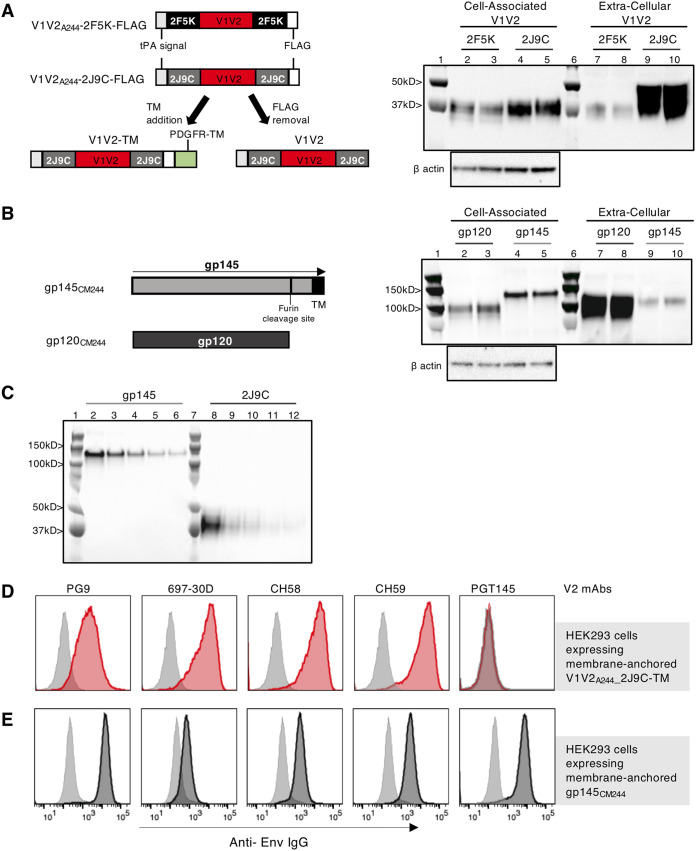
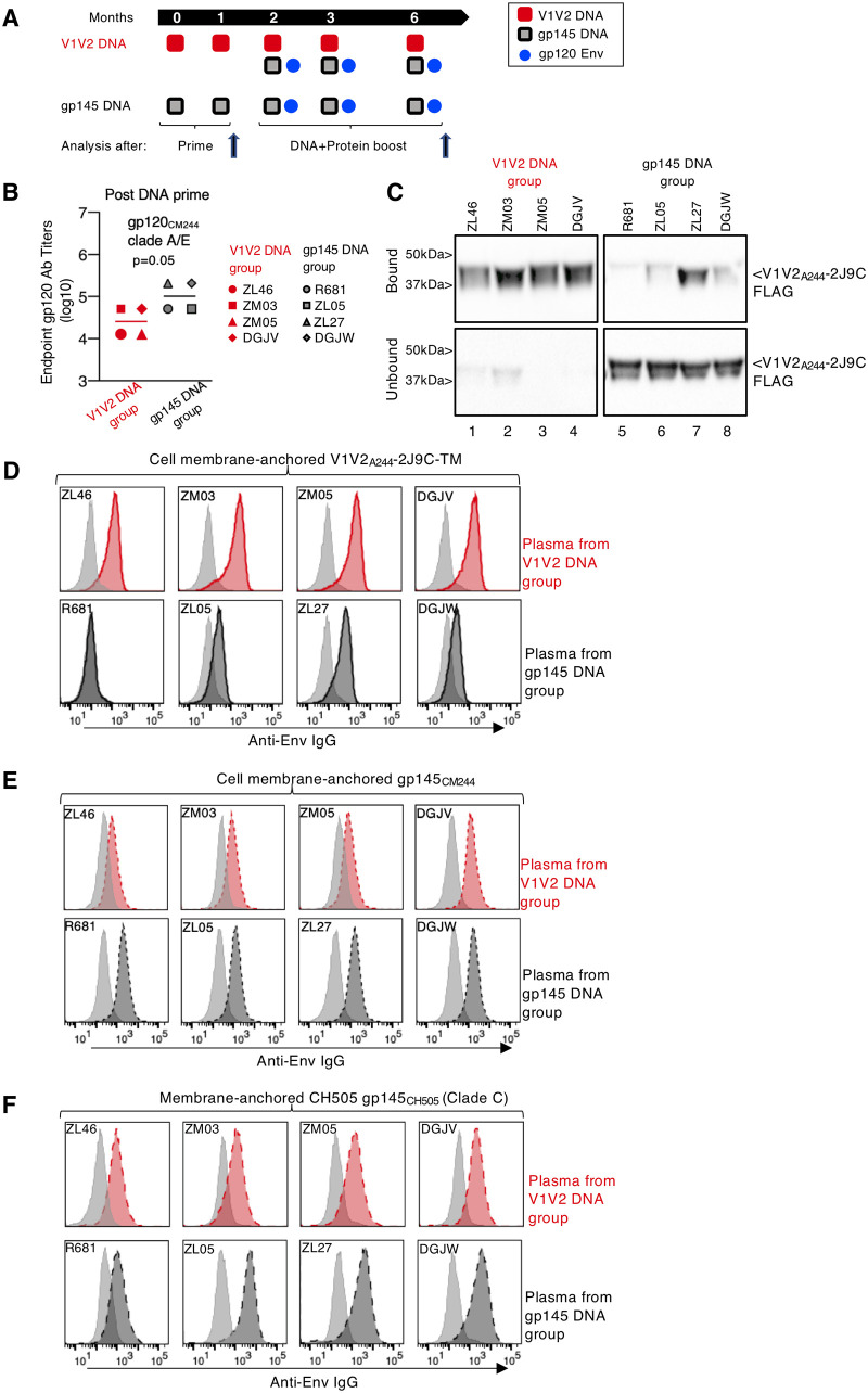
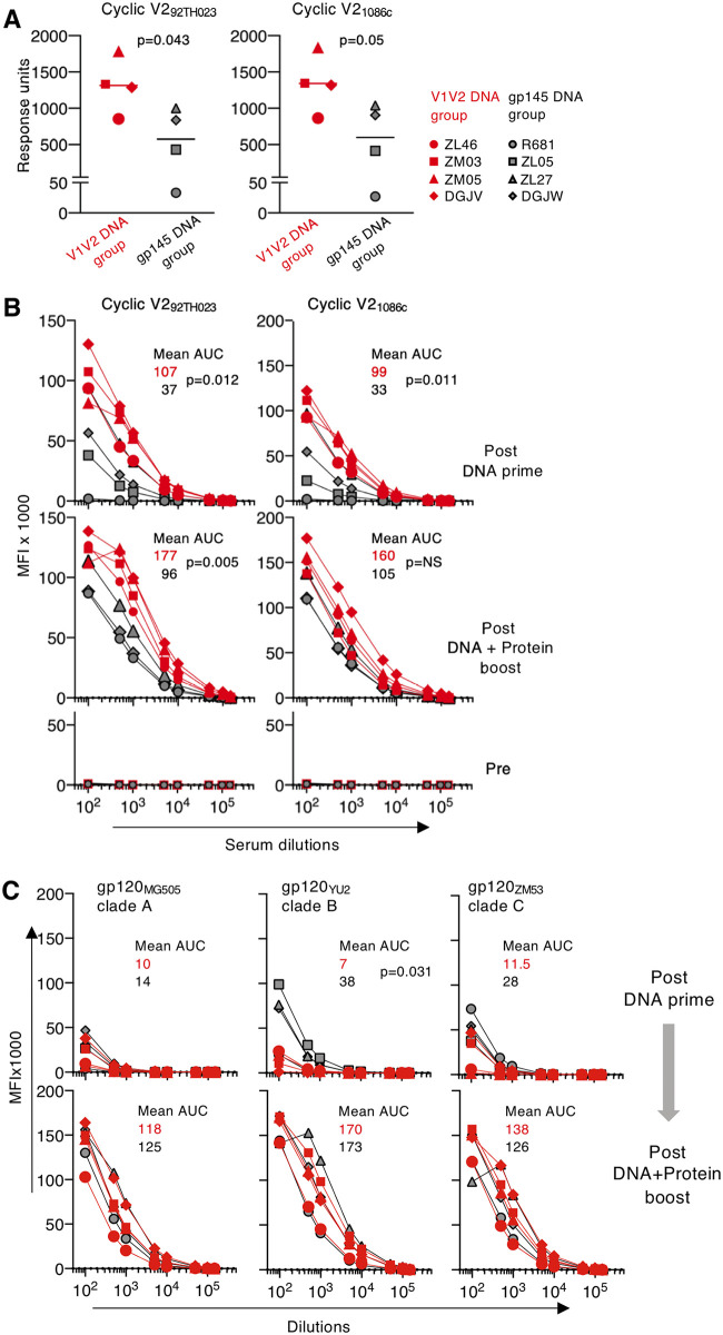
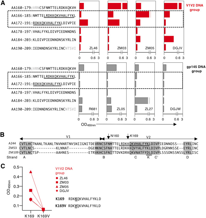
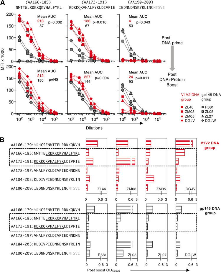
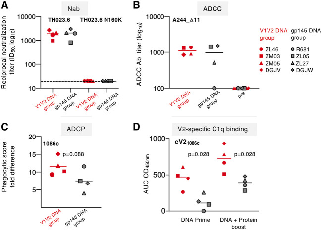
The RV144 vaccine trial revealed a correlation between reduced risk of HIV infection and the level of nonneutralizing-antibody (Ab) responses targeting specific epitopes in the second variable domain (V2) of the HIV gp120 envelope (Env) protein, suggesting this region as a target for vaccine development. To favor induction of V2-specific Abs, we developed a vaccine regimen that included priming with DNA expressing an HIV V1V2 trimeric scaffold immunogen followed by booster immunizations with a combination of DNA and protein in rhesus macaques. Priming vaccination with DNA expressing the HIV recombinant subtype CRF01_AE V1V2 scaffold induced higher and broader V2-specific Ab responses than vaccination with DNA expressing CRF01_AE gp145 Env. Abs recognizing the V2 peptide that was reported as a critical target in RV144 developed only after the priming immunization with V1V2 DNA. The V2-specific Abs showed several nonneutralizing Fc-mediated functions, including ADCP and C1q binding. Importantly, robust V2-specific Abs were maintained upon boosting with gp145 DNA and gp120 protein coimmunization. In conclusion, priming with DNA expressing the trimeric V1V2 scaffold alters the hierarchy of humoral immune responses to V2 region epitopes, providing a method for more efficient induction and maintenance of V2-specific Env Abs associated with reduced risk of HIV infection.IMPORTANCE The aim of this work was to design and test a vaccine regimen focusing the immune response on targets associated with infection prevention. We demonstrated that priming with a DNA vaccine expressing only the HIV Env V1V2 region induces Ab responses targeting the critical region in V2 associated with protection. This work shows that V1V2 scaffold DNA priming immunization provides a method to focus immune responses to the desired target region, in the absence of immune interference by other epitopes. This induced immune responses with improved recognition of epitopes important for protective immunity, namely, V2-specific humoral immune responses inversely correlating with HIV risk of infection in the RV144 trial.
Keywords: ADCC; ADCP; C1q; DNA vaccine; Env; HIV; NAb; V1V2; antibody; cyclic V2; gp145; linear peptide; prime-boost; rhesus macaque.
Copyright © 2020 Devasundaram et al.
Figures
Similar articles
-
A Trimeric HIV-1 Envelope gp120 Immunogen Induces Potent and Broad Anti-V1V2 Loop Antibodies against HIV-1 in Rabbits and Rhesus Macaques.J Virol. 2018 Feb 12;92(5):e01796-17. doi: 10.1128/JVI.01796-17. Print 2018 Mar 1. J Virol. 2018. PMID: 29237847 Free PMC article.
-
Rationally Designed Vaccines Targeting the V2 Region of HIV-1 gp120 Induce a Focused, Cross-Clade-Reactive, Biologically Functional Antibody Response.J Virol. 2016 Nov 28;90(24):10993-11006. doi: 10.1128/JVI.01403-16. Print 2016 Dec 15. J Virol. 2016. PMID: 27630234 Free PMC article.
-
HIV-1 gp120 and Modified Vaccinia Virus Ankara (MVA) gp140 Boost Immunogens Increase Immunogenicity of a DNA/MVA HIV-1 Vaccine.J Virol. 2017 Nov 30;91(24):e01077-17. doi: 10.1128/JVI.01077-17. Print 2017 Dec 15. J Virol. 2017. PMID: 29021394 Free PMC article.
-
The HIV-1 gp120 V1V2 loop: structure, function and importance for vaccine development.Expert Rev Vaccines. 2014 Dec;13(12):1489-500. doi: 10.1586/14760584.2014.951335. Epub 2014 Aug 28. Expert Rev Vaccines. 2014. PMID: 25163695 Review.
-
Vaccine-induced V1V2-specific antibodies control and or protect against infection with HIV, SIV and SHIV.Curr Opin HIV AIDS. 2019 Jul;14(4):309-317. doi: 10.1097/COH.0000000000000551. Curr Opin HIV AIDS. 2019. PMID: 30994501 Free PMC article. Review.
Cited by
-
Vaccination with immune complexes modulates the elicitation of functional antibodies against HIV-1.Front Immunol. 2023 Oct 3;14:1271686. doi: 10.3389/fimmu.2023.1271686. eCollection 2023. Front Immunol. 2023. PMID: 37854587 Free PMC article.
-
Differential V2-directed antibody responses in non-human primates infected with SHIVs or immunized with diverse HIV vaccines.Nat Commun. 2022 Feb 16;13(1):903. doi: 10.1038/s41467-022-28450-1. Nat Commun. 2022. PMID: 35173151 Free PMC article.
-
A Pentavalent HIV-1 Subtype C Vaccine Containing Computationally Selected gp120 Strains Improves the Breadth of V1V2 Region Responses.Vaccines (Basel). 2025 Jan 28;13(2):133. doi: 10.3390/vaccines13020133. Vaccines (Basel). 2025. PMID: 40006680 Free PMC article.
-
Control of SARS-CoV-2 infection after Spike DNA or Spike DNA+Protein co-immunization in rhesus macaques.PLoS Pathog. 2021 Sep 22;17(9):e1009701. doi: 10.1371/journal.ppat.1009701. eCollection 2021 Sep. PLoS Pathog. 2021. PMID: 34551020 Free PMC article.
-
Identification and validation of ferroptosis-related genes in patients infected with dengue virus: implication in the pathogenesis of DENV.Virus Genes. 2023 Jun;59(3):377-390. doi: 10.1007/s11262-023-01985-1. Epub 2023 Mar 27. Virus Genes. 2023. PMID: 36973608 Free PMC article.
References
-
- Rerks-Ngarm S, Pitisuttithum P, Nitayaphan S, Kaewkungwal J, Chiu J, Paris R, Premsri N, Namwat C, de Souza M, Adams E, Benenson M, Gurunathan S, Tartaglia J, McNeil JG, Francis DP, Stablein D, Birx DL, Chunsuttiwat S, Khamboonruang C, Thongcharoen P, Robb ML, Michael NL, Kunasol P, Kim JH, MOPH-TAVEG Investigators. 2009. Vaccination with ALVAC and AIDSVAX to prevent HIV-1 infection in Thailand. N Engl J Med 361:2209–2220. doi:10.1056/NEJMoa0908492. - DOI - PubMed
-
- Haynes BF, Gilbert PB, McElrath MJ, Zolla-Pazner S, Tomaras GD, Alam SM, Evans DT, Montefiori DC, Karnasuta C, Sutthent R, Liao HX, DeVico AL, Lewis GK, Williams C, Pinter A, Fong Y, Janes H, DeCamp A, Huang Y, Rao M, Billings E, Karasavvas N, Robb ML, Ngauy V, de Souza MS, Paris R, Ferrari G, Bailer RT, Soderberg KA, Andrews C, Berman PW, Frahm N, De Rosa SC, Alpert MD, Yates NL, Shen X, Koup RA, Pitisuttithum P, Kaewkungwal J, Nitayaphan S, Rerks-Ngarm S, Michael NL, Kim JH. 2012. Immune-correlates analysis of an HIV-1 vaccine efficacy trial. N Engl J Med 366:1275–1286. doi:10.1056/NEJMoa1113425. - DOI - PMC - PubMed
-
- Rolland M, Edlefsen PT, Larsen BB, Tovanabutra S, Sanders-Buell E, Hertz T, deCamp AC, Carrico C, Menis S, Magaret CA, Ahmed H, Juraska M, Chen L, Konopa P, Nariya S, Stoddard JN, Wong K, Zhao H, Deng W, Maust BS, Bose M, Howell S, Bates A, Lazzaro M, O'Sullivan A, Lei E, Bradfield A, Ibitamuno G, Assawadarachai V, O'Connell RJ, deSouza MS, Nitayaphan S, Rerks-Ngarm S, Robb ML, McLellan JS, Georgiev I, Kwong PD, Carlson JM, Michael NL, Schief WR, Gilbert PB, Mullins JI, Kim JH. 2012. Increased HIV-1 vaccine efficacy against viruses with genetic signatures in Env V2. Nature 490:417–420. doi:10.1038/nature11519. - DOI - PMC - PubMed
-
- Karasavvas N, Billings E, Rao M, Williams C, Zolla-Pazner S, Bailer RT, Koup RA, Madnote S, Arworn D, Shen X, Tomaras GD, Currier JR, Jiang M, Magaret C, Andrews C, Gottardo R, Gilbert P, Cardozo TJ, Rerks-Ngarm S, Nitayaphan S, Pitisuttithum P, Kaewkungwal J, Paris R, Greene K, Gao H, Gurunathan S, Tartaglia J, Sinangil F, Korber BT, Montefiori DC, Mascola JR, Robb ML, Haynes BF, Ngauy V, Michael NL, Kim JH, de Souza MS, MOPH TAVEG Collaboration. 2012. The Thai Phase III HIV Type 1 Vaccine trial (RV144) regimen induces antibodies that target conserved regions within the V2 loop of gp120. AIDS Res Hum Retroviruses 28:1444–1457. doi:10.1089/aid.2012.0103. - DOI - PMC - PubMed
Publication types
MeSH terms
Substances
Grants and funding
LinkOut - more resources
Full Text Sources
Medical
