

Reverse Genetics Approach for Developing Rotavirus Vaccine Candidates Carrying VP4 and VP7 Genes Cloned from Clinical Isolates of Human Rotavirus
- PMID: 33087468
- PMCID: PMC7944460
- DOI: 10.1128/JVI.01374-20
Reverse Genetics Approach for Developing Rotavirus Vaccine Candidates Carrying VP4 and VP7 Genes Cloned from Clinical Isolates of Human Rotavirus
Abstract
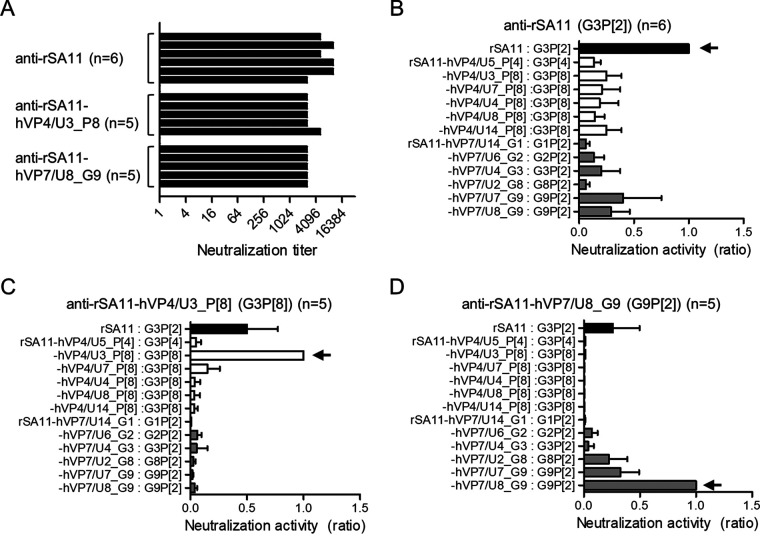
Species A rotaviruses (RVs) are a leading cause of severe acute gastroenteritis in infants and children younger than 5 years. Currently available RV vaccines were adapted from wild-type RV strains by serial passage of cultured cells or by reassortment between human and animal RV strains. These traditional methods require large-scale screening and genotyping to obtain vaccine candidates. Reverse genetics is a tractable, rapid, and reproducible approach to generating recombinant RV vaccine candidates carrying any VP4 and VP7 genes that provide selected antigenicity. Here, we developed a vaccine platform by generating recombinant RVs carrying VP4 (P[4] and P[8]), VP7 (G1, G2, G3, G8, and G9), and/or VP6 genes cloned from human RV clinical samples using the simian RV SA11 strain (G3P[2]) as a backbone. Neutralization assays using monoclonal antibodies and murine antisera revealed that recombinant VP4 and VP7 monoreassortant viruses exhibited altered antigenicity. However, replication of VP4 monoreassortant viruses was severely impaired. Generation of recombinant RVs harboring a chimeric VP4 protein for SA11 and human RV gene components revealed that the VP8* fragment was responsible for efficient infectivity of recombinant RVs. Although this system must be improved because the yield of vaccine viruses directly affects vaccine manufacturing costs, reverse genetics requires less time than traditional methods and enables rapid production of safe and effective vaccine candidates.IMPORTANCE Although vaccines have reduced global RV-associated hospitalization and mortality over the past decade, the multisegmented genome of RVs allows reassortment of VP4 and VP7 genes from different RV species and strains. The evolutionary dynamics of novel RV genotypes and their constellations have led to great genomic and antigenic diversity. The reverse genetics system is a powerful tool for manipulating RV genes, thereby controlling viral antigenicity, growth capacity, and pathogenicity. Here, we generated recombinant simian RVs (strain SA11) carrying heterologous VP4 and VP7 genes cloned from clinical isolates and showed that VP4- or VP7-substituted chimeric viruses can be used for antigenic characterization of RV outer capsid proteins and as improved seed viruses for vaccine production.
Keywords: reverse genetics; rotavirus; vaccine.
Copyright © 2020 American Society for Microbiology.
Figures

Similar articles
-
A rotavirus VP4 or VP7 monoreassortant panel identifies genotypes that are less susceptible to neutralization by systemic antibodies induced by vaccination or natural infection.mBio. 2025 Jul 9;16(7):e0089725. doi: 10.1128/mbio.00897-25. Epub 2025 May 30. mBio. 2025. PMID: 40444468 Free PMC article.
-
Establishment of a Reverse Genetics System for Rotavirus Vaccine Strain LLR and Developing Vaccine Candidates Carrying VP7 Gene Cloned From Human Strains Circulating in China.J Med Virol. 2024 Dec;96(12):e70065. doi: 10.1002/jmv.70065. J Med Virol. 2024. PMID: 39610277
-
Vaccine-derived NSP2 segment in rotaviruses from vaccinated children with gastroenteritis in Nicaragua.Infect Genet Evol. 2012 Aug;12(6):1282-94. doi: 10.1016/j.meegid.2012.03.007. Epub 2012 Apr 2. Infect Genet Evol. 2012. PMID: 22487061 Free PMC article.
-
Genetic diversity of G1P[8] rotavirus VP7 and VP8* antigens in Finland over a 20-year period: No evidence for selection pressure by universal mass vaccination with RotaTeq® vaccine.Infect Genet Evol. 2013 Oct;19:51-8. doi: 10.1016/j.meegid.2013.06.026. Epub 2013 Jul 4. Infect Genet Evol. 2013. PMID: 23831933 Review.
-
Circulating rotavirus strains in children with acute gastroenteritis in Iran, 1986 to 2023 and their genetic/antigenic divergence compared to approved vaccines strains (Rotarix, RotaTeq, ROTAVAC, ROTASIIL) before mass vaccination: Clues for vaccination policy makers.Virus Res. 2024 Aug;346:199411. doi: 10.1016/j.virusres.2024.199411. Epub 2024 Jun 3. Virus Res. 2024. PMID: 38823689 Free PMC article. Review.
Cited by
-
Rotavirus epidemiology adjusted pattern in a tropical setting: mathematical correction for false positive problem relating to primary immunochromatography test surveillance.Int J Biochem Mol Biol. 2022 Dec 15;13(6):54-59. eCollection 2022. Int J Biochem Mol Biol. 2022. PMID: 36721841 Free PMC article.
-
Rotavirus Reverse Genetics Systems and Oral Vaccine Delivery Vectors for Mucosal Vaccination.Microorganisms. 2025 Jul 4;13(7):1579. doi: 10.3390/microorganisms13071579. Microorganisms. 2025. PMID: 40732089 Free PMC article. Review.
-
Characterization of viroplasm-like structures by co-expression of NSP5 and NSP2 across rotavirus species A to J.J Virol. 2024 Sep 17;98(9):e0097524. doi: 10.1128/jvi.00975-24. Epub 2024 Aug 28. J Virol. 2024. PMID: 39194242 Free PMC article.
-
Development of viral infectious clones and their applications based on yeast and bacterial artificial chromosome platforms.Mol Biomed. 2025 Apr 29;6(1):26. doi: 10.1186/s43556-025-00266-7. Mol Biomed. 2025. PMID: 40295404 Free PMC article. Review.
-
The Role of the VP4 Attachment Protein in Rotavirus Host Range Restriction in an In Vivo Suckling Mouse Model.J Virol. 2022 Aug 10;96(15):e0055022. doi: 10.1128/jvi.00550-22. Epub 2022 Jul 12. J Virol. 2022. PMID: 35862708 Free PMC article.
References
-
- Gentsch JR, Laird AR, Bielfelt B, Griffin DD, Banyai K, Ramachandran M, Jain V, Cunliffe NA, Nakagomi O, Kirkwood CD, Fischer TK, Parashar UD, Bresee JS, Jiang B, Glass RI. 2005. Serotype diversity and reassortment between human and animal rotavirus strains: implications for rotavirus vaccine programs. J Infect Dis 192(Suppl 1):S146–S159. doi:10.1086/431499. - DOI - PubMed
-
- Banyai K, Laszlo B, Duque J, Steele AD, Nelson EA, Gentsch JR, Parashar UD. 2012. Systematic review of regional and temporal trends in global rotavirus strain diversity in the pre rotavirus vaccine era: insights for understanding the impact of rotavirus vaccination programs. Vaccine 30(Suppl 1):A122–A130. doi:10.1016/j.vaccine.2011.09.111. - DOI - PubMed
Publication types
MeSH terms
Substances
LinkOut - more resources
Full Text Sources
Miscellaneous
