. 2020 Oct 21;12(20):20152-20162.
doi: 10.18632/aging.103729. Epub 2020 Oct 21.

Polymerase I and transcript release factor transgenic mice show impaired function of hematopoietic stem cells

Affiliations

Polymerase I and transcript release factor transgenic mice show impaired function of hematopoietic stem cells

Lin Bai et al. Aging (Albany NY). .

Abstract

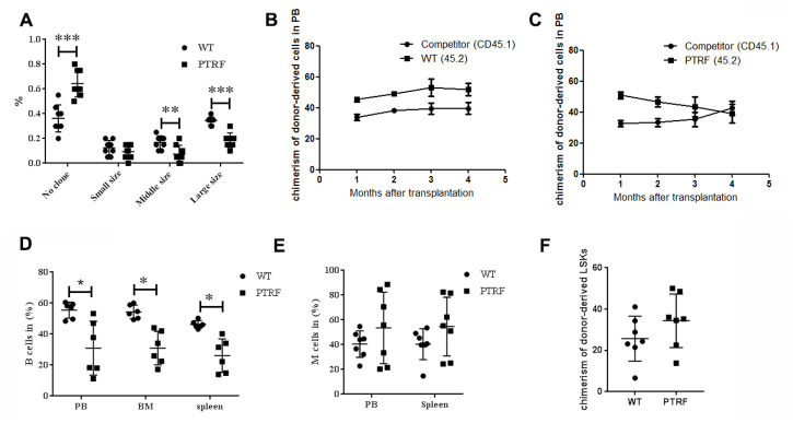
The age-dependent decline in stem cell function plays a critical role in aging, although the molecular mechanisms remain unclear. PTRF/Cavin-1 is an essential component in the biogenesis and function of caveolae, which regulates cell proliferation, endocytosis, signal transduction and senescence. This study aimed to analyze the role of PTRF in hematopoietic stem cells (HSCs) senescence using PTRF transgenic mice. Flow cytometry was used to detect the frequency of immune cells and hematopoietic stem/progenitor cells (HSCs and HPCs). The results showed than the HSC compartment was significantly expanded in the bone marrow of PTRF transgenic mice compared to age-matched wild-type (WT) mice, and exhibited the senescent phenotype characterized by G1 cell cycle arrest, increased SA-β-Gal activity and high levels of reactive oxygen species (ROS). The PTRF-overexpressing HSCs also showed significantly lower self-renewal and ability to reconstitute hematopoiesis in vitro and in vivo. Real-time PCR was performed to analyze the expression levels of senescence-related genes. PTRF induced HSCs senescence via the ROS-p38-p16 and caveolin-1-p53-p21 pathways. Furthermore, the PTRF+cav-1-/- mice showed similar HSCs function as WT mice, indicating that PTRF induces senescence in HSCs partly through caveolin-1. Thus PTRF impaired HSCs aging partly via caveolin-1.

Keywords: HSCs; PTRF; caveolin-1.


Conflict of interest statement

CONFLICTS OF INTEREST: The authors declare that they have no conflicts of interest.

Figures

Figure 1
Figure 1
PTRF expression increased during aging and skewed differentiation potential of HSCs. (A) PTRF mRNA levels in the bone marrow of young (2-mo) and old mice (20-mo), n=3. (B) Immunoblot showing PTRF protein expression in the bone marrow (BM) of young and old mice, n=3. (C) PTRF mRNA levels in the BM of 2-month old PTRF transgenic and wild-type (WT) mice, n=3. (D) Immunoblot showing PTRF protein expression in the BM of young PTRF and WT mice, n=3. (EG) The percentage of B cells (E), M cells (F) and T cells (G) in the peripheral blood, BM and spleen of young PTRF transgenic mice. Data represent the mean ± SD of three independent experiments. *, P < 0.05; **, P < 0.01. Data were normalized against GAPDH expression.
Figure 2
Figure 2
PTRF overexpression alters the hematopoietic stem/progenitor cell compartments in mice. (A) Representative flow cytometry plots showing HSCs and progenitor populations in the BM. (BI) The percentage of LSKs, LT-HSCs, ST-HSCs, MPP, CLP, CMPs, GMPs and MEPs cells in the BM from 2, 6 and 12-month old mice. n=5 mice per group. Data represent the mean ± SD of three independent experiments. *, P < 0.05.
Figure 3
Figure 3
PTRF impairs HSCs function. (A) Frequency of colonies generated by LT-HSCs from PTRF and WT mice (n=3). (B) The percentage of CD45.2+ cells in the PB of irradiated recipient mice 4-, 8-, 12- and 16 weeks after transplantation with WT HSCs. (C) The percentage of CD45.2+ cells in the PB of irradiated recipient mice 4-, 8-, 12- and 16 weeks after transplantation with PTRF-overexpressing HSCs. (D, E) The percentage of donor-derived B (D) and M cells (E) from PTRF or WT mice in the BM of recipient mice at 16 weeks post-transplantation. (F) The percentage of donor-derived LSKs in the BM of recipient mice at 16 weeks post-transplantation. n=9 mice per group. Data represent the mean ± SD. *, P < 0.05.
Figure 4
Figure 4
PTRF overexpression induced cell cycle arrest at G1 phase and accelerated cellular senescence. (A) Flow cytometry plots showing cell cycle distribution of LSKs from PTRF and WT mice. (B) The percentage of cells in each phase of the cell cycle. (C) Flow cytometry plots showing viable and apoptotic LSKs from PTRF and WT mice. (D) The percentage of apoptotic LSK cells. (E) Flow cytometry plots showing C12FDG levels in the LSKs from PTRF and WT mice. (F) The percentage of SA-β-gal-positive LSK cells. n=4 or 5 mice per group. (G) Flow cytometry plots showing ROS producing cells stained with the DCFH probe. (H) The percentage of ROS-positive LSK cells. Data represent the mean ± SD. *, P < 0.05; **, P < 0.01.
Figure 5
Figure 5
PTRF altered the expression pattern of senescence-associated genes in LSKs. (AC) RT-PCR analysis of the indicated genes in sorted LSK cells from PTRF and WT mice. Data represent the mean ± SD of three independent experiments. *, P < 0.05; **, P < 0.01. Data were normalized against GAPDH expression.
Figure 6
Figure 6
PTRF impaired HSCs function partly through Cav-1. (A) Number of colonies generated by LT-HSCs from PTRF+Cav-1-/- and WT mice (n=3). (B) The percentage of donor-derived cells in the PB of PTRF+Cav-1-/- and WT mice at 4, 8, and 12 weeks post-transplantation. (C, D) The percentage of donor-derived B (C) and M cells (D) from PTRF+Cav-1-/- and WT mice in the PB at 4, 8, and 12 weeks post-transplantation. n=9 mice per group. Data represent the mean ± SD.

Similar articles

Cited by

References

    1. Sperka T, Wang J, Rudolph KL. DNA damage checkpoints in stem cells, ageing and cancer. Nat Rev Mol Cell Biol. 2012; 13:579–90. 10.1038/nrm3420 - DOI - PubMed
    1. Florian MC, Nattamai KJ, Dörr K, Marka G, Uberle B, Vas V, Eckl C, Andrä I, Schiemann M, Oostendorp RA, Scharffetter-Kochanek K, Kestler HA, Zheng Y, Geiger H. A canonical to non-canonical Wnt signalling switch in haematopoietic stem-cell ageing. Nature. 2013; 503:392–96. 10.1038/nature12631 - DOI - PMC - PubMed
    1. Pang WW, Price EA, Sahoo D, Beerman I, Maloney WJ, Rossi DJ, Schrier SL, Weissman IL. Human bone marrow hematopoietic stem cells are increased in frequency and myeloid-biased with age. Proc Natl Acad Sci USA. 2011; 108:20012–17. 10.1073/pnas.1116110108 - DOI - PMC - PubMed
    1. Sudo K, Ema H, Morita Y, Nakauchi H. Age-associated characteristics of murine hematopoietic stem cells. J Exp Med. 2000; 192:1273–80. 10.1084/jem.192.9.1273 - DOI - PMC - PubMed
    1. Schwamborn JC, Müller M, Becker AH, Püschel AW. Retraction: ‘Ubiquitination of the GTPase Rap1B by the ubiquitin ligase Smurf2 is required for the establishment of neuronal polarity’. EMBO J. 2014; 33:3012. 10.15252/embj.201470130 - DOI - PMC - PubMed

Publication types

MeSH terms