. 2020 Nov 15;60(11):553-562.
doi: 10.2176/nmc.oa.2020-0060. Epub 2020 Oct 21.

Impact of Intraoperative 3-Tesla MRI on Endonasal Endoscopic Pituitary Adenoma Resection and a Proposed New Scoring System for Predicting the Utility of Intraoperative MRI

Affiliations

Impact of Intraoperative 3-Tesla MRI on Endonasal Endoscopic Pituitary Adenoma Resection and a Proposed New Scoring System for Predicting the Utility of Intraoperative MRI

Masahiro Tanji et al. Neurol Med Chir (Tokyo). .

Abstract

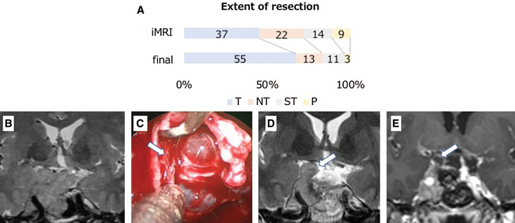
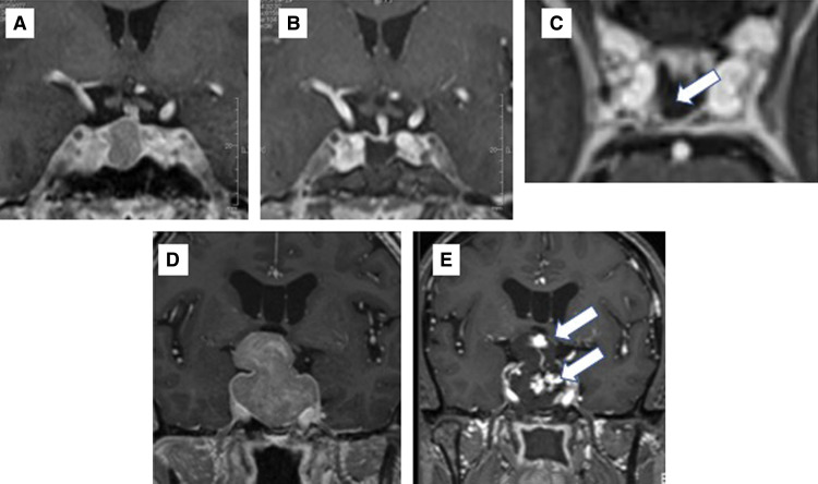
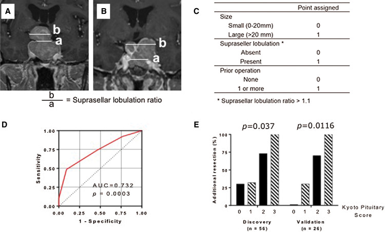
The aim of this study was to evaluate the impact of 3-Tesla intraoperative high-field magnetic resonance imaging (3T-iMRI) for pituitary adenoma resection, and to propose a new scoring system for predicting the utility of 3T-iMRI. This retrospective study evaluated 82 patients with pituitary adenoma who underwent purely endoscopic endonasal resection with 3T-iMRI between 2015 and 2019. 3T-iMRI revealed unexpected residual tumor in 39 cases (47.6%), which led to further resection and contributed to upgrading of the resection level in 28 cases (34.1%), which led to gross total resection rates (GTRs) of 67.1% and near total resection of 15.9%. To construct a new scoring system, patients were divided into a discovery cohort (56 patients) and a validation cohort (26 patients). Three variables for the scoring system were selected according to a univariate analysis of the discovery cohort: the size of the tumor (>20 mm: 1 point), the presence of suprasellar tumor lobulation (1 point) and the history of previous operations (1 point). The risk of additional resection after iMRI was well stratified by this scoring system (range 0-3; p = 0.0037 for trend). Robustness of the system was confirmed in the validation cohort (0 points, 0%; 1 point, 30.8%; 2 points, 70.0%; 3 points, 100%; p = 0.0116 for trend). These results indicate that 3T-iMRI optimized the extent of resection, even with the use of an endoscope, and that the proposed scoring system is useful for predicting whether 3T-iMRI is likely to be of value for a particular patient.

Keywords: Knosp grade; intraoperative MRI; pituitary score; pituitary surgery; transsphenoidal surgery.


Conflict of interest statement

Conflicts of Interest Disclosure

All authors declare that they have no conflict of interest.

Figures

Fig. 1
Fig. 1
(A) The extent of pituitary adenoma resection following intraoperative MRI and final MRI (n = 82). T = total resection, 100%; NT = near total resection, 95% or greater; ST = subtotal resection, 80%–95%; P = partial resection of less than 80%. (B–E) Illustrative cases of iMRI images for multi-lobular recurrent adenoma. Recurrent non-functioning lobulated pituitary adenoma with planned subtotal resection (B) was removed by an endonasal endoscopic approach. After the initial resection, the descent of arachnoid was confirmed but the fibrous septum created by the previous operation was found on the right side (arrow) (C). 3T-iMRI showed residual tumor in the cavernous sinus and subdural space under the septum (arrow) (D) requiring second-look surgery. The second iMRI showed additional removal of the tumor (E). 3T-iMRI: 3-Tesla intraoperative high-field magnetic resonance imaging.
Fig. 2
Fig. 2
(A–C) Illustrative cases of iMRI images for small residual tumor. A GHoma (A) appeared to be successfully removed following iMRI (B), but the axial image revealed a residual mass behind the ICA (arrow) (C). The neuronavigation images were updated and the residual tumor was precisely located, with total removal achieved. (D–E) Illustrative cases of early hematoma detection. Preoperative MRI of a lobulated tumor is shown in (D). Intraoperative MRI showed contrast agent extravasation in the removal cavity, indicating hematoma formation (E). ICA: internal carotid artery.
Fig. 3
Fig. 3
(A) Measurement of suprasellar lobulation. Suprasellar lobulation ratio was defined as a ratio between the maximal suprasellar horizontal diameter and the diaphragm. The maximal horizontal diameter of the tumor in the suprasellar area was measured with coronal preoperative contrast-enhanced T1-weighted imaging (b). The diameter of the diaphragm defect was also measured on coronal slices. When it was not apparent, the measurement was performed at the level of the most proximal C2 portion of the carotid on contrast-enhanced T1-weighted images (a). When there was no suprasellar extension, the ratio was defined as 1. (B) Measurement of multi-lobular adenoma. For multi-lobular tumor, the maximal suprasellar horizontal diameter was defined as the maximal distance between the farthest lobulation. (C) Kyoto Pituitary Score. The new scoring system (range: 0–4) is the summation of three variables: the maximum diameter of the tumor (>20 mm: 1 point, ≤20 mm: 0 points), suprasellar tumor lobulation (suprasellar lobulation ratio >1.1: 1 point, ≤1.1: 0 points) and the presence of history of prior surgeries (1 point). (D) ROC analysis for the Kyoto Pituitary Score. The AUROC was 0.732 (p = 0.0003). (E) Rates of additional resection following 3T-iMRI stratified by the Kyoto Pituitary score in the discovery cohort (0 points, 30.8%; 1 point, 32.0%; 2 points, 73.3%; 3 points, 100%; p = 0.0037 for trend) and validation cohort (0 points, 0%;1 point, 30.8%; 2 points, 70.0%; 3 points,100%; p = 0.0116 for trend). AUROC: area under the ROC curve, ROC: receiver operating characteristic, 3T-iMRI: 3-Tesla intraoperative high-field magnetic resonance imaging.

Similar articles

Cited by

References

    1. Kassam AB, Prevedello DM, Carrau RL, et al. : Endoscopic endonasal skull base surgery: analysis of complications in the authors’ initial 800 patients. J Neurosurgery 114: 1544–1568, 2011 - PubMed
    1. de Divitiis E, Laws ER, Giani U, Iuliano SL, de Divitiis O, Apuzzo ML: The current status of endoscopy in transsphenoidal surgery: an international survey. World Neurosurg 83: 447–454, 2015 - PubMed
    1. Soneru CP, Riley CA, Hoffman K, Tabaee A, Schwartz TH: Intra-operative MRI vs endoscopy in achieving gross total resection of pituitary adenomas: a systematic review. Acta Neurochir (Wien) 161: 1683–1698, 2019 - PubMed
    1. Fahlbusch R, Golby A, Prada F, Zada G: Utility of intraoperative imaging. Neurosurg Focus 40: 1610, 2016 - PubMed
    1. Zaidi HA, De Los Reyes K, Barkhoudarian G, et al. : The utility of high-resolution intraoperative MRI in endoscopic transsphenoidal surgery for pituitary macroadenomas: early experience in the advanced multimodality image guided operating suite. Neurosurg Focus 40: E18, 2016 - PMC - PubMed